Media matters! Alterations in the loading and release of Histoplasma capsulatum extracellular vesicles in response to different nutritional milieus
Funding information: Conselho Nacional de Desenvolvimento Científico e Tecnológico, Grant/Award Numbers: 301304/2017-3, 311179/2017-7, 405520/2018-2, 408711/2017-7, 440015/2018-9; Foundation for the National Institutes of Health, Grant/Award Numbers: R01AI052733, R21AI124797; Fundação Oswaldo Cruz, Grant/Award Numbers: VPPCB-007-FIO-18-2-57, VPPIS-001-FIO-18-66
Abstract
Histoplasma capsulatum is a dimorphic fungus that most frequently causes pneumonia, but can also disseminate and proliferate in diverse tissues. Histoplasma capsulatum has a complex secretion system that mediates the release of macromolecule-degrading enzymes and virulence factors. The formation and release of extracellular vesicles (EVs) are an important mechanism for non-conventional secretion in both ascomycetes and basidiomycetes. Histoplasma capsulatum EVs contain diverse proteins associated with virulence and are immunologically active. Despite the growing knowledge of EVs from H. capsulatum and other pathogenic fungi, the extent that changes in the environment impact the sorting of organic molecules in EVs has not been investigated. In this study, we cultivated H. capsulatum with distinct culture media to investigate the potential plasticity in EV loading in response to differences in nutrition. Our findings reveal that nutrition plays an important role in EV loading and formation, which may translate into differences in biological activities of these fungi in various fluids and tissues.
1 INTRODUCTION
Extracellular vesicles (EVs) have been identified in all biological kingdoms. Fungal EVs were first described in Cryptococcus neoformans, in 2007, as a trans-cell wall transport mechanism that effectively delivers macromolecules to the extracellular environment (Rodrigues et al., 2007). The following year, this process was extended to several additional fungi, which demonstrates that this remarkable process is ancient, given that both Basidiomycota and Ascomycota generated EVs (Albuquerque et al., 2008). Furthermore, research on fungal EVs reveals that their contents contain diverse components associated with fungal virulence, and are recognised by individuals previously infected with the specific fungus (Albuquerque et al., 2008; Rodrigues et al., 2007). Since then, EVs have been shown to modify host effector responses, including altering macrophage activation, macrophage polarisation and host susceptibility to fungal challenge (Zamith-Miranda, Nimrichter, Rodrigues, & Nosanchuk, 2018). Histoplasmosis, caused by the dimorphic fungus Histoplasma capsulatum, is a significant cause of mortality and morbidity on the global stage, and it is the most common cause of fungal pneumonia in the United States (Chu, Feudtner, Heydon, Walsh, & Zaoutis, 2006; Cleare, Zamith-Miranda, & Nosanchuk, 2017). Thermally dimorphic fungi are remarkable among fungal pathogens because they cause disease in humans with normal or impaired immune defences (Gauthier, 2017). Histoplasma capsulatum persists in soil as well as bird and bat excretes (Prevention CfDCa, 2014). Disruption of soil by natural events or by human activities, such as construction, leads to aerosolisation of conidia and hyphal fragments. When inhaled into the lungs of a mammalian host (37°C), these infectious propagules convert into pathogenic yeast to cause pneumonia. Once infection is established in the lungs, the yeast can widely disseminate and produce EVs into the host environment (Baltazar et al., 2018). We have previously demonstrated that H. capsulatum produces EVs containing a vast array of proteins, lipids and carbohydrates (Albuquerque et al., 2008). Remarkably, the packaging and release of H. capsulatum EVs can be dynamically regulated by the binding of antibody to the cell surface of this yeast (Baltazar et al., 2018; Matos Baltazar et al., 2016) which suggests that other stressors may dramatically impact EV biogenesis and, consequently, composition.
Histoplasma capsulatum is commonly cultivated in several different rich or nutritionally restrictive media. However, during infection, the fungus can reside and flourish in fluid and tissue environments throughout the body, ranging from the blood and cerebral spinal fluid to the lung and brain, and variables such as the pH, oxygen content and available nutrients are different in these diverse locations. In this study, we examined the impact of three standard growth media on EVs content loading, which we hypothesized would alter EVs characteristics and payloads. Given that there are significantly different nutritional conditions that H. capsulatum encounters during disseminated disease, exploring variations in EV production in different nutritional environments may lead to insights into EV plasticity and pathogenesis.
2 RESULTS
2.1 Protein and ergosterol content and size of EV
EVs isolated from H. capsulatum cultivated in BHI, Ham's F-12, and RPMI are referred hereafter as EV-BHI, EV-Ham's and EV-RPMI, respectively. EV-Ham's presented the highest yield of both total protein and ergosterol. EV-BHI and EV-RPMI had comparable amounts of protein and ergosterol, which were less than that observed for EV-Ham's (Figure 1a, b).
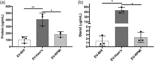
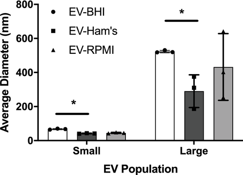
Figure 2 presents the size of the EVs isolated from each medium. DLS analysis of fungal EVs reveals two population sizes of EVs in the three conditions, which is similar to prior reports for H. capsulatum and other fungi (Joffe, Nimrichter, Rodrigues, & Del Poeta, 2016). The small EVs population range from 40 to 70 nm, and the larger from 200 to 500 nm. There was a difference in population sizes of the small and large EVs between EV-BHI and EV-Ham's, otherwise all EVs are within range of previously described fungal EVs.
2.2 Proteomic analysis of EVs
Fungal EVs contain diverse proteins in their cargo, including molecules that act as virulence factors (Rodrigues, Godinho, Zamith-Miranda, & Nimrichter, 2015). Hence, we analysed the protein content of EVs released from H. capsulatum cultured in distinct media conditions to determine if differences in nutrition altered the presence of potentially biologically active proteins. The proteomic analysis identified approximately 2,000 proteins, and 270 were significantly altered among the distinct growth conditions. Some of these proteins were found to be involved in key pathways, particularly cellular product synthesis and metabolism. Table 1 shows proteins included in important KEGG pathways, classified by fold enrichment, some of which we will refer to later in the text.
| Map description | Fold enrichment | Fisher test |
|---|---|---|
| Other types of O-glycan biosynthesis | 25.59 | 9.7E-05 |
| Fatty acid biosynthesis | 10.24 | 2.4E-03 |
| Fatty acid degradation | 10.04 | 9.1E-05 |
| Pyruvate metabolism | 9.75 | 8.6E-07 |
| Arginine biosynthesis | 9.48 | 1.2E-04 |
| Citrate cycle (TCA cycle) | 7.58 | 9.6E-05 |
| Alanine, aspartate and glutamate metabolism | 7.06 | 1.5E-04 |
| Oxidative phosphorylation | 6.52 | 5.3E-10 |
| Amino sugar and nucleotide sugar metabolism | 6.46 | 7.4E-05 |
| Fatty acid metabolism | 6.32 | 8.8E-04 |
| 2-Oxocarboxylic acid metabolism | 5.17 | 2.2E-03 |
| Propanoate metabolism | 5.12 | 1.7E-02 |
| Glyoxylate and dicarboxylate metabolism | 5.06 | 6.4E-03 |
| Ribosome | 4.88 | 7.4E-07 |
| Peroxisome | 4.88 | 4.4E-04 |
| Starch and sucrose metabolism | 4.88 | 7.3E-03 |
| Valine, leucine and isoleucine degradation | 4.71 | 8.2E-03 |
| Glycolysis/gluconeogenesis | 4.27 | 1.1E-02 |
| Phenylalanine metabolism | 4.27 | 2.7E-02 |
| Carbon metabolism | 4.22 | 2.0E-05 |
| Aminoacyl-tRNA biosynthesis | 4.16 | 5.4E-03 |
| Endocytosis | 4.14 | 5.3E-04 |
| Protein processing in endoplasmic reticulum | 4.10 | 2.6E-04 |
| Biosynthesis of antibiotics | 3.94 | 6.5E-09 |
| Biosynthesis of amino acids | 3.86 | 4.7E-05 |
| Biosynthesis of secondary metabolites | 3.73 | 1.5E-10 |
| Metabolic pathways | 3.49 | 3.9E-23 |
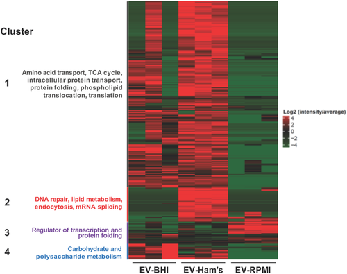
The proteins were grouped into four different clusters according to their abundance in BHI [group 4], RPMI [group 3], Ham's [group 2] and BHI and Ham's [group 1]. There were more abundant proteins in EV-Ham's, and a greater variance in EV-BHI within the replicates.
EV-BHI and EV-Ham's presented similar protein yields in group 4, which contains most of the protein yields. However, the majority of altered proteins were significantly abundant in EV-Ham's compared to EV-BHI and EV-RPMI. The most variation in protein alteration between the EV-BHI, EV-Ham's, and EV-RPMI occurred in group 1. Proteins from EV-RPMI were most abundant in group 2 (Figure 3).
The proteins abundant in both EV-Ham's and EV-BHI were in the largest cluster in the heatmap (Figure 3). In this cluster, important proteins involved with chitin metabolism were found, including chitin synthase (C0NZK3, C0NSP8 and C0NDJ0) and chitinase (C0NSB6). Also, enzymes involved with host response modulation were detected, such as peroxidase (C0NND7) and alkaline phosphatase (C0NBI7). Lipid metabolism enzymes were also abundant in these EVs, such as carnitine (C0NWZ6), long-chain-fatty-acid-CoA ligase (C0P0B7), acetyl-CoA carboxylase (C0NM24). EV-BHI presented an abundance of sugar metabolism-related enzymes, including alpha-glucosidase (C0NEI5) and glucosidase I (C0NHJ7). Interestingly, EV-RPMI did not present abundance of enzymes involved in cell wall remodelling and the only lipid-related abundant protein in EV-RPMI was the Inositol-3-phosphate synthase (C0NLK4). Among the proteins exported in EV-Ham's, the high affinity copper transporter (C0NSW0), abundant only in EV-Ham's. EV-Ham's had the highest abundance of siderochrome-iron uptake transporter (C0NX70), as well as V-type proton ATPase subunit A (C0NJV7).
2.3 Lipidomic analysis of EVs
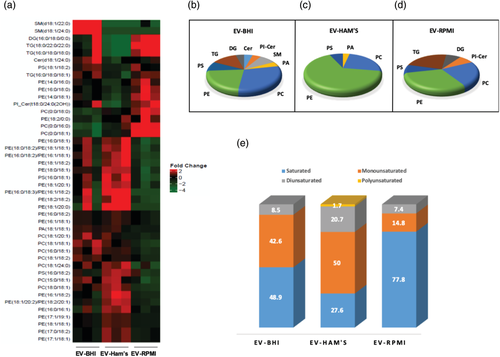
The lipidomic analysis detected 100 lipids, with significant alterations in 44 lipids among the different media conditions. Among the significantly altered lipids, the greatest variety was found in EV-Ham's, in mostly species of phosphatidylethanolamine (PE) and phosphatidylcholine (PC). Figure 4a shows a heatmap with the significantly altered lipids. The heatmap shows 2 distinct clusters of altered lipids. The biggest cluster shows lipids that were abundant in EV-Ham's, and the smaller cluster shows EV-RPMI abundant lipids. Lipids abundant in EV-BHI are present in both clusters, except for the sphingomyelins, which were abundant only in EV-BHI.
EV-BHI presented the largest variety in class of lipids among the tested conditions, including several species of sphingomyelin, one ceramide, and a phosphoinositol ceramide. Phosphoinositol ceramide was also abundant in both EV-BHI and EV-RPMI. EV-Ham's yielded significantly lower abundances of triacylglycerides (TG) and diacylglycerides (DG) compared to EV-BHI and EV-RPMI (Figure 4b, c). Most of the abundant lipids found in EV-RPMI (approximately 78%) had saturated fatty acids (FA), while approximately 70% of the FA found in lipids abundant in EV-Ham's were unsaturated. Regarding the presence of unsaturated lipids, EV-BHI's lipids were in an intermediary level between the other conditions. (Figure 4e).

Lysophospholipids were more abundant in EV-RPMI, especially lysophosphatidylcholine (LysoPC). LysoPCs were more abundant in EV-RPMI and EV-Ham's compared to EV-BHI (Figure 5). One species of lysophosphatidylethanolamine (LysoPE) was present in EV-RPMI and EV-Ham's. The few odd-chain FA found to be abundant in our analysis were present mostly in EV-Ham's, and none of them were lysophospholipids.
2.4 Metabolomic analysis of EVs
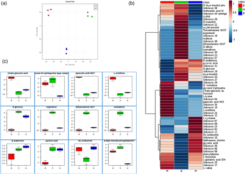
Carbohydrate metabolites vary in the EVs from each condition. EV-Ham's presents the most abundant D-glucose content (Figure 6); however, the amount of D-glucose present in the media BHI and RPMI was not translated in the abundance of this carbohydrate in EV generated in their presence. Carbohydrates such as maltose, isomaltose, D-allose, and pyruvic acid, also associated with energy storage and production, were most abundantly found in EV-Hams. Although EV-Ham's presents a majority of the carbohydrate metabolites, EV-BHI presents more abundance of trehalose and fructose. EV-BHI presents the most abundance of trehalose, and the least abundance of glucose and pyruvic acid compared to EV-Hams and EV-RPMI. EV-BHI had the highest abundance of L-ornithine even though the abundance of arginase was the same among all tested conditions. The high abundance of ethanolamine in EV-Ham's reflects the lipidomic profile of these EV, where phospholipids containing ethanolamine represent more than half of the lipid classes found in this experimental condition.
3 DISCUSSION
Compositional analyses of fungal EVs are essential to the understanding of how these extracellular compartments exert their functions. For instance, there is now concrete evidence that fungal EVs modulate virulence transfer between distinct fungal strains (Bielska et al., 2018) activation of immune cells (Bitencourt et al., 2018; Colombo et al., 2019; Peres da Silva et al., 2015; Vargas et al., 2015), prion transfer (Liu, Hossinger, Hofmann, Denner, & Vorberg, 2016), and drug resistance in biofilms (Zarnowski et al., 2018) among other functions. However, in the absolute majority of the studies showing fundamental biological functions of EVs, the bioactive vesicular components remained unknown. Therefore, knowledge on the compositional diversity of fungal EVs directly impact the understanding of their functions in nature, but also the design of EV-based biotechnological tools, including vaccines and therapeutic agents.
The initial description of EVs in C. neoformans has been followed by numerous interesting reports of EVs produced in other important human pathogenic fungi. Although EVs can also be released from non-pathogenic fungi, as well as in organisms from other kingdoms, their release by pathogenic fungi has called the attention of microbiologists mainly because these EVs can carry virulence factors (Rodrigues et al., 2015). Although many aspects about fungal EV biology and their effects on hosts have been addressed to date, it is still poorly understood whether environmental conditions modulate EV cargo. Previously we partially explored this question, comparing EVs from H. capsulatum released in the presence or absence of monoclonal antibodies against a heat-shock protein exposed on the surface of H. capsulatum and found that antibody dynamically altered EV cargo in a dose-dependent manner (Guimaraes, Frases, Gomez, Zancope-Oliveira, & Nosanchuk, 2009). In the current work we analysed the content of EV released from H. capsulatum cultivated in distinct media conditions, to evaluate EV plasticity regarding their cargo and constitution in response to nutrition.
Each of the medium tested in our study has a different chemical composition. BHI is a nutritious, buffered culture medium that contains infusions of brain and heart tissue and peptones to supply protein and other nutrients necessary to support the growth of fastidious and non-fastidious microorganisms (Rosenow, 1919). Ham's F-12 was designed for serum-free single-cell plating of Chinese Hamster Ovary (CHO) cells, and has been used for a wide range of mammalian cells as well as bacteria and fungi. Ham's F-12 Nutrient Mixture medium supplements were referenced from a defined medium called Histoplasma macrophage medium aforementioned. RPMI is unique from other medium because it contains the reducing agent glutathione and high concentrations of vitamins, inositol and choline. A full description of media components has been included in Table S1. Nutridoma, was added to RPMI as a supplement. It consists as a biochemically defined replacement for Foetal Bovine Serum (FBS). We observed the growth of H. capsulatum based on OD and were able to determine when the fungus reached stationary phase growth. This stage of growth was determined to be 96 hr after initial inoculation in each of the three media.
Proteins and lipids, among other organic molecules, are present in H. capsulatum EVs, some of which are closely linked to virulence during infection and disease. EV-Ham's had the highest amount of both protein and ergosterol. However, these EVs were the smallest in size among the tested conditions, being smaller than EV-BHI and statistically similar to EV-RPMI, suggesting that EV-Ham's are either produced in higher amounts than the other conditions, or that they are “denser” than EVs from the other media. Application of methods that could directly measure EV counts in each medium could answer this question in the future. Following the total protein content, the proteomic analysis showed that most of the altered proteins were abundantly found in EV-Ham's, as EV-RPMI had only a few proteins significantly more abundant than the other conditions. Interestingly, EV-RPMI had lower abundance of proteins associated with carbohydrate and polysaccharide metabolism in Cluster 4 (Figure 3) even though RPMI medium contains higher levels of glucose (10%) compared to Ham's F-12. EV-BHI showed some inconsistencies between its replicates, probably because of the nature of the medium, where different animals' brain and heart material are present, so variations between distinct lots might occur. It may be possible that a few of the proteins analysed were directly from the media itself. However, when we subjected the different media without fungal inoculation to our EV isolation protocol and tested residual fluid by DLS, there were no EVs detected (data not shown). In comparison, Ham's F-12 medium was the most consistent condition where the yeast cells exported, through EVs, the largest variety of proteins. This feature might reflect how suitable this medium is on the growth of H. capsulatum for experiments that involve host-pathogen interactions. Copper and some other micronutrients in the host environment can become toxic to host pathogens. Among the proteins exported in EV-Ham's, the high affinity copper transporter (C0NSW0), abundant only in EV-Ham's, is an important immune subversion protein mechanism as one of the strategies used by host phagocytes to inhibit the viability of intracellular H. capsulatum is to reduce the access of copper into the phagosome (Shen, Beucler, Ray, & Rappleye, 2018). Therefore, the presence of this metal transporter can protect the host pathogen by inhibiting the use of copper in host defence cells. The presence of this metal transporter can be due to the fact that Ham's F-12 medium contains the micronutrient metal cupric sulphate, while the other media do not. EV-Ham's had the highest abundance of siderochrome-iron uptake transporter (C0NX70), a protein essential for the fungus to survive in environments with low iron availability. Iron acquisition in H. capsulatum also relies on the cooperative action of proton ATPases as the V-type proton ATPase subunit A (C0NJV7), which is also consistently abundant in EV-Ham's.
The lipid profile of EV cultivated in distinct media was remarkably distinct. EV-BHI incorporated or synthesised different classes of lipids, as ceramides and sphingomyelins were present almost in an exclusive way in this condition. Ham's F12 and RPMI are more defined media, while BHI has brain and heart material different animals, and it is the only one of these media that actually carries lipids, with the exception of linoleic acid present in Ham's. Sphingomyelins are not synthesised by yeast cells due to the lack of sphingomyelin synthase in fungi (Christie, 2019), but they are probably up taken from media and were loaded into EVs. Nevertheless, polyunsaturated FA were consistently found only in EV-Ham's, as well as the highest abundance of diunsaturated FA. The condition with fewer unsaturated FA was also found in EV-Ham's. Almost 80% of the abundant FA in EV-RPMI were unsaturated, and accordingly the ergosterol presence in these EV was quite low. EV-Hams having fewer unsaturated FA is presumably balanced by the highest amount of ergosterol, limiting the EVs membrane fluidity. EV-RPMI also presented the highest abundance of lysophospholipids, despite the equal abundance of phospholipases in all the conditions. This data suggests that in this condition, the lysis of phospholipids would take place before the assembling of EV by intracellular phospholipases, or even in the extracellular milieu by secreted phospholipases non-associated with EVs. In any case, differences in lipid composition induced by growth conditions might impact virulence and/or immunological potential. For instance, fungal EVs enriched with the immunogenic glycolipid ergosterylglucoside induced protection of the invertebrate host Galleria mellonella against a lethal challenge with C. neoformans (Colombo et al., 2019).
Carbohydrate metabolites found within the EVs were also interesting. The glucose in each medium is described as follows: BHI medium provides 3 g/L of dextrose, Ham's medium is supplemented with 1.82 g/L of glucose, and RPMI 1640 has 2 g/L of glucose. EV-Ham's presented the highest amount of carbohydrates associated with energy production and storage. This could possibly be due to the high amounts received from the medium. Besides energy production, EV-RPMI also presented high amounts of carbohydrates associated with energy production. Trehalose, a stress tolerance related carbohydrate (Estruch, 2000; François & Parrou, 2001), had increased abundance in EV-BHI. It has also been reported to be involved in mechanisms like substituting water molecules in cell membrane to protect from desiccation and freezing damage, conferring thermotolerance by stabilising proteins during heat shock, and acting as a compatible osmolyte (Hare, Cress, & Van Staden, 1998). Other functions of trehalose include being an alternative carbon source for filamentous fungi, especially in glycogen deficient environments (Thammahong, Puttikamonkul, Perfect, Brennan, & Cramer, 2017). The high abundance of trehalose in EV-BHI can be related to the medium composition of mammalian ingredients, compounded with host temperate growth conditions. The presence of L-ornithine in EV-BHI does not reflect the abundance of arginase in these EVs, as this enzyme was equally abundant among all conditions. This pattern suggests that although it is possible to have L-ornithine synthesis inside EVs, the differential abundance found in EV-BHI was probably due to an increased synthesis of this metabolite inside the yeast cells or even a differential sorting during EV biogenesis.
Fungal EV cargo can be modulated by host factors, such as specific antibody binding to fungal cell surface proteins (Matos Baltazar et al., 2016). However, the impact of media condition on EV biogenesis has not been examined. With this study, we demonstrate remarkable alterations of fungal EV cargo loading and EV release based on the available nutrients for fungal growth, and begin to reveal how these conditions can be related to cell-host interactions. These findings provide a strong proof of concept that that different host and environmental conditions, in this case media, can induce changes in the EV composition that may potentially lead to a more or less virulent state of a fungus.
4 EXPERIMENTAL PROCEDURES
4.1 Fungal culture
The H. capsulatum wild-type strain G217B (ATCC 26032) was used for all experiments. The strain has been maintained at −80°C and cultivated in liquid medium at 37°C prior to use. The media used for H. capsulatum cultivation were: brain heart infusion (BHI; BD Diagnostic System), Ham's F-12 Nutrient Mixture (Thermofisher) supplemented with glucose (1.82 g/L), glutamic acid (1 g/L), HEPES (6.5 g/L) and L-Cysteine (8.4 mg/L) referenced from a defined medium called Histoplasma macrophage medium (Worsham & Goldman, 1988), and Roswell Park Memorial Institute 1640 (RPMI; Corning Cellgro) supplemented with 1% Nutridoma (Roche Applied Science). BHI and Ham's F12 are generally considered rich nutritional growth media whereas RPMI is a standard medium used for cell culture.
4.2 EV isolation
Histoplasma capsulatum G217B was inoculated into Ham's F-12 medium and incubated at 37°C with rotary agitation at 150 rpm. Fifty millilitres of yeast cell culture in each media were adjusted to 0.05 Optical Density (OD) and further cultivated for 4 days at 37°C with rotary agitation. Culture supernatants were collected by centrifugation for 10 minutes at 4,000×g for 20 min at 4°C. The supernatants were then filtered through a 0.45 μm filter, then concentrated (approximately 15 times) in an Amicon filtration system with a 100-kDa cutoff membrane (Millipore). The collected concentrates were then subjected to 150,000×g ultracentrifugation (Beckman Coulter Optima TLX) with a TLA 100.3 rotor at 4°C, (Beckman Coulter) 2 times for 1 hr each, with a suspension step after the first hour. EV pellets were suspended in 200 μl of sterile PBS.
4.3 EV analysis
After isolation, EVs were quantified for protein and ergosterol by indirect methods. Protein quantification was determined by BCA Protein Assay Reagent kit (Pierce) and sterol quantification determined by Amplex Red Sterol assay kit (Invitrogen). EVs were analysed by Dynamic Light Scattering (DLS) for vesicle size (Albuquerque et al., 2008).
4.4 Multi-omics analysis
EVs were submitted to Metabolite, Protein and Lipid Extraction (MPLEx) and analysed well-established a protocol as previously tested for different samples, including EVs and fungal cells (Coelho et al., 2019; Nakayasu et al., 2016; Nicora et al., 2020; Zamith-Miranda et al., 2019). Briefly, EVs were re-suspended in 25 μl of 50 mM NH4HCO3 and extracted with 5 volumes of chloroform: methanol solution (2:1, v:v). Samples were vortexed, incubated on ice for 5 min and centrifuged for 5 min at 12,000×g at 4°C to separate the extracts into different solvent layers. Metabolites, proteins and lipids were collected into separate vials and submitted to specific mass spectrometry analysis pipelines. The hydrophilic metabolite layer was dried in a vacuum centrifuge prior to be derivatised with methoxyamine and N-methyl-N-(trimethylsilyl)trifluoroacetamide, and analysed in a GC 7890A coupled with a single quadrupole MSD 5975C (Agilent Technologies, Inc) (Ansong et al., 2013). Data files were processed with Metabolite Detector (Hiller et al., 2009) for extracting peaks and deconvoluting the fragmentation spectra. Identifications were done by matching against the Fiehn Metabolomics Retention Time Locked (RTL) Library (Kind et al., 2009). Lipids were dried in a vacuum centrifuged and dissolved in pure methanol before being analysed by liquid chromatography–tandem mass spectrometry (LC–MS/MS) on an Orbitrap mass spectrometer (Thermo Fisher Scientific) and identifications were done with the LIQUID software (Kyle et al., 2017). Peak areas of manually validated lipid species were extracted with MZmine 2.0 (Pluskal, Castillo, Villar-Briones, & Oresic, 2010). The protein layer is reduced with dithiothreitol, alkylated with iodoacetamide and digested with trypsin (Matos Baltazar et al., 2016). Digested peptides were analysed LC–MS/MS on a Q-Exactive mass spectrometer (Thermo Fisher Scientific). Collected data was processed with MaxQuant (v1.5.5.1) (Tyanova, Temu, & Cox, 2016) by searching against the H. capsulatum G186AR sequences downloaded from the Uniprot Knowledge Base on August 15, 2016. Methionine oxidation and protein N-terminal acetylation were considered as variable modifications, while cysteine carbamidomethylation was set as fixed modifications. The label-free quantification (LFQ) values were used for quantitative analysis.
4.5 Statistical analysis
Statistical analyses were done by using one-way ANOVA, followed by Tukey's comparison test and analysis. Function-enrichment analysis was done by annotating H. capsulatum sequences by blasting against the KEGG database (Kanehisa, Sato, & Morishima, 2016) testing for enrichment using Fisher's exact test. Lipid ontology analysis was done using Lipid Mini-On (Clair et al., 2019). Asterisks presented in figures represent p-values, one asterisk for a value <.05, and two asterisks for a value <0.01.
ACKNOWLEDGEMENTS
J.D.N. was supported in part by NIH R01AI052733 and R21AI124797. E.S.N. was partially supported by NIH R21AI124797. Parts of this work were performed in the Environmental Molecular Science Laboratory, a U.S. Department of Energy (DOE) national scientific user facility at Pacific Northwest National Laboratory (PNNL) in Richland, WA. MLR and LN are supported by grants from the Brazilian agency Conselho Nacional de Desenvolvimento Científico e Tecnológico (CNPq, grants 405520/2018-2, 440015/2018-9, and 301304/2017-3 to M.L.R.; 311179/2017-7 and 408711/2017-7 to L.N.) and Fiocruz (grants VPPCB-007-FIO-18-2-57 and VPPIS-001-FIO-18-66 to M.L.R,). We also acknowledge support from the Instituto Nacional de Ciência e Tecnologia de Inovação em Doenças de Populações Negligenciadas (INCT-IDPN). M.L.R. is currently on leave from the position of Associate Professor at the Microbiology Institute of the Federal University of Rio de Janeiro, Brazil.
CONFLICT OF INTEREST
We declare that there are no conflicts of interest.
AUTHOR CONTRIBUTIONS
Joshua D. Nosanchuk conceived the presented idea. Levi G. Cleare carried out the experiments to isolate extracellular vesicles. Levi G. Cleare and Daniel Zamith characterized the vesicles. Heino M. Heyman, Sneha P. Couvillion, and Ernesto S. Nakayasu performed omic analysis. Leonardo Nimrichter, Marcio L. Rodrigues, and Ernesto S. Nakayasu contributed to the interpretation of results. Levi G. Cleare and Daniel Zamith wrote the manuscript with input from all authors.


