Extracellular vesicles as trans-genomic agents: Emerging roles in disease and evolution
Funding Information
This work was supported in part by Japan Agency for Medical Research and Development (AMED) and the New Energy and Industrial Technology Development Organization (NEDO).
Abstract
The composition of genetic material in extracellular vesicles (EV) has sparked interest particularly in the potential for horizontal gene transfer by EV. Although the RNA content of EV has been studied extensively, few reports have examined the DNA content of EV. It is still unclear how DNA is packaged inside EV, and whether they are functional in recipient cells. In this review, we describe the biological significance of genetic material in EV and their possible impacts in recipient cells, with focus on DNA from cancer cell-derived EV and the potential roles they may play in the cancer microenvironment. Another important feature of the genetic content of EV is the presence of retrotransposon elements. In this review, we discuss the possibility of an EV-mediated mechanism for the dispersal of retrotransposon elements, and their potential involvement in the development of genetically influenced diseases. In addition to this, we discuss the potential involvement of EV in the transfer of genetic material across species, and their possible impacts in modulating genome evolution.
It has been long regarded that the transfer of genetic material occurs through vertical transmission from parent to offspring, or through horizontal transmission by vectors and infectious agents. In recent years, accumulating evidence points toward a new mode of genetic material transfer involving EV. The term EV is used to describe a class of membrane-bound vesicles that are released from cells, including exosomes, shedding vesicles, microparticles, retroviral-like particles, ectosomes, microvesicles, oncosomes, and apoptotic bodies.1, 2 The classification of EV is a subject of debate; however, it is generally regarded that these membrane-bound vesicles are produced by either the release of intraluminal vesicles contained inside the endosome-derived multivesicular body (MVB) by fusion with the plasma membrane, or by direct assembly and budding from the plasma membrane.2 The origin of EV in addition to their clear distinction from other vesicles is not defined in many studies; therefore, for consistency, the term EV will be used to refer to a heterogeneous group of vesicles released from cells.
EV released from donor cells (EV-donor cells) can be taken up by neighboring cells or carried in extracellular fluid such as blood and interstitial fluid and delivered to EV-acceptor cells at distant sites in the body.3 They contain a variety of cellular components including lipids, metabolites, nucleic acids, and proteins, which are reflective of the donor cell (Fig. 1). Based on their composition, EV have been shown to cause a variety of phenotypic changes in recipient cells, and are suggested to play diverse roles in various biological processes. Within the context of cancer biology, it is suggested that alterations in their composition could promote tumor growth and metastasis by contributing to the cancer microenvironment. As demonstrated by many studies, it is becoming increasingly evident that they are key mediators in intercellular communication.

Do EV Contain DNA?
The RNA content of EV has been studied extensively; however, only a handful of reports have characterized the DNA content of EV. The reason for this could be attributed to gaps in the scientific literature in understanding how DNA could be packaged inside EV. Nevertheless, several groups have reported the presence of mtDNA, ssDNA, and dsDNA from EV from various sources (Table 1). The presence of mtDNA, but not of genomic DNA, was detected from EV from glioblastoma cells and astrocytes.4 However, upon DNase treatment of EV, it was found that a significant portion (~95%) of mtDNA detected was shown to be free DNA attached to the surface of EV.4 In another study, EV from malignant tumor cells (glioblastoma, medulloblastoma, atypical teratoid rhabdoid tumor, and melanoma cells) were found to contain ssDNA, including genomic DNA and retrotransposon elements.5 The ssDNA detected from cancer cell-derived EV was found to contain high levels of the c-Myc oncogene. Others have reported the presence of dsDNA in EV from various cancer cell lines (melanoma, breast, lung, prostate, pancreatic cancers), demonstrating that dsDNA in EV could be a common feature of cancer cell-derived EV that may be representative of the whole genomic DNA.6
| EV source | Type of DNA detected | Characteristics of DNA extracted from EV | Reference |
|---|---|---|---|
| Astrocytes, glioblastomas | mtDNA | Majority on outer surface of EV | Guescini et al.4 2010 |
| Medulloblastomas | ssDNA | Amplified c-Myc oncogene sequences | Balaj et al.5 2011 |
| Various cancer cell lines (melanoma, pancreatic, breast, lung, and leukemia) | dsDNA | Identification of driver mutations such as BRAF and EGFR | Thakur et al.6 2014 |
| Ras-driven cancer cells | dsDNA | H-ras DNA, histone-bound | Lee et al.23 2014 |
| Ras-driven cancer cells | dsDNA | No permanent transformation of recipient cells | Lee et al.24 2016 |
| hMSC transduced with Arabidopsis thaliana DNA | dsDNA | Majority on outer surface of EV, horizontal transfer of Arabidopsis thaliana DNA | Fischer et al.8 2016 |
- EV, extracellular vesicle.
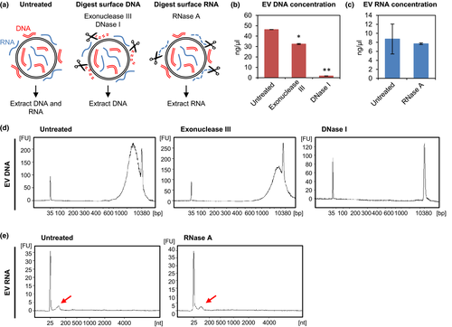
The presence of DNA in EV has sparked considerable interest in EV as novel mediators of horizontal gene transfer (HGT). However, very little is known about the origin and biological significance of DNA in EV. The mechanisms of DNA packaging in EV are poorly understood and whether DNA is packaged within the membrane-bound space of EV remains unclear. Recent studies have revealed that DNA is not contained within the membrane-enclosed space of EV, but is largely attached to the outer surface of EV.7, 8 Indeed, treatment of EV with nucleases such as DNase I and Exonuclease III before DNA extraction significantly reduces the overall DNA detected from cancer cell-derived EV (Yumi Kawamura and Takahiro Ochiya, 2017, personal observation), demonstrating that DNA is mainly associated with the outer EV membrane (Fig. 2). As with cellular membranes, the adhesive properties of EV are governed in part by their structural composition. It has been suggested that the outer surface of EV are capable of interacting with various proteins, nucleic acids, and other surface membranes regulating their aggregation, motility in the circulation, and functionality in targeted sites.9-11 Therefore, it is likely that DNA associated with EV (termed EV-DNA) may have some physiological significance in recipient cells and may play an important role, particularly in the cancer microenvironment.

Possible Origins of EV-DNA and Their Implications in the Cancer Microenvironment
Possible origins for EV-DNA include cell-free DNA (cfDNA) found in apoptotic bodies,12, 13 vesicles released by apoptotic cells,14 or cfDNA attached to the surface of EV.7, 15 Circulating cfDNA is mostly double-stranded, ranging from frequently observed size fragments of 180 bp to fragments larger than 10 000 bp.16 It is generally regarded that cfDNA is released from dying cells during the process of apoptosis or necrosis, from both tumor and non-tumor cells. Studies have shown that the concentration of cfDNA is increased in patients with cancer, in comparison to healthy individuals.17 Interestingly, reduced DNase activity was observed from plasma samples of cancer patients, which may indicate impaired degradation and clearance of cfDNA in cancer.18 Taken together, reduced DNase activity in cancer could also be related to higher DNA concentrations detected from cancer cell-derived EV. Given that cfDNA from cancer patients can induce the oncogenic transformation of normal cells,19 cfDNA on EV may carry substantial biological relevance in the tumor setting.
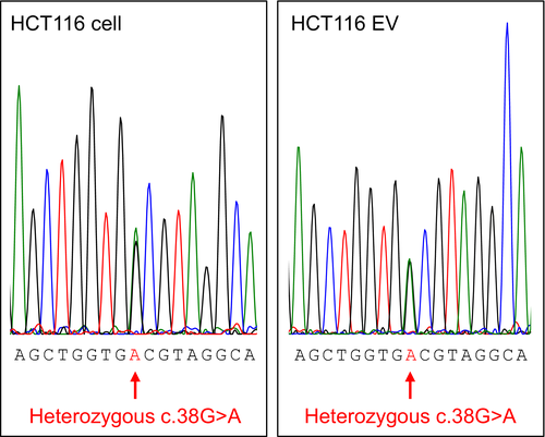
Previous reports have shown that exogenous DNA associated with EV is taken up more efficiently than free DNA, thereby suggesting that DNA associated with EV may be involved in their uptake at recipient cells.20 In addition to this, it has been shown that cancer cell-derived EV take part in pre-metastatic niche formation21 and show organotropism towards potential metastatic sites.22 These findings lead to an intriguing hypothesis that cancer cell-derived EV-DNA including oncogenes could be transported to potential sites of metastasis by EV, inducing tumor-favorable effects (pro-metastatic effects) such as genetic instability in recipient cells, eventually leading to the modulation of the cancer microenvironment and pre-metastatic niche formation. In fact, we have seen that DNA associated with cancer cell-derived EV contains the oncogenic KRAS mutation, which is reflective of the parental cell line (Fig. 3). Although it remains unclear whether DNA associated with cancer cell-derived EV is functional in EV-recipient cells, under certain conditions, these oncogenic mutations may have the potential to transform normal cells.

Lee et al. have assessed the ability of HGT by EV containing oncogenic sequences. In 2014, they showed that dsDNA fragments including full-length H-ras could be incorporated in EV from H-ras transformed cell lines.23 However, a follow-up study by the same group demonstrated that EV containing oncogenic H-ras failed to permanently transform immortalized fibroblasts.24 These data indicate cellular defense mechanisms that prevent the delivery and integration of EV-DNA into the genome of normal cells. As previous studies have shown that apoptotic bodies can horizontally transfer oncogenes such as H-ras and c-Myc to neighboring cells deficient of p53,13, 25 the integration of oncogenic sequences carried by EV could also be regulated by similar cellular defense mechanisms. Deciphering the cellular conditions that would allow the integration of exogenous DNA into the host genome would be important in understanding the extent to which EV can mediate the HGT of oncogenes and other exogenous DNA to EV-recipient cells.
The disparities in DNA detected from EV reflect the complexity and heterogeneity of EV populations. However, given the importance of EV in intercellular communication and their ability to transport genetic material to neighboring and distant cells, determining the origin and physiological significance of EV-DNA could be relevant in understanding their functions in living systems. Finally, the detection of DNA from cancer cell-derived EV may have potential uses as circulating biomarkers for detecting tumor DNA from body fluids such as blood, urine, and saliva.
Retrotransposon Elements Are Found in Cancer Cell-Derived EV
Another important feature of cancer cell-derived EV is the discovery of retrotransposon elements. In 2011, it was reported that cancer cell-derived EV are highly enriched in retrotransposon elements including LINE-1 (L1), ALU and human endogenous retroviruses (HERV).5 This is an intriguing discovery as it indicates a new modality for retrotransposons to spread horizontally from one cell to another. Retrotransposon elements, also known as “jumping genes,” are mobile genetic elements that replicate through RNA intermediates that are reverse transcribed and inserted into new genomic locations. Following the complete sequencing of the human genome, it has been shown that more than 40% of the human genome is derived from retrotransposon elements.26 Retrotransposons are capable of altering the genome by causing mutations, deletions and rearrangements, and their effects range from local genetic instability to large-scale genomic variation. In somatic cells, these elements are silenced by epigenetic and post-transcriptional mechanisms. However, active retrotransposition has been implicated in various diseases, and more than 70 diseases have been documented to be associated with heritable and somatic retrotransposition events.27, 28
Although the ability of retrotransposons to replicate and integrate into the host genome is well studied, the ability to transfer from one cell to another remains largely unknown. As these elements themselves are not infectious, it has been suggested that horizontal transmission would be unlikely without the involvement of a vector. In addition to this, others have pointed out the instability and rapid degradation of RNA intermediates in the extracellular environment.29 The discovery of retrotransposon RNA in EV may indicate an alternative mechanism for the intercellular transfer of retrotransposon elements. Considering the protective features of EV and their ability to carry RNA to neighboring and distant cells, this finding has important implications in understanding the mechanisms of somatic retrotransposition and the involvement of EV containing active retrotransposon elements in the development and spread of genetically influenced diseases.
Possible Contributions of EV-Mediated Retrotransposition in Cancer Development
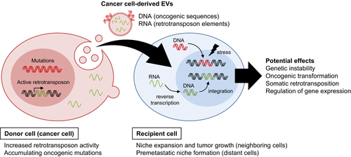
Retrotransposon insertions are frequently observed in cancers. In particular, somatic L1 insertions have been found in the APC tumor suppressor locus in colorectal cancer,30 and within the MYC locus in breast cancer.31 Since then, whole-genome and exome sequencing studies have identified somatic retrotransposon insertions from colorectal, lung squamous, head and neck, and endometrial carcinomas.32, 33 As indicated by these studies, somatic retrotransposon insertions are believed to play an important role in the tumorigenesis of various cancers. Balaj et al. showed that EV can transfer retrotransposon RNA to normal cells.5 However, the functional impact of retrotransposon transfer in EV-recipient cells has not been reported. It has been speculated that EV-mediated delivery would allow the “Trojan Horse-like” delivery of retrotransposons to distant sites, thereby avoiding immune-recognition in a tumor setting.34 Given that active retrotransposon elements can yield extensive genomic insertions, further analysis will be important in observing the effects and activities of EV-mediated transfer of active retrotransposon elements in recipient cells. Understanding this new biological concept will elucidate the spread of retrotransposons by EV and their potential involvement in the formation of genetic instability in target cells (Fig. 4).

Implications of EV-Mediated Retrotransposon Transfer in Other Diseases
Retrotransposition events in neural progenitor cells have been shown to be important in the generation of genetic mosaicism that contributes to neuronal diversity and brain plasticity.35 However, accumulating evidence shows that the deregulation of retrotransposons may cause deleterious effects in neuronal genomes, which may lead to the development of neurodegenerative diseases. Altered levels of retrotransposon expression and increased retrotransposition insertions have been observed in neurodegenerative and neuropsychiatric diseases. For instance, hyperactive retrotransposition and increased L1 copies have been reported in schizophrenia, Alzheimer's disease (AD) and Parkinson's disease (PD). In addition to this, active HERV have been identified in neurons of patients with amyotrophic lateral sclerosis (ALS), and have been associated with neuronal injury and death of motor neurons.36, 37 However, it is still unclear how the deregulation of retrotransposon elements can cause neurodegenerative diseases.
Neurons are particularly vulnerable to the accumulation of DNA damage as a result of their limited capacity for cell replacement in adulthood. The accumulation of DNA lesions is considered to be a major contributor to age-associated neurodegenerative diseases such as AD, as well as PD.38-40 In addition to this, impaired DNA damage repair has been documented in many neurodegenerative diseases and is considered to be an important indication preceding neurodegeneration.(39, 41 Multiple DNA repair molecules have been found to be mutated or deregulated in AD patients.41 DNA lesions and double strand breaks (DSB) are particularly vulnerable to retrotransposon insertions,42 and are thought to be associated with increased retrotransposition events in neurodegenerative diseases.43, 44
EV have been suggested to play a significant role in the dissemination of protein aggregates in the brain, such as β-amyloid and tau in AD, and α-synuclein in PD.45, 46 The accumulation of pathogenic proteins has been associated with the development of neurodegenerative diseases (reviewed in47). Given that increased retrotransposition insertions in neurons are prominent in various neurodegenerative diseases, EV may also be responsible for the dissemination of not only deleterious proteins but also of genetic material such as deregulated retrotransposon elements. Furthermore, accumulating evidence suggests that distinct mechanisms might be involved in the onset of disease, spread, and neuronal death.48, 49 Therefore, the development of neurological diseases could be modulated by various factors not limited to pathogenic protein aggregation in the brain.
Clinical studies have shown that the spread of neurodegenerative diseases follows stereotypical patterns, and may derive from a focus of genetically altered cells.50 The idea that the disease is initiated in a focal area suggests that somatic mosaicism may be responsible for the genetic changes in initiating cells. Indeed, it has been suggested that somatic retrotransposition in neurons can cause mutations that may be involved in the onset of neurodegenerative diseases.44 However, the mechanisms that underlie the spread of the disease from the initial focus to other regions of the brain remain to be fully determined. The role of EV in the spread of deleterious cargo may explain the anatomical spread of neurodegenerative diseases from one brain region to an adjacent region. Considering that retrotransposons can act as insertional mutagens that can target DNA DSB, it is plausible to hypothesize that the effects of EV containing active retrotransposon elements can exacerbate the spread of insertional mutagenesis in affected neurons, which may play a role in the potential spread of neurodegeneration.
Although the involvement of retrotransposons in EV and their activity in the progression of neurodegenerative disease has not been demonstrated so far, this finding would be important in understanding the spread of somatic retrotransposition in the brain. The analysis of EV-mediated transfer of active retrotransposons will add to the current understanding of the role of EV not only in the degeneration of neurons but also in the development and spread of various human diseases associated with deregulated retrotransposition events.
Evolutionary Impacts of HGT by EV
The discovery of genetic elements in EV, in particular retrotransposon elements, signifies notable importance in understanding the mechanisms of genome evolution by exogenous genetic elements carried by EV. The horizontal transfer of retrotransposons has been critical in generating genome diversity that has led to the evolution of various organisms. Genome-wide comparisons of humans and other primate genomes have revealed the magnitude and significance of retrotransposon elements in human evolution.27 An example of horizontal transfer of retrotransposons is the case of BovB, an L1 retrotransposon that has been found to be widespread among mammalian species.51 However, the precise mechanisms regarding the transfer of retrotransposons between species remain largely unknown.
The successful horizontal transfer of a retrotransposon requires the delivery of the RNA intermediate, reverse transcription and integration into the host genome. It has been regarded that retrotransposons do not have the ability to infect other cells, and therefore would not be able to disperse horizontally across species without a vector involved. Retroviruses, which have the ability to assemble virus particles that can exit the cell, have been proposed as potential vectors for the horizontal transfer of retrotransposons. Recently, it was shown that EV are able to transfer HERV-K HML2 RNA to normal cells, suggesting that a functional envelope gene may not be required for HERV to infect other cells.5 This discovery raises the possibility of an EV-mediated mechanism for the dispersal of retrotransposons, and opens a new door into understanding their basic nature and scope in interspecies transfer.
A recent review by Nolte-'t Hoen et al. discusses the many aspects in which EV resemble retroviruses. Both EV and retroviruses share similar structural aspects including size and buoyancy, but can also be similar in functional aspects, such as virus propagation. EV generated from virus-infected cells (termed virus-induced EV) have been shown to incorporate viral proteins and fragments of the viral genome, which can be transferred to target cells.52 The authors argue that virus-induced EV and virus-like particles are intermediate entities that are indistinguishable from one another. These intriguing similarities with retroviruses raise the possibility of EV as potential transporters of retrotransposon elements. Moreover, intercellular communication by EV is not unique to eukaryotic cells. The release of membrane vesicles is an evolutionarily conserved mechanism in eukaryotes, bacteria, and archaea, and is a basic feature in many organisms.53 In addition to this, the mechanistic strategies of EV release are similar, typically involving the highly conserved endosomal sorting complex required for transport (ESCRT) machinery, which are key components in the excision of membrane necks in the biogenesis of MVB, cytokinesis, and budding of enveloped viruses.54-56 The conserved feature of EV production and transmission may indicate their potential ability for interspecies transfer of genetic material.
Several reports have demonstrated the transfer of EV across species and their involvement in interspecies communication. The delivery of infectious agents by EV has been reported in host–pathogen interactions by intracellular and extracellular eukaryotic parasites57 and in human–fungal interactions.58 EV have also been implicated in plant–fungal interactions, in the secretion of virulence and defense molecules by both fungi and plants.59 Furthermore, preliminary studies have shown that EV from food sources are taken up by intestinal macrophages and stem cells in humans, inducing the expression of target genes in recipient cells.60 The ability of EV to carry genetic material across species suggests their potential role in the transfer of exogenous genetic material into the human genome. Further studies examining the distribution of genetic elements such as retrotransposons by EV-mediated mechanisms may consequently open up new ways of considering the functional impacts of EV in genome modulation, and will provide additional evidence for external influence on the evolution of the human genome.
Conclusion
The presence of genetic material in EV has heightened considerable interest in their potential as mediators of HGT not only between cells in a single living organism, but also across different biological species. Further studies assessing the functionality of genetic material in EV-recipient cells will help to better define their roles as trans-genomic agents and their involvement in modulating various cellular responses. EV are packaged with abundant cellular information; however, the mechanisms that regulate EV cargo selection is still poorly understood. A deeper understanding of the biogenesis and cargo selection of EV may elucidate many of the biological processes that are regulated by EV, permitting an understanding of inter- and intracellular communication by EV.
Acknowledgments
This work was supported in part by Japan Agency for Medical Research and Development (AMED) and the New Energy and Industrial Technology Development Organization (NEDO).
Disclosure Statement
The authors have no conflict of interest.


