Propagation of elite rescue dogs by somatic cell nuclear transfer
Abstract
The objective of the present study was to compare the efficiency of two oocyte activation culture media to produce cloned dogs from an elite rescue dog and to analyze their behavioral tendencies. In somatic cell nuclear transfer procedure, fused couplets were activated by calcium ionophore treatment for 4 min, cultured in two media: modified synthetic oviduct fluid (mSOF) with 1.9 mmol/L 6-dimethylaminopyridine (DMAP) (SOF-DMAP) or porcine zygote medium (PZM-5) with 1.9 mmol/L DMAP (PZM-DMAP) for 4 h, and then were transferred into recipients. After embryo transfer, pregnancy was detected in one out of three surrogate mothers that received cloned embryos from the PZM-DMAP group (33.3%), and one pregnancy (25%) was detected in four surrogate mothers receiving cloned embryos from the SOF-DMAP group. Each pregnant dog gave birth to one healthy cloned puppy by cesarean section. We conducted the puppy aptitude test with two cloned puppies; the two cloned puppies were classified as the same type, accepting humans and leaders easily. The present study indicated that the type of medium used in 6-DMAP culture did not increase in cloning efficiency and dogs cloned using donor cells derived from one elite dog have similar behavioral tendencies.
Introduction
Dogs are man's best friends. Dogs and humans have had a close relationship for thousands of years and during that time their bonds have become greatly strengthened. The dog is one of the most intelligent and thoughtful animals. It also exhibits some outstanding abilities, for example, the sense of smell in dogs is on average 10,000 to 100,000 times better than in humans (Walker et al. 2003); even the best odor detection machines cannot outperform elite dogs. Because of such attributes, dogs have been bred for many useful purposes, such as: (i) assisting humans in hunting and driving livestock; (ii) guarding humans from dangerous predators; (iii) being a close companion; and (iv) being a valuable tool in society as service animals (Stafford 2006; Walsh 2009). Service dogs have special roles to provide security, detect drugs and explosives, assist the blind and save human lies (Maejima et al. 2007; Walsh 2009; McKeown 2012). However, it is difficult to produce dogs that are completely suitable as service animals because of the extensive training period and costs, so that the production efficiency of general service dogs is about 30% (Maejima et al. 2007).
In the Korean National Emergency Management Agency, there is a retired veteran rescue dog named Baekdu that performed lifesaving activities worldwide for 6 years. Baekdu demonstrated excellent discretion in the International Rescue Dog Organization as well as in domestic/international disaster situations. Here, we considered canine somatic cell nuclear transfer (SCNT) for the preservation and propagation of Baekdu's abilities. Canine SCNT is a useful assisted reproductive technique for producing pet dogs (Jang et al. 2008; Park et al. 2011), endangered canids (Kim et al. 2007; Oh et al. 2008) and transgenic dogs (Hong et al. 2009; Kim et al. 2011; Oh et al. 2011), but the overall efficacy of the process is still low. In order to improve this low efficiency, two basic activation media used for reconstructed oocytes were compared in terms of in vivo development of cloned embryos. The first medium is modified synthetic oviduct fluid (mSOF) which is a culture medium containing BSA (bovine serum albumin) which a protein preparation that has variable, undefined functions and is used in our laboratory for dog cloning (Oh et al. 2011; Kim et al. 2014). The second medium is porcine zygote medium (PZM-5) which is a chemically defined medium with a synthetic polymer, PVA (polyvinyl alcohol) (Bavister 1981; Garcia-Mengual et al. 2008). Thus, the purpose of the present study was: (i) to clone Baekdu and to compare in vivo development of canine cloned embryos produced using two oocyte activation media and (ii) behavioral analysis of Baekdu clones through the puppy aptitude test which is performed primarily to evaluate basic aptitudes of puppies as service dogs (Pfaffenberger 1976; Weiss 2002).
Materials and Methods
Chemicals
Chemicals were purchased from Sigma Chemical Co. (St. Louis, MO, USA) unless otherwise stated.
Animals
In the study, 30 kg mixed origin, large-breed bitches (12 oocyte donor dogs and 7 embryo transfer recipients) between 1 and 5 years of age were used. All animal care and experiments were carried out in accordance with the Guide for the Care and Use of Laboratory Animals established by the Institutional Animal Care and Use Committee of Seoul National University (Approval number; SNU-121123-13).
Preparation of donor fibroblasts
Ear skin tissue was collected from a 10-year-old male German shepherd, a veteran rescue dog, Baekdu. The tissue was washed three times using PBS (Dulbecco's phosphate-buffered saline; Invitrogen, Carlsbad, CA, USA), minced, cultured in Dulbecco's modified Eagle's medium (DMEM; Invitrogen) supplemented with 10% (v/v) fetal bovine serum (Invitrogen) at 38°C in a humidified atmosphere of 5% CO2 and 95% air. After 7 days of incubation, a fibroblast monolayer was established, passaged, cryopreserved in 10% dimethyl sulfoxide (DMSO) and stored in liquid nitrogen. Cells from passages 3 to 5 were used as donor cells for SCNT.
Somatic cell nuclear transfer
Canine in vivo matured oocytes were recovered by aseptic surgical procedures 70–76 h after the day of ovulation which was considered as the day when the serum progesterone concentration reached 4.0–9.9 ng/mL as previously described (Kim et al. 2012). Oocytes surrounded by cumulus cell layers were denuded by repeated pipetting in HEPES-buffered tissue culture medium (TCM)-199 supplemented with 0.1% (w/v) hyaluronidase. The first polar body and metaphase II spindle of denuded oocytes were removed under an inverted microscope equipped with fluorescence. One donor somatic cell was injected into the perivitelline space of each enucleated oocyte, then fused with electric stimulation using two pulses of direct current of 72 V for 15 µsec with an Electro-Cell Fusion apparatus (NEPA GENE Co., Chiba, Japan). The fused couplets were activated by calcium ionophore treatment for 4 min, and then cultured in mSOF medium supplemented with 1.9 mmol/L 6-dimethylaminopyridine (DMAP) (SOF-DMAP), or in PZM-5 supplemented with 1.9 mmol/L DMAP (PZM-DMAP) for 4 h.
Embryo transfer and pregnancy diagnosis
After activation, SCNT couplets were immediately surgically transferred using a 3.5-Fr Tom Cat Catheter (Sherwood, St. Louis, MO, USA) into the ampullary portion of the oviducts of naturally synchronous recipients. Recipients were prepared by predicting ovulation time based on serum progesterone concentrations. Pregnancy diagnoses were assessed with a SONOACE 9900 ultrasound machine (Medison, Seoul, Korea) approximately 31 days after embryo transfer.
Puppy aptitude test
The puppy aptitude test (PAT) of this experiment used Volhard PAT (The American Kennel Club AkC 1985) with modification. The PAT used a scoring system from 1-6 and consisted of 10 tests: Social attraction, Following, Restraint, Social Dominance, Elevation Dominance, Retrieving, Touch Sensitivity, Sound Sensitivity, Sight Sensitivity, and Stability. The tests were performed sequentially and in the order listed. The tests were carried out in an area (3 m x 3 m) unfamiliar to the puppy. At age 7 weeks, a puppy got each subtest for about 3 sec, counting only the first response. There were no other dogs or people, except the tester, in the testing area. The tester was a stranger to the puppy and four scorers evaluated the puppy from outside the test area through a window at the same time. Each test was scored separately, and interpreted on its own merits. In order to preclude the nursing and raising environment, the cloned puppies were brought up suckling breast milk of the same recipient (recipient 1) in the same condition. The puppies stayed with recipient 1 till 7 weeks of age when they were weaned.
Microsatellite analysis for genotyping
Parentage analysis was performed using genomic DNA from the oocyte donor dog, donor cells, cloned puppies and recipients. Genomic DNA was extracted from blood or tissue samples or cell pellets according to the instructions provided with the G-spin Genomic DNA Extraction Kit (Intron Biotechnology, Seongnam-si, Gyeonggi-do, Korea), and analyzed using microsatellite assays with canine-specific markers (Lee et al. 2005; Oh et al. 2009). Based on the complete nucleotide sequence of canine mitochondrial DNA (mtDNA; GenBank accession number U96639), oligonucleotide primers were synthesized for the hypervariable region as described in previous studies (Lee et al. 2005; Oh et al. 2009).
Statistical analysis
Statistical analysis was performed with GraphPad Prism 5 software (GraphPad, San Diego, CA, USA). Statistical significance of in vivo development of canine cloned embryos was analyzed using Column Statistics. Dog behavior data was analyzed by two-way analysis of variance (ANOVA) with the Bonferroni post-test. Significance level was considered as P < 0.05.
Results
In vivo development of canine cloned embryos using two different activation protocols
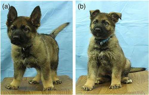
A total of 56 and 64 activated, cloned embryos in the PZM-DMAP and SOF-DMAP groups were transferred into three and four female recipient dogs, respectively. Pregnancy was detected in one out of the three surrogate mothers for the PZM-DMAP group (33.3%), and one pregnancy (25%) was detected in the four surrogate mothers of the SOF-DMAP group (Table 1). The two pregnant dogs each gave birth to one healthy cloned puppy by cesarean section (Fig. 1). The cloned dogs and the donor cells had identical microsatellite patterns for all loci (Table 2), and the mtDNA sequences of the cloned dogs were identical to those of the oocyte donor dog (Table 3).
| Group | No. of transferred embryos | No. of recipient females | No. of pregnancies (pregnancies/ recipients) | No. of cloned pups (births/embryos transferred) | Clone ID | Status of cloned pups |
|---|---|---|---|---|---|---|
| SOF-DMAP | 64 | 4 | 1 (25.0%) | 1 (1.6%) | Clone 1 | Live |
| PZM-DMAP | 56 | 3 | 1 (33.0%) | 1 (1.8%) | Clone 2 | Live |
- SOF-DMAP, modified synthetic oviduct fluid with 1.9 mmol/L 6-DMAP; PZM-DMAP, porcine: zygote medium with 1.9 mmol/L 6-DMAP.

| Name | Donor cell | Clone 1 | Recipient 1 | Clone 2 | Recipient 2 | |||||
|---|---|---|---|---|---|---|---|---|---|---|
| PEZ1 | 123 | 119 | 123 | 119 | 123 | 119 | 123 | 119 | 115 | 115 |
| FHC2054 | 180 | 176 | 180 | 176 | 155 | 155 | 180 | 176 | 151 | 151 |
| FHC2010 | 236 | 224 | 236 | 224 | 232 | 228 | 236 | 224 | 224 | 224 |
| PEZ5 | 103 | 103 | 103 | 103 | 111 | 111 | 103 | 103 | 111 | 111 |
| PEZ20 | 180 | 176 | 180 | 176 | 196 | 188 | 180 | 176 | 177 | 173 |
| PEZ12 | 268 | 268 | 268 | 268 | 283 | 283 | 268 | 268 | 276 | 264 |
| PEZ3 | 128 | 128 | 128 | 128 | 124 | 124 | 128 | 128 | 131 | 124 |
| PEZ6 | 188 | 176 | 188 | 176 | 183 | 183 | 188 | 176 | 184 | 184 |
| PEZ8 | 226 | 226 | 226 | 226 | 238 | 233 | 226 | 226 | 233 | 229 |
| FHC2079 | 274 | 270 | 274 | 270 | 274 | 270 | 274 | 270 | 278 | 274 |
| Sample ID | Nucleotide positions* | |||||||||||||||
|---|---|---|---|---|---|---|---|---|---|---|---|---|---|---|---|---|
| Reference (U96639 v.2) | 15435 | 15518 | 15526 | 15595 | 15612 | 15632 | 15639 | 15643 | 15652 | 15800 | 15814 | 15815 | 15912 | 15955 | 16003 | 16083 |
| G | A | C | C | T | C | T | A | G | T | C | T | C | C | A | A | |
| Donor cell | G | A | C | C | T | C | T | A | G | T | C | T | C | C | A | A |
| Clone 1 | A | C | T | T | C | T | G | G | A | C | T | C | T | T | G | G |
| Oocyte donor 1 | A | C | T | T | C | T | G | G | A | C | T | C | T | T | G | G |
| Clone 2 | G | A | C | C | T | C | T | A | G | T | C | T | C | C | A | A |
| Oocyte donor 2-1 | A | C | T | T | C | T | G | G | A | C | T | C | T | T | G | G |
| Oocyte donor 2-2 | G | A | C | C | T | C | T | A | G | T | C | T | C | C | A | A |
- * The nucleotide positions were numbered from those of GenBank accession no U96639 v.2, and 661bases (from 15431 to 16091) were examined.
Puppy aptitude test of the two cloned puppies
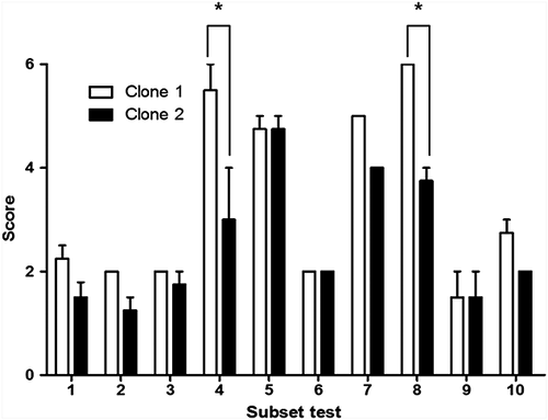
PAT was conducted to assess each puppy's temperament. A score of 1 to 6 was used to evaluate the response of each subtest. Scores of social dominance and sound sensitivity were different between the two cloned dogs (P < 0.001), but there was no difference in the other tests (Fig. 2). From the interpretation of all 10 subtests, the two cloned puppies were classified as being the same type 3 (dogs of this type accept humans and leaders easily).

Discussion
Cloning or SCNT is one of the assisted reproductive technologies (ARTs) currently used in animal reproduction. Although SCNT is inefficient compared with other ARTs such as in vitro fertilization or artificial insemination (Oback & Wells 2007), SCNT and banking of somatic cells for SCNT are still utilized to multiply elite animals with desired phenotypic traits and to produce genetically modified animals (Brophy et al. 2003; Faber et al. 2004; Yonai et al. 2005; Hoshino et al. 2009).
For animal cloning, embryo activation is a key step in the development of cloned embryos (Alberio et al. 2001). Correct activation is essential to support normal development of cloned embryos. Generally, the combination of ionomycin or ionophore and 6-DMAP is a routine tool for the activation of cloned embryos. Ionomycin or ionophore are widely used to increase intracellular Ca2+ levels in activated oocytes (Morgan & Jacob 1994). 6-DMAP, a protein serine/threonine kinase inhibitor (Neant & Guerrier 1988), contributes to DNA synthesis (Ledda et al. 1996) and accelerates pronucleus formation in activated, reconstructed embryos. Many studies have been reported for establishing a species-specific activation protocol because optimal treatment time and concentration of 6-DMAP is critical for this step (Choi et al. 2004; Lan et al. 2005). However, during 6-DMAP treatment, little attention has been paid to effects of culture medium on embryo development. Therefore, the present study investigated the effects of two different culture media used for 6-DMAP treatment after calcium ionophore treatment of fused couplets on SCNT efficiency. With respect to full-term development of cloned embryos, there was no difference between oocytes activated in SOF-DMAP and PZM-DMAP. This result shows that the type of medium used in 6-DMAP culture has no effect on dog cloning efficiency.
Next, we investigated whether the resulting cloned dogs have potential as candidates for rescue dogs. Our study compared the behavioral patterns of the two cloned dogs using PAT, which can predict the performance of a puppy and its expected temperament they will have in adulthood. Also, specific tests predictive of their future employment as adult service dogs have been developed (Slabbert & Odendaal 1999). The necessity of applying the PAT at an early age has been emphasized because early prediction of a dog's adult behavior could save costs of training and reduce time wasted to train doubtful puppies. The present study was carried out using the Volhard puppy behavioral test with modification (The American Kennel Club AkC 1985; Burghardt 2003). Although the two cloned dogs did not show a perfectly identical score throughout the 10 subtests, overall they showed a similar tendency. Interpretation of the PAT scores indicated that the two cloned dogs mostly belong to type 3, that is, the dog accepts human leaders easily, adapts well to new situations and is generally good with children and elderly people, as well as making a good obedience prospect, and it usually has a common sense approach to life (The American Kennel Club AkC 1985). Thus, the two cloned dogs were evaluated as possessing the appropriate temperament for rescue training.
Also, we performed PAT test using 7-week-old shepherds (control) born by natural mating. Six controls were littermates, and grew up in the same environment. The result was interesting as follows: two belonged to type 2 and two belonged to type 3. The other two controls showed a result that is type 4 (data not shown). In other words, the control group derived from natural mating showed various types in PAT test, not the same as the cloned animals.
In conclusion, the present study demonstrates that dogs cloned using donor cells derived from one elite rescue dog have similar behavioral characteristics. The numbers of elite working dogs in diverse fields can be increased by using the SCNT technique with donor cells derived from a small piece of tissue from elite working dogs; this tissue fragment can be collected once only, cultured, passaged and frozen for many future cloning applications. Further monitoring of the similarities of the performance of cloned dogs in lifesaving and security activities is required and is ongoing.
Acknowledgments
We thank Dr. Barry D. Bavister for his valuable editing of the manuscript. This study was supported by RDA (PJ010928032015), IPET (#311062-04-3SB010), Research Institute for Veterinary Science, Nestle′ Purina PetCare and the BK21 plus program.


