Estimation of beef marbling in the Longissimus muscle with computer image analysis of ultrasonic pictures of the Iliocostalis muscle area
Abstract
This study investigated an objective method for estimating beef marbling using ultrasonic images of the Iliocostalis muscle and the Lomgissimus muscle area sections. Thirty-one Japanese Black cattle steers were used in this study. The end of the left side shoulder blade bone was scanned using an ultrasonic device. Ultrasonic images were captured of the Longissimus muscle area and that around the Iliocostalis muscle area. Twenty items were measured in the two images using computer image analysis software. The level of beef marbling was measured according to the Beef Marbling Standard (BMS) for carcass grading, and the percentage of ether-extractable fat content in the Longissimus muscle (EE). The difference in the gray level between the Iliocostalis muscle and intermuscular fat (X10) was used to estimate the BMS and the EE, which were highly correlated (r2 = 67.72% and 61.30%). An equation was developed using four parameters from the two ultrasonic images, which could estimate the BMS (r2 = 85.88%). This equation could also estimate the EE (r2 = 68.98%). The equations used to estimate beef marbling were based on one to four parameters that included X10. Thus, ultrasonic images of the Iliocostalis muscle area section are important for estimating beef marbling.
Introduction
Estimating the potential quality of beef carcass using a live animal can increase profits because the appropriate fattening period can be assessed more accurately, and the feeding management can be improved. Thus, ultrasonic techniques are being explored and used increasingly in the beef industry. This technique can effectively improve the breeding of beef cattle. Recently, Japanese Black cattle (Wagyu) have been bred for high marbling and a high fat content. A technique based on ultrasonic imaging would be useful for estimating highly marbled beef cattle. The level of beef marbling has been subjectively evaluated based on the line below the Longissimus muscle shape and the difference in the gray level between the Longissimus muscle and the surrounding area in ultrasonic images (Harada 1982; Nade et al. 2007). However, the subjective evaluation of beef cattle with highly marbled beef using ultrasonic techniques is difficult because the gray level, which is the shade of white to black, is uniform in the Longissimus muscle area in ultrasonic images. Thus, some Japanese researchers have subjectively evaluated the marbling level based on the difference in the gray level of the intermuscular fat and the Iliocostalis muscle in the field. The difference in the gray level of the intermuscular fat and the Iliocostalis muscle is easier to determine compared with that in the Longissimus muscle area. However, ultrasonic images should be objectively evaluated to facilitate the accurate and consistent improvement of beef cattle and their feeding management based on computer image analysis (CIA).
In the present study, we investigated the relationship between the CIA of ultrasonic images of the Longissimus muscle area, a section around the Iliocostalis muscle, and the actual beef marbling to develop an objective method for estimating beef marbling.
Materials and Methods
The animals used in this study comprised 32 fattened Wagyu steers aged approximately 28 months. The animals were scanned during the 2-week period before slaughtering. Ultrasonic images of the Longissimus muscle area and around the Iliocostalis muscle area were captured using an ultrasonic device (HS-2000; Honda Electric Company Ltd, Aichi, Japan). Hair at the end of the left side shoulder blade bone was shaved, and oil was applied before the area was scanned. The scanned area corresponded to the designated carcass grading part between the sixth and seventh rib bones (Japan Meat Grading Association 1988). The dynamic range was 55, and the total gain was 60. The ultrasonic images were stored on a magnetic optical disk and analyzed using CIA software (WinRoof ver 6.5; MITANI Ltd, Tokyo, Japan). The shade of white to black in the ultrasonic image was calculated using CIA software to convert 256 levels (0 to 255) into a number measured as the gray level. In this study, the average gray level, standard deviation and value which subtracted the minimum from the maximum of the gray level of the principal muscle in the ultrasonic image was analyzed using CIA software because the ultrasonic technicians subjectively evaluate the difference and roughness of gray level in the ultrasonic image. Furthermore, the difference of gray level between the principal muscles was also subjectively evaluated. However, since the points evaluated by each technician may differ, many variables were measured in this study.
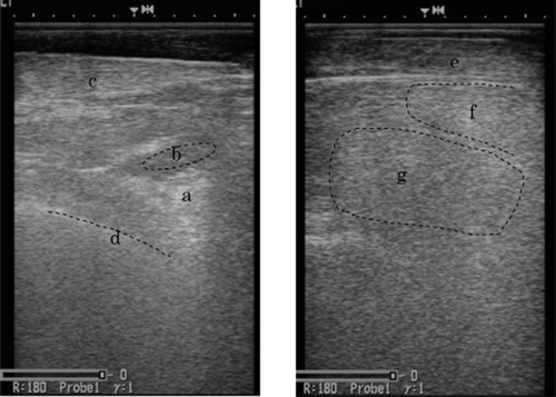
The areas and the data used for developing the prediction equation measured by CIA (Fig. 1) are as follows:

Areas of ultrasonic images used for computer image analysis. (a) Iliocostalis muscle, (b) intermuscular fat, (c) Latissimus muscle, (d) rib bone, (e) Trapezius muscle, (f) Spinalis muscle, (g) Longissimus muscle.
X1: average gray level of the Iliocostalis muscle,
X2: standard deviation of the gray level of the Iliocostalis muscle,
X3: value which subtracted the minimum from the maximum of the gray level of the Iliocostalis muscle,
X4: average gray level of the intermuscular fat,
X5: standard deviation of the gray level of the intermuscular fat,
X6: value which subtracted the minimum from the maximum of the gray level of the intermuscular fat,
X7: average gray level between the Latissimus muscle and rib bone,
X8: standard deviation of the gray level between the Latissimus muscle and rib bone,
X9: value which subtracted the minimum from the maximum of the gray level between the Latissimus muscle and rib bone,
X10: value which subtracted X4 from X1,
X11: average gray level of the Longissimus muscle,
X12: standard deviation of the gray level of the Longissimus muscle,
X13: value which subtracted the minimum from the maximum of the gray level of the Longissimus muscle,
X14: average gray level of the Trapezius muscle,
X15: standard deviation of the gray level of the Trapezius muscle,
X16: value which subtracted the minimum from the maximum of the gray level of the Trapezius muscle,
X17: average gray level of the Spinalis muscle,
X18: standard deviation of the gray level of the Spinalis muscle,
X19: value which subtracted the minimum from the maximum of the gray level of the Spinalis muscle,
X20: value which subtracted X11 from X17.
The animals were slaughtered at approximately 28 months of age and the Beef Marbling Standard (BMS) of the carcasses were graded based on the Japan Meat Grading Association (JMGA) standard (1988). The Longissimus muscle was removed from the carcass to measure its fat content, and the ether-extractable (EE) fat content of the muscle was measured using the Soxhlet method (AOAC 1990).
Statistical anaysis
The relationships between the beef marbling that was measured using the post-slaughter BMS, the EE fat content, and the parameters extracted by the CIA of the ultrasonic images of live animals were investigated using the REG procedure in SAS (ver.9.0; SAS Institute Inc., Cary, NC, USA). A prediction equation was constructed for each rib-eye area section, each Iliocostalis muscle area section, and the two sections combined. Combinations of parameters were calculated using one to four parameters. We determined equations that yielded the highest correlations (r2) with the BMS and the EE fat content of the Longissimus muscle. We included combinations that had strong relationships between the independent variables; the correlation coefficient was > 0.60 or the P-value of the relationship between the independent variables was < 0.01.
Results and Discussion
Measurements of beef parameters
Table 1 shows the BMS of the carcasses, the EE fat contents of the Longissimus muscles, and the measurements made by CIA of the ultrasonic images. The BMS ranged from 5 to 10, and the average was 6.7. The BMS values were approximately the same or higher than the standard grade of Wagyu. The EE fat content of the Longissimus muscle ranged from 31.0% to 54.9%, and the average was 43.8%.
| Average | Standard deviation | Coefficient of variation (%) | |
|---|---|---|---|
| BMS (No.) | 6.7 | 1.6 | 23.6 |
| Ether extract (%) | 43.75 | 6.28 | 14.36 |
| X1 | 132.42 | 10.25 | 7.74 |
| X2 | 15.37 | 1.89 | 12.28 |
| X3 | 97.61 | 10.98 | 11.25 |
| X4 | 116.28 | 7.06 | 6.07 |
| X5 | 14.02 | 1.69 | 12.03 |
| X6 | 82.13 | 10.33 | 12.57 |
| X7 | 130.79 | 6.36 | 4.86 |
| X8 | 17.08 | 2.24 | 13.09 |
| X9 | 120.32 | 12.60 | 10.47 |
| X10 | 16.14 | 7.15 | 44.32 |
| X11 | 113.79 | 11.08 | 9.74 |
| X12 | 19.54 | 1.89 | 9.66 |
| X13 | 128.65 | 10.26 | 7.97 |
| X14 | 89.37 | 10.31 | 11.54 |
| X15 | 20.22 | 1.79 | 8.83 |
| X16 | 130.13 | 10.91 | 8.38 |
| X17 | 132.94 | 10.56 | 7.94 |
| X18 | 17.09 | 2.34 | 13.71 |
| X19 | 106.00 | 12.97 | 12.23 |
| X20 | 19.15 | 10.60 | 55.38 |
- BMS, Beef Marbling Standard Japan Meat Grading Association; X1, average gray level of the Iliocostalis muscle; X2, standard deviation of the gray level of the Iliocostalis muscle; X3, value which subtracted the minimum from the maximum of the gray level of the Iliocostalis muscle; X4, average gray level of the intermuscular fat; X5, standard deviation of the gray level of the intermuscular fat; X6, value which subtracted the minimum from the maximum of the gray level of the intermuscular fat; X7, average gray level between the Latissimus muscle and rib bone; X8, standard deviation of the gray level between the Latissimus muscle and rib bone; X9, value which subtracted the minimum from the maximum of the gray level between the Latissimus muscle and rib bone; X10, value which subtracted X4 from X1; X11, average gray level of the Longissimus muscle; X12, standard deviation of the gray level of the Longissimus muscle; X13, value which subtracted the minimum from the maximum of the gray level of the Longissimus muscle; X14, average gray level of the Trapezius muscle, X15, standard deviation of the gray level of the Trapezius muscle; X16, value which subtracted the minimum from the maximum of the gray level of the Trapezius muscle; X17, average gray level of the Spinalis muscle; X18, standard deviation of the gray level of the Spinalis muscle; X19, value which subtracted the minimum from the maximum of the gray level of the Spinalis muscle; X20, value which subtracted X11 from X17.
The coefficients of variation (CV) for X10 and X20 were 44.32% and 55.38%, respectively. The CVs of the other parameters were < 13.71%, which were low. The CVs of X10 and X20 were higher than the other parameters.
Estimation of the BMS based on images of the Iliocostalis muscle area section
Table 2 shows the equations for estimating the BMS based on the images of the Iliocostalis muscle area section. X10 and BMS had a high correlation (r2 = 67.72%). The relationship between the BMS and X10 was negative. Therefore, a low BMS was estimated when the difference in the gray level between the Iliocostalis muscle and intermuscular fat was high. The maximum residual error was 2.02. This residual error was for BMS no. 5, which had the highest level of marbling, while the other residual errors for the BMS ranged from 1.18 to 1.54. The second highest coefficient of determination was X1, although the correlation was not strong (r2 = 39.77%). A prediction equation based on the two parameters was constructed using X8 and X10 or X9 and X10, and the coefficients of determination were approximately 70%. The correlation for the prediction equation formulated using X3, X8 and X10 was 74.22%. The correlation of the prediction equation based on four parameters was higher than that using these three parameters. Therefore, X10, which was measured by subtracting the gray level of the intermuscular fat from the gray level of the Iliocostalis muscle, was selected for various equations because this information was important for estimating the BMS of carcasses.
| Number of variables | Equation | r2 (%) | Error | ||
|---|---|---|---|---|---|
| AVG | RSD | Max | |||
| One | 9.607 − 0.182 (X10) | 67.72 | 0.70 | 0.54 | 2.02 |
| 19.541 − 0.097 (X1) | 39.77 | 0.99 | 0.69 | 2.40 | |
| Two | 7.616 + 0.119 (X8) − 0.184 (X10) | 70.56 | 0.63 | 0.57 | 1.96 |
| 7.457 + 0.019 (X9) − 0.187 (X10) | 69.87 | 0.66 | 0.55 | 2.00 | |
| Three | 9.347 − 0.032 (X3) + 0.193 (X8) − 0.177 (X10) | 74.22 | 0.60 | 0.51 | 1.70 |
| 8.614 − 0.030 (X3) + 0.031 (X9) − 0.182 (X10) | 73.42 | 0.63 | 0.51 | 1.85 | |
| Four | 9.938 − 0.028 (X3) − 0.017 (X6) + 0.206 (X8) − 0.164 (X10) | 74.90 | 0.62 | 0.48 | 1.63 |
| 9.006 − 0.033 (X3) + 0.146 (X8) + 0.011 (X9) − 0.178 (X10) | 74.44 | 0.59 | 0.53 | 1.72 | |
- BMS, Beef Marbling Standard Japan Meat Grading Association; r2, coefficient of determination; AVG, average absolute value of the residual error; RSD, residual standard deviation; Max, maximum absolute value of the residual error. X1, average gray level in Iliocostalis muscle; X3, value which subtracted the minimum from the maximum of the gray level of the Iliocostalis muscle; X6, value which subtracted the minimum from the maximum of the gray level of the intermuscular fat; X8, standard deviation of the gray level between the Latissimus muscle and rib bone; X9, value which subtracted the minimum from the maximum of the gray level between the Latissimus muscle and rib bone; X10, value which subtracted X4 from X1 (X4, average gray level in intermuscular fat).
Estimation of the percentage of EE fat content of the Longissimus muscle based on images of the Iliocostalis muscle area section
Table 3 shows the equations used to estimate the percentage of EE fat content from the images of the Iliocostalis muscle area section. X10 had a high correlation with the percentage of EE fat content (r2 = 61.30%). When the equations were formulated using two to four parameters, the equations using X1, X4 and X10 were created. However, the correlation coefficient among X1, X4 and X10 was high, and there was a multicollinearity. The multiple regression function using X1, X4 and X10 was rejected because the reliability of the equation was low. Using the two parameters of X8 and X10, the r2 was 62.15%. Using the three parameters of X2, X8 and X10, the r2 was 65.13%. Therefore, X10 was the most important parameter for estimating of the percentage of EE fat content of the Longissimus muscle.
| Number of variables | Equation | r2 (%) | Error | ||
|---|---|---|---|---|---|
| AVG | RSD | Max | |||
| One | 54.851 − 0.687 (X10) | 61.30 | 3.29 | 2.02 | 9.50 |
| 97.970 − 0.409 (X1) | 44.63 | 3.70 | 2.78 | 11.55 | |
| Two | 50.529+0.259(X8) − 0.693(X10) | 62.15 | 3.24 | 2.02 | 9.81 |
| Three | 54.583 − 0.805(X2)+0.710(X8) − 0.655(X10) | 65.13 | 2.95 | 2.19 | 8.53 |
- r2, coefficient of determination; AVG, average absolute value of the residual error; RSD, residual standard deviation; Max, maximum absolute value of the residual error. X1, average gray level in the Iliocostalis muscle; X2, standard deviation of the gray level in the Iliocostalis muscle; X8, standard deviation of the gray level between the Latissimus muscle and rib bone; X10, value which subtracted X4 from X1 (X4, average gray level in intermuscular fat).
Estimation of the BMS using images of the Longissimus muscle area section
Table 4 shows the equations used to predict the BMS based on images of the Longissimus muscle area section. X20 and BMS had a high coefficient of determination (r2 = 50.46%). The parameter with the second highest r2 was X11. The equations formulated using two parameters from X11 and X17 or X11 and X20 had an r2 of 61.56%. An equation was formulated using three parameters, which included X13 in addition to the equation with the two parameters. The r2 values were 65.25% and 65.24%. The r2 values of equations formulated with four parameters (r2 = 66.42%) were increased by 1.2%, so the improvement in r2 was small. Therefore, X11, the average gray level of the Longissimus muscle, and X20, the difference in the gray level of the Longissimus muscle minus that of the Spinalis muscle, were the most important parameters for estimating the BMS using ultrasonic images of the Longissimus muscle area section. The ultrasonic images with a lower X11 or a higher X20 correlated with a higher BMS.
| Number of variables | Equation | r2 (%) | Error | ||
|---|---|---|---|---|---|
| AVG | RSD | Max | |||
| One | 4.652 + 0.106 (X20) | 50.46 | 0.88 | 0.66 | 2.87 |
| 17.332 − 0.094 (X11) | 43.19 | 0.90 | 0.76 | 3.11 | |
| Two | 11.594 − 0.131 (X11) + 0.075 (X17) | 61.56 | 0.78 | 0.58 | 2.05 |
| 11.594 − 0.056 (X17) + 0.131 (X20) | 61.56 | 0.78 | 0.58 | 2.05 | |
| Three | 14.800 − 0.136 (X11) − 0.031 (X13) + 0.086 (X17) | 65.25 | 0.77 | 0.50 | 2.25 |
| 14.800 − 0.031 (X13) − 0.051(X17) + 0.086 (X20) | 65.24 | 0.77 | 0.50 | 2.25 | |
| Four | 12.834 − 0.142 (X11) − 0.045(X13) + 0.105 (X17) + 0.110 (X18) | 66.42 | 0.73 | 0.53 | 2.24 |
| 12.834 − 0.045 (X13) − 0.037(X17) + 0.110 (X18) + 0.142(X20) | 66.42 | 0.73 | 0.53 | 2.24 | |
- BMS, Beef Marbling Standard Japan Meat Grading Association; r2, coefficient of determination; AVG, average absolute value of the residual error; RSD, residual standard deviation; Max, maximum absolute value of the residual error. X11, average gray level in Longissimus muscle; X13, value which subtracted the minimum from the maximum of the gray level in Longissimus muscle; X17, average gray level in Spinalis muscle; X18, standard deviation of the gray level in Spinalis muscle; X20, value which subtracted X11 from X17.
Estimating the percentage of EE fat content of the Longissimus muscle using images of Longissimus muscle area section
Table 5 shows the equations used to predict the percentage of EE fat content of the Longissimus muscle based on images of the Longissimus muscle area section. The relationship between the EE fat content of the Longissimus muscle and X11 was the strongest using a single parameter, although r2 was not high (13.85%). Equations were formulated using two, three and four parameters, which comprised X11, X13, X14, X16, X18 and X19. X11 was the most important parameter and it had a negative correlation. However, the r2 value of the equation formulated using X11, X14, X18 and X19 was 31.41%. The residual error was also high. Therefore, the estimation of the EE fat content of the Longissimus muscle was less precise using the rib-eye area section parameters.
| Number of variables | Equation | r2 (%) | Error | ||
|---|---|---|---|---|---|
| AVG | RSD | Max | |||
| One | 67.756 − 0.211 (X11) | 13.85 | 4.84 | 3.13 | 10.89 |
| 39.869 + 0.203 (X20) | 11.73 | 4.91 | 3.15 | 10.75 | |
| Two | 57.482 − 0.290 (X11) + 0.215 (X14) | 24.42 | 4.41 | 3.11 | 12.04 |
| 85.003 − 0.239 (X11) − 0.133 (X19) | 21.10 | 4.51 | 3.18 | 10.12 | |
| Three | 71.546 − 0.297 (X11) + 0.179 (X14) − 0.095 (X19) | 27.83 | 4.35 | 2.98 | 11.86 |
| 66.264 − 0.289 (X11) − 0.066 (X13) + 0.212 (X14) | 25.58 | 4.38 | 3.08 | 11.89 | |
| Four | 70.800 − 0.296 (X11) + 0.186 (X14) + 1.176 (X18) − 0.294 (X19) | 31.41 | 4.23 | 2.92 | 10.26 |
| 74.478 − 0.291 (X11) + 0.183 (X14) − 0.036 (X16) − 0.088 (X19) | 28.18 | 4.40 | 2.89 | 11.69 | |
- r2, coefficient of determination; AVG, average absolute value of the residual error; RSD, residual standard deviation; Max, maximum absolute value of the residual error. X11, average gray level in Longissimus muscle; X13, value which subtracted the minimum from the maximum of the gray level of the Longissimus muscle; X14, average gray level in Trapezius muscle; X16, value which subtracted the minimum from the maximum of the gray level of the Trapezius muscle; X18, standard deviation of the gray level in Spinalis muscle; X19, value which subtracted the minimum from the maximum of the gray level of the Spinalis muscle; X20, X17 − X11 (X17, average gray level in Spinalis muscle).
Estimating the BMS using images of the Iliocostalis muscle and the Longissimus muscle area sections
Table 6 shows the equations used to estimate the percentage of EE fat content in the Longissimus muscle based on images of the Iliocostalis muscle and the Longissimus muscle area sections. The r2 of the equation that comprised X10 and X20 was 79.47%, which was higher than the equations based on one parameter. The r2 of the equation formulated using X10 and X11 was 77.92%. The equation formulated using three parameters, which included X17 or X20 in addition to the equation with two parameters, had an r2 value of 83.56%. The equation formulated with X10, X12, X18 and X20 had an r2 of 85.88%. The absolute average and the maximum residual error were 0.51 and 1.04, respectively. The equation formulated with X10, X3, X8 and X11 had an r2 of 85.88%. The absolute average and the maximum residual error were 0.46 and 1.41, respectively. Therefore, the equation formulated with four parameters was highly precise.
| Number of variables | Equation | r2 (%) | Error | ||
|---|---|---|---|---|---|
| AVG | SD | Max | |||
| One | 9.607 − 0.182 (X10) | 67.72 | 0.70 | 0.54 | 2.02 |
| 4.652 + 0.106 (X20) | 50.46 | 0.88 | 0.66 | 2.87 | |
| Two | 7.767 − 0.138 (X10) + 0.059 (X20) | 79.47 | 0.58 | 0.41 | 1.45 |
| 14.837 − 0.146 (X10) − 0.051 (X11) | 77.92 | 0.55 | 0.49 | 1.78 | |
| Three | 11.818 − 0.124 (X10) − 0.079 (X11) + 0.044 (X17) | 83.56 | 0.52 | 0.36 | 1.35 |
| 11.818 − 0.124(X10) − 0.035(X17)+0.079(X20) | 83.56 | 0.52 | 0.36 | 1.35 | |
| Four | 9.083 − 0.132 (X10) − 0.265 (X12) + 0.192 (X18) + 0.085 (X20) | 85.88 | 0.51 | 0.29 | 1.04 |
| 15.814 − 0.035 (X10) − 0.041 (X3) + 0.195 (X8) − 0.055 (X11) | 85.88 | 0.46 | 0.37 | 1.41 | |
- BMS, Beef Marbling Standard Japan Meat Grading Association; r2, coefficient of determination; AVG, average absolute value of the residual error; RSD, residual standard deviation; Max, maximum absolute value of the residual error. X3, value which subtracted the minimum from the maximum of the gray level of the Iliocostalis muscle; X8, standard deviation of the gray level between the Latissimus muscle and rib bone; X10, value which subtracted X4 from X1; X11, average gray level of Longissimus muscle; X12, standard deviation of the gray level of Longissimus muscle; X17, average gray level of Spinalis muscle; X18, standard deviation of the gray level of the Spinalis muscle; X20, value which subtracted X11 from X17 (X17, average gray level of spinalis muscle).
Estimation of the EE fat content of the Longissimus muscle using parameters derived from ultrasonic images of the Iliocostalis muscle and the Longissimus muscle area sections
Table 7 shows the equations used to predict the percentage of EE fat content of the Longissimus muscle based on images of the Iliocostalis muscle and the Longissimus muscle area sections. The equation with the highest r2 using two parameters comprised X10 and X16 (r2 = 64.82%). Equations using three parameters were formulated with X10, X12 and X16 (r2 = 66.13%). The r2 value of the equation formulated using four parameters, X10, X16, X18 and X19, was 68.98%. The absolute average and the maximum residual error were 3.08% and 6.73%, respectively. Therefore, a combination of parameters from the Iliocostalis muscle and the Longissimus muscle area sections increased the accuracy when estimating beef marbling. The r2 value of the equation formulated using data from two ultrasonic images was approximately 4% higher compared with that formulated using images of the Iliocostalis muscle area section alone.
| Number of variables | Equation | r2 (%) | Error | ||
|---|---|---|---|---|---|
| AVG | SD | Max | |||
| One | 54.851 − 0.687 (X10) | 61.30 | 3.29 | 2.02 | 9.50 |
| 97.970 − 0.409 (X1) | 44.63 | 3.70 | 2.78 | 11.55 | |
| Two | 69.060 − 0.696 (X10) − 0.108 (X16) | 64.82 | 3.25 | 1.71 | 7.09 |
| 64.608 − 0.700 (X10) − 0.489 (X12) | 63.44 | 3.13 | 2.07 | 9.52 | |
| Three | 74.321 − 0.704(X10) − 0.360(X12) − 0.093(X16) | 65.92 | 3.14 | 1.80 | 7.43 |
| Four | 73.036 − 0.686(X10) − 0.114(X16)+1.209(X18) − 0.227(X19) | 68.98 | 3.07 | 1.59 | 6.73 |
| 68.506 − 0.795(X2)+0.743(X8) − 0.665(X10) − 0.111(X16) | 68.85 | 2.89 | 1.91 | 7.36 | |
- r2, coefficient of determination; AVG, average absolute value of the residual error; RSD, residual standard deviation; Max, maximum absolute value of the residual error. X1, average gray level in Iliocostalis muscle; X2, standard deviation of the gray level of the Iliocostalis muscle; X8, standard deviation of the gray level between the Latissimus muscle and rib bone; X10, value which subtracted X4 from X1; X12, standard deviation of the gray level of Longissimus muscle; X16, value which subtracted the minimum from the maximum of gray level in Trapezius muscle; X18, standard deviation of the gray level of the Spinalis muscle; X19, value which subtracted the minimum from the maximum of the gray level of the Spinalis muscle.
The equation formulated using the parameters derived from the Iliocostalis muscle area section had a higher r2 when estimating the BMS and percentage of EE fat content of the Longissimus muscle than that from the Longissimus muscle area section. Thus, the Iliocostalis muscle area section was important for estimating beef marbling. In particular, X10 that was derived by subtracting the gray level of the Iliocostalis muscle from that of the intermuscular fat were the most important parameters. X10 was primarily affected by the Latissimus muscle because the Iliocostalis muscle and intermuscular fat are under the Latissimus muscle (Fig. 1). The EE fat content of the Latissimus muscle was correlated with that of the Longissimus muscle. If the Latissimus muscle has a high fat content, the ultrasound will be scattered and refracted into the Latissimus muscle, so it will be weakened before reaching the intermuscular fat and the Iliocostalis muscle. Thus, the gray level of the Iliocostalis muscle and the difference of the gray level in the Iliocostalis muscle subtracted from that of the intermuscular fat were lower in the Longissimus muscle with a high fat content. X10 has a high CV, so this parameter can be determined relatively easily in ultrasonic images.
Most of the parameters formulated to estimate beef marbling in this study were the values which were used to evaluate the difference and roughness of gray level in the ultrasonic image. The subjective evaluating method in the field was objectively confirmed using the CIA.
In this study, the r2 of the equation formulated using data from the Longissimus muscle area section was lower than that using data from the Iliocostalis muscle. Ozutsumi et al. (1988, 1996), Bandojima et al. (2007) and Kawada et al. (2008) reported that data derived from analyzing the Longissimus muscle area section by CIA could be used to estimate beef marbling with high precision. Fukuda et al. (2012) reported CIA of the Longissimus muscle area section using a neural network approach. All of these previous reports detected a relationship between data derived from the Longissimus muscle area section and beef marbling. However, it was difficult to trace the circumference of the Longissimus muscle. Kawada et al. (2008) reported that data derived from the Iliocostalis muscle in the Longissimus muscle area section was correlated with the BMS. However, the data derived from the Iliocostalis muscle in images of the Longissimus muscle area section were too weak for a large Longissimus muscle. Bandojima et al. (2007) and Kawada et al. (2008) reported that the precision when estimating highly marbled beef was lower compared with that of less marbled beef. In this study, the correlations between the residual errors of all the equations used to predict beef marbling, BMS and the EE fat content were around 0.1. There was no correlation between the residual error and the beef marbling level.
Furthermore, farmers want to know the carcass quality and the present condition of animals in the field. However, there is a situation in which the ultrasonic measurement technician cannot make estimates using computer image analysis. Based on the present study, the difference in the gray level between the Iliocostalis muscle and the intermuscular fat in ultrasonic images is an important parameter which can be used by technicians to estimate beef marbling in the field.


