Detection of early visual impairment in patients with epiretinal membrane
Abstract.
Purpose: Patients with epiretinal membrane sometimes complain of impaired central visual function, despite good best corrected visual acuity (BCVA), as measured by visual acuity (VA) charts. Here, we evaluate early epiretinal membrane–induced changes in central VA.
Methods: Subjects were 72 eyes of 36 patients with epiretinal membrane in only one eye and a BCVA in each eye better than 1.0, as measured by conventional Landolt C chart, at the Retina Division Clinic of the Department of Ophthalmology, Keio University Hospital, between December 2010 and November 2011. The conventional Landolt VA, functional VA (FVA) and contrast VA measurements were taken after a general eye examination. For the FVA, Landolt optotypes were sequentially displayed every 2 seconds, which size was changed according to the correctness of the answer. To exclude the influence of other diseases, a standard Schirmer test was performed to diagnose dry eye, and corneal and lens densities were evaluated.
Results: Average BCVA measured by Landolt C chart was not changed between affected and unaffected fellow eyes. However, the affected eyes showed a poorer FVA score (0.21 ± 0.12, affected; 0.09 ± 0.12, fellow) and visual maintenance ratio (VMR) (0.90 ± 0.04, affected; 0.94 ± 0.04, fellow), measured by the FVA system, and contrast VA score (0.35 ± 0.11, affected; 0.25 ± 0.14, fellow) than fellow eyes. The FVA and contrast VA values were correlated with the presence of epiretinal membrane, but not with the presence of dry eye, cataract and corneal densities.
Conclusion: FVA and contrast VA results reflected early changes in central visual function caused by epiretinal membrane, which were not detected by conventional Landolt BCVA.
Introduction
Central visual function is usually evaluated by visual acuity charts, such as the Landolt C chart and Snellen chart. However, patients with epiretinal membrane sometimes complain of impaired central visual function, despite having good best corrected visual acuity (BCVA) as measured by a chart. While epiretinal membrane is known to cause impaired vision, the inconvenience experienced by these patients may be underestimated by the results of a chart examination.
One such disease that may result in only slight impairment of the central visual function, as measured objectively, while the patient has a subjective experience of a marked impairment, involves dry eye. For such diseases, we previously reported that the functional visual acuity system (FVA system) is useful for detecting the impairment (Ishida et al. 2005; Kaido et al. 2007). In this system, Landolt optotypes are sequentially presented for 2 seconds, to detect changes in the continuous VA. The presentations are performed automatically, thus avoiding any influence on the measurement by the examiner. The FVA system seems to correlate well with individuals’ performance of certain daily activities, such as reading, driving and video display terminal work (Goto et al. 2002; Kaido et al. 2007). This method has also been used to measure visual function in anterior eye diseases, such as mild cataract (Yamaguchi et al. 2011) or posterior capsule opacification after cataract surgery (Wakamatsu et al. 2011); however, it has not been used for posterior eye diseases.
In this study, we measured central visual function using the FVA system in patients with epiretinal membrane in one eye, but not the other, and with a BCVA in each eye that was better than 1.0 (better than 20/20 in converted Snellen VA), ascertained by conventional Landolt VA. We analysed whether the FVA system could detect early impairment in central visual function caused by epiretinal membrane, and compared the results with those from contrast visual acuity (contrast VA), an approved method for evaluating poor VA levels (Ridder et al. 2009; Gualtieri et al. 2011; Nielsen & Hjortdal 2012; Hashemi et al. 2012).
Methods
This study followed the tenets of the Declaration of Helsinki and was approved by the Ethics Committee of the Keio University School of Medicine.
Subjects
Subjects were 72 eyes of 36 patients (18 male and 18 female patients; average age 63.3 ± 8.4 years; range, 44–80) who had one eye with epiretinal membrane and a healthy fellow eye, and for whom, the BCVA of both eyes measured by conventional Landolt C chart was better than 1.0 (20/20 in converted Snellen VA), at the Retina Division Clinic of the Department of Ophthalmology, Keio University Hospital, between December 2010 and November 2011. The diagnosis of presence or absence of epiretinal membrane was based on the fundus examination by six retina specialists in the Retina Division (HS, AU, TK, HM, NN and YO) and cross-sectional optical coherence tomography (OCT) images. Patients with other ocular or systemic diseases were excluded.
Eye examinations
All the subjects underwent a Landolt BCVA measurement, slit-lamp examination and binocular indirect ophthalmoscopy after pupil dilation with 0.5% tropicamide. The baseline conventional Landolt BCVA was 1.0 or better in all patients.
The FVA was measured using an FVA measurement system (Nidek, Tokyo, Japan) with best correction, which is described elsewhere (Kaido et al. 2007). Briefly, the device consisted of three parts: a hard disk, a monitor and a joystick. Landolt optotypes were presented on the monitor, and their size changed every 2 seconds depending on the correctness of the responses. The optotypes were displayed automatically, starting with conventional Landolt rings of the size reflecting the patient’s BCVA. If the response was correct, smaller optotypes were presented as the next one; if the response was incorrect, then larger optotypes were presented. When there was no response within the set display time, the answer was taken to be incorrect, and the next optotype was automatically enlarged. The sequential displays of optotype in every 2 seconds last for 60 seconds.
The outcome parameters of the FVA measurement system were the FVA score and visual maintenance ratio (VMR). The FVA score was defined as the average of the FVA measured every 2 seconds during 60 seconds under a natural blinking state. To compare the changes in VA over time, the VMR was determined. The VMR is an objective index calculated as the logMAR values of the FVA scores over the testing interval divided by the logMAR baseline visual acuity score; VMR = (lowest logMAR VA score – FVA at 60 seconds)/(lowest logMAR VA score – baseline VA). Thus, VMR represents integral of the FVA in each time-point. Repeatability of this method was confirmed previously (Kaido et al. 2011). In the previous study, FVA score was measured on two different occasions, 1 hr apart, and no difference was observed between the scores.
The contrast VA was tested with best correction using CSV-1000 LanC charts (VectorVision, Inc., Greenville, OH, USA), and the values were recorded in log scale. This test was performed monocularly in eyes with undilated pupils, at a testing distance of 2.5 m, under best spectacle correction. Background illumination of the translucent chart was provided by a fluorescent source in the instrument and was automatically calibrated to 85 cd/m2.
The standard Schirmer test was performed without topical anaesthesia. Standardized strips of filter paper (Showa Yakuhin, Tokyo, Japan) were placed in the lateral canthus away from the cornea and left in place for 5 min with the eyes closed. Values of measurement were reported as millimetres of wetting after 5 min.
The corneal and cataract densities were measured using an Oculus Pentacam (Oculus, Inc., Wetzlar, Germany). The corneal and lens density measurements were automatically changed into a numeric value between 0 and 100 as a standardized grey scale. The corneal and lens densities were taken as the peak value on the images.
Statistical analysis
The values of affected eyes were compared with those of healthy eyes by the Wilcoxon signed-rank test. Parameters were compared using Spearman’s correlation analyses. The statistical analyses were performed using Statcel software version 3 (OMS, Saitama, Japan). Statistical significance was determined at p < 0.05.
Results
Table 1 shows the average of each examination value for the eyes affected with epiretinal membrane and the healthy fellow eyes. There was no difference in the average conventional Landolt BCVA between the two groups; however, both FVA values, that is, the FVA score and the VMR, and the contrast VA were significantly reduced in the affected eyes compared with the fellow eyes. The results of the Schirmer test, corneal density and cataract density were not different between the affected and fellow eyes.
| Fellow eyes (n = 36) | Affected eyes (n = 36) | p-value | |
|---|---|---|---|
| Conventional Landolt VA | −0.07 ± 0.02 | −0.06 ± 0.03 | 0.09 |
| FVA | 0.09 ± 0.12 | 0.21 ± 0.12 | < 0.001* |
| VMR | 0.94 ± 0.04 | 0.90 ± 0.04 | < 0.001* |
| Contrast VA | 0.25 ± 0.14 | 0.35 ± 0.11 | < 0.001* |
| Schirmer test (mm) | 8.56 ± 6.55 | 9.39 ± 7.31 | 0.19 |
| Density of cornea | 19.07 ± 3.29 | 19.12 ± 2.94 | 0.99 |
| Density of cataract | 14.97 ± 4.43 | 15.15 ± 4.25 | 0.92 |
- VA = visual acuity, FVA = functional visual acuity, VMR = visual maintenance ratio that shows integral of the FVA in each time-point.
- Average ± SD. *p < 0.05.
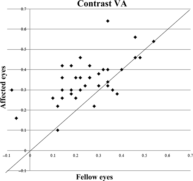
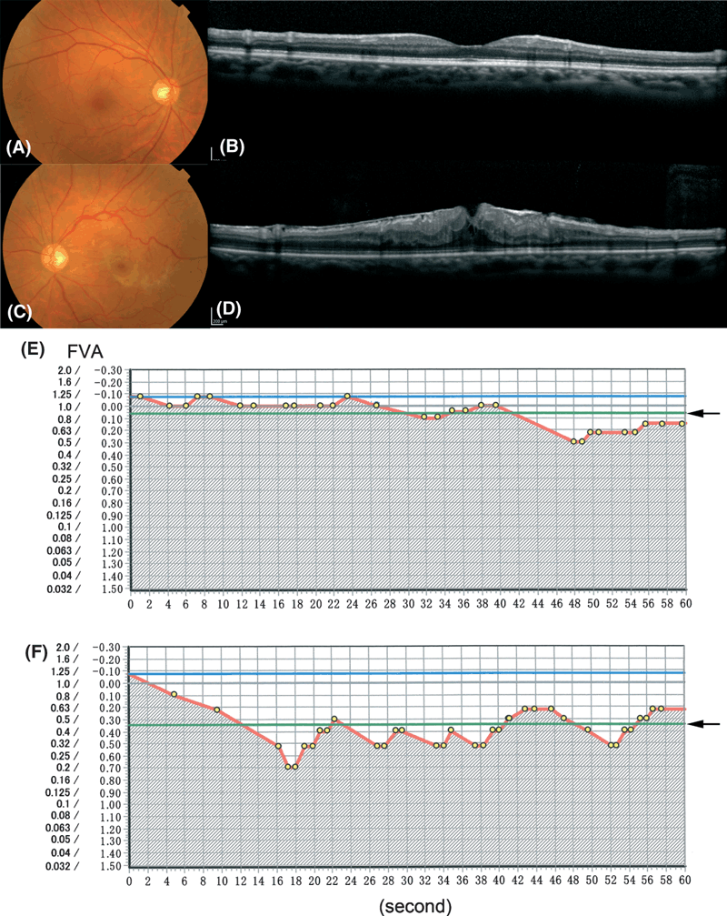
1-3 show the comparison of the FVA score, VMR and contrast VA values between the two eyes of each patient. In most cases, the value was inferior in the affected eye compared with the fellow eye of the same patient. Figure 4 shows a representative case whose conventional Landolt BCVA in each eye was 1.2. FVA scores of affected and fellow eyes were 0.35 and 0.06, and VMRs were 0.85 and 0.95, respectively. Contrast VA was 0.64 in the affected eye and 0.34 in the fellow eye.

Comparison of the functional visual acuity (FVA) score between the affected and fellow eye in each patient. The FVA score for the affected eye versus the fellow eye was plotted for each patient. The FVA score in the affected eye was inferior to the fellow eye in most patients. FVA: functional visual acuity.

Comparison of the visual maintenance ratio (VMR) value between the affected and fellow eye in each patient. The VMR score for the affected eye versus the fellow eye was plotted for each patient. VMR in the affected eye was inferior to that in the fellow eye in most patients. FVA: functional visual acuity. VMR: visual maintenance ratio that shows integral of the FVA in each time-point.

Comparison of the contrast visual acuity (VA) value between the affected and fellow eye in each patient. The contrast VA score for the affected eye versus the fellow eye was plotted for each patient. The contrast VA in the affected eye was inferior to the fellow eye in most patients. VA: visual acuity.

Fundus photograph, optical coherence tomography (OCT) image and FVA record of a representative case. Fundus photograph, OCT and FVA record of the fellow eye (A, B, E), and the affected eye (C, D, F), in the same patient. FVA was shown in a Landolt dot score/logMAR score. Arrows showed FVA score which is the average of FVA in each time-point plotted in the record. Shadow area divided by FVA score (red line) beneath the starting VA (blue line) showed VMR which is the integral of the FVA in each time-point. OCT: optical coherence tomography. VA: visual acuity. FVA: functional visual acuity. VMR: visual maintenance ratio that shows integral of the FVA in each time-point.
Table 2 shows the relationships between the parameters. There was significant correlation between each FVA value and the respective contrast VA, as well as between the FVA score and VMR. In contrast, there was no correlation between each FVA value or contrast VA, and the results of the Schirmer test, corneal density or cataract density.
| Fellow eyes (n = 36) | Affected eyes (n = 36) | |||
|---|---|---|---|---|
| CC | p-value | CC | p-value | |
| FVA-VMR | −0.98 | < 0.001* | −0.92 | < 0.001* |
| FVA-contrast VA | 0.56 | < 0.001* | 0.40 | 0.02* |
| VMR-contrast VA | −0.55 | < 0.001* | −0.37 | 0.02* |
| FVA-Schirmer test | −0.08 | 0.57 | −0.21 | 0.19 |
| FVA-corneal density | −0.09 | 0.60 | −0.01 | 0.94 |
| FVA-lens density | 0.04 | 0.81 | 0.15 | 0.38 |
| VMR-Schirmer test | 0.08 | 0.69 | 0.26 | 0.13 |
| VMR-corneal density | 0.11 | 0.53 | 0.01 | 0.97 |
| VMR-lens density | 0.04 | 0.83 | −0.18 | 0.30 |
| Contrast VA-Schirmer test | 0.12 | 0.49 | 0.10 | 0.58 |
| Contrast VA-corneal density | −0.12 | 0.49 | −0.13 | 0.47 |
| Contrast VA-lens density | 0.17 | 0.31 | 0.14 | 0.39 |
- VA = visual acuity, FVA = functional visual acuity, VMR = visual maintenance ratio that shows integral of the FVA in each time-point, CC = correlation coefficient.
- * p < 0.05.
Discussion
We demonstrated that the FVA values, that is, the FVA score and VMR, and contrast VA were lower on average in eyes affected with epiretinal membrane compared with unaffected fellow eyes. Therefore, these values reflected early changes in central visual function caused by epiretinal membrane. In contrast, no change was detected by conventional Landolt BCVA. The FVA values correlated with each other and with the contrast VA. On the other hand, all three values were independent of dry eye, as judged by the Schirmer test, and of opacities of the cornea or lens, in this study.
The FVA system has been used to measure central visual function in patients with anterior eye diseases and conditions, such as Sjögren syndrome (Goto et al. 2002), Stevens–Johnson syndrome (Kaido et al. 2006), post-LASIK (Tanaka et al. 2004), mild cataract (Yamaguchi et al. 2011) and posterior capsule opacification after cataract surgery (Wakamatsu et al. 2011). The FVA system is sensitive enough to detect a difference in central visual function between patients with Stevens–Johnson syndrome, which has more severe dry eye findings (Kaido et al. 2006), and those with Sjögren syndrome. It also detects the improvement after the removal of mild cataract with a preoperative BCVA > 20/25, even when the conventional Landolt BCVA does not change (Yamaguchi et al. 2009, 2011).
In this study, the FVA score and VMR reflected the presence of epiretinal membrane independent of the condition of the anterior eye. Thus, the FVA system was also applicable to the posterior eye disease.
The FVA value reflects the temporal change in continuous VA, but because it uses a brief (2-second) display of the optotype, it also reflects the subject’s quick recognition of the target. The impairment measured by FVA in dry eye is explained by the temporary reduction in ocular surface smoothness during gaze, due to tear-film destruction; in dry eye patients, the VMR impairment is obvious (Kaido et al. 2006). However, in patients with epiretinal membrane, the changes in FVA value may reflect an impairment in patients’ ability to quickly identify the optotypes caused by irregularity of the neural retina related to the epiretinal membrane. When the patients do not make a judgment within 2 seconds, the FVA score is reduced; therefore, VMR, the integral of the FVA score, is also reduced.
Contrast VA is a traditional method for detecting slight changes in visual acuity, and like the Landolt BCVA chart, the contrast VA chart can be stared at for a long period. Therefore, the use of this test controls for the patients’ ability to recognize the target during allowed time to examine it (in contrast to the 2-second limit in the FVA test). The FVA values were correlated with the contrast VA results; therefore, this finding supports the idea that the FVA values represent changes in visual function and not simply a slow response time on the part of the subjects. In addition, patients with epiretinal membrane sometimes have difficulty reading. Because reading requires strings of characters to be recognized in a very short time, this problem may be reflected in the FVA system rather than in the contrast VA. Further observation will be required to assess the meaning of each method’s results.
Several methods for evaluating visual function in patients with epiretinal membrane have been previously reported. M-charts (Matsumoto 2003; Arimura et al. 2011), PreView PHP (Arimura et al. 2011), the SLO procedure for recording the area of distorted vision (Arndt et al. 2007) and the Amsler chart (Shinoda et al. 2000) are used to evaluate the metamorphopsia. These methods could be useful when the metamorphopsia is obvious; however, the influence of the epiretinal membrane on visual function may also cause blurred vision. Both distorted vision and blurred vision are reflected by the FVA system and contrast VA values.
Microperimetry 1 (MP1) (Jiang et al. 2011) and two electrophysiological methods, multifocal visual evoked potential (mfVEP) (Jiang et al. 2011) and multifocal electroretinogram (mfERG) (Shimada et al. 2011; Kim et al. 2012), all of which are used to measure focal visual function, have been examined for their ability to assess the visual impairment in patients with epiretinal membrane. However, when the BCVA is relatively good, MP1 is not sensitive enough to detect slight changes, and mfVEP has large fluctuations (Jiang et al. 2011), so these methods are unlikely to be useful for detecting early changes in visual function caused by epiretinal membrane. Whether or not mfERG is valuable for patients with rather good visual function, such as BCVA ≥ 1.0, is currently an open question.
Whether and when to use surgery to treat patients with epiretinal membrane is now mainly determined by the results of conventional Landolt BCVA. Thus, patients with notable distorted and/or blurred vision sometimes do not undergo vitreous surgery, because of their good conventional Landolt BCVA results, and are kept waiting for the surgery until their conventional VA gets worse. However, with recent significant progress in vitreous surgical techniques, surgery may be used in earlier and milder cases. Moreover, preoperative visual function is reported to be correlated with visual prognosis (Lim & An 2011), which supports the idea of earlier indication of surgery. The FVA system and/or contrast VA may be useful for determining the timing of surgical treatment, because they more sensitively detect the impairment of visual quality, although further studies are required.
Recent views emphasize the importance of quality of vision for an individual’s quality of life. Our results indicate that the FVA system and/or contrast VA would be valuable for detecting early and mild changes in central visual function due to retinal diseases as well as anterior eye diseases.
Acknowledgements
We thank Dr. Minako Kaido for technical advice and the orthoptists and medical staff of our clinic for technical assistance.


