A study on the morphoquantitative and cytological characteristics of the bulbar conjunctiva of the maned wolf (Chrysocyon brachyurus; Illiger, 1815)
Abstract
The maned wolf, Chrysocyon brachyurus, is a near-threatened carnivore inhabiting the Brazilian Cerrado. Few studies have been conducted on this species, and even fewer have explored its ophthalmological characteristics. Vision is critical to wild canids; thus, this study aimed to provide a morphoquantitative description of the bulbar conjunctiva of the maned wolf using cytological and histological analyses. Ten healthy maned wolves from a conservational centre, including 4 females and 6 males aged 1–12 years (6.5 ± 2.8), were included in the study. The samples for cytological analysis were collected from the inferior conjunctival sac using a cytobrush, and conjunctival tissue was collected for histological analysis from the temporal canthus zone. The cytological samples were stained using the Papanicolaou method, and the histological sections were stained using haematoxylin and eosin, Periodic acid–Schiff, picrosirius red and Masson's trichrome stains. The cytological samples were studied for stain quality, and the different cell types were counted. Histological examination was used to determine tissue types in the conjunctiva and their proportions. Analyses revealed a stratified squamous epithelium with some goblet cells and eventual pigmentation in the basal layer. Loose connective tissue with the presence of some mononuclear and polymorphonuclear cells was also observed. The epithelium of the maned wolf's bulbar conjunctiva resembles that of dogs and other carnivore species; furthermore, its physiological and pathological responses were similar to those of other carnivore species.
1 INTRODUCTION
The maned wolf (Chrysocyon brachyurus) belongs to the Carnivora order and Canidae family, the same as those of the domestic dog. This is an omnivore species and frequently consumes both the wolf's plant (Solanum lycocarpum) and small mammals. The maned wolf has crepuscular habits, including walking as long as 8 hr per night and resting during the day in proximity to streams and creeks. The wolf is mostly a solitary species and is only seen accompanied by other wolves during their reproduction period or when assisting their puppies (Gomes, 2007; Massara et al., 2012; Melo et al., 2007).
Currently, due to human activity, the maned wolf is considered a near-threatened species (Cáceres et al., 2010; Paula and DeMatteo, 2017). In Brazil, it is not uncommon to find this species in captivity or conservation centres for the purpose of protection. In the wild, the maned wolf is primarily found in the Brazilian Cerrado, which is its favoured habitat for reproduction and hunting. Occasionally, it may inhabit Atlantic forest regions, close to the rocky fields that resemble the Brazilian Cerrado (Coelho et al., 2008; Massara et al., 2012). Moreover, the animal can be found in urban regions looking for easier access to food in response to loss of its natural habitat caused by deforestation and urban growth (Coelho et al., 2008). Migration of the wolf to urban regions facilitates contact with domestic animals and increases the risk of infectious and parasitic diseases (Silveira et al., 2016).
Vision is an important sense of survival for many species. Vision quality, effectiveness and adaptability may vary according to the position of the species in the trophic network (Beauchamp, 2013; Bhattacharyya et al., 2017). The conjunctiva is an ocular structure that maintains visual integrity.
The conjunctiva is a semi-mobile membrane that is anatomically divided into palpebral, forniceal and bulbar. It is covered by the epithelium, varying from prismatic to squamous, depending on its location, and is supported by loose connective tissue (Latkovic & Nilsson, 1978; Bolzan et al., 2005). The conjunctiva participates in ocular surface defence (Knop & Knop, 2000), acting as a mechanical barrier to external agents, and promotes stability of the tear film, reflecting presence of focal aggregations of lymphoid tissue (Maggs et al., 2013; Samuelson, 2014).
Few studies on the vision and ocular structures of maned wolves are available. Consequently, ophthalmic parameters of domestic species are extrapolated to wild species; however, each species has its morphofunctional peculiarities. Schirmer tear test 1 and intraocular pressure of maned wolves were discussed by Honsho et al. (2016); however, there are no published data on the cytological and histologic features of its conjunctiva. Thus, this study aimed to describe the cytological and histological features of the bulbar conjunctival layers in maned wolves.
2 MATERIALS AND METHODS
2.1 Ethics and animals
This study was conducted in accordance with the guidelines set by the Association for Research in Vision and Ophthalmology for the Use of Animals (ARVO, 2018). All protocols were approved by the Ethics Committee on the Use of Animals of the University of Franca (protocol number: 031/13). Twenty eyes of 10 healthy maned wolves, including 4 females and 6 males aged 1–12 years (6.5 ± 2.8), were studied. The animals were procured from an environmental development centre, belonging to the Brazilian company of metallurgy and mining, located in the city of Araxá, MG, Brazil.
All maned wolves were systemically anaesthetized (Dias et al., 2015), and their ocular surfaces were desensitized using proxymetacaine (Anestalcon®-Alcon S/A, 04575–060, São Paulo, Brazil) and evaluated for health.
2.2 Collection, preparation and analyses of conjunctival samples
Epithelial cells were collected using one sterile cervical cytobrush (Endobrush®-Alamar Tecno Cient. Ltda., 09961–720, Diadema, Brazil) that was introduced into the medial lower conjunctival sac and turned clockwise 6 times. Tissue fragments of the bulbar conjunctiva were collected from the temporal canthus of both eyes and placed on filter paper (Whatman® Filter Paper N02SPS4700; Sigma-Aldrich, St. Louis, MO, USA) inside individual bottles filled with buffered formalin (Synth Formaldeído solução P.A.®-Labsynth Prod. Lab. Ltda., 09990–080, Diadema, Brazil) and routinely embedded in paraffin.
The cytological preparations (n = 20) were stained using the Papanicolaou method (Papanicolaou®-Newprov Prod. Lab. Ltda., 83323–020, Pinhais, Brazil). Histologic slides (n = 24) were prepared using haematoxylin and eosin (HE), Periodic acid–Schiff (PAS), Masson's trichrome (MT) and picrosirius red stains.
The cytological samples were evaluated for quantitative (Table 1) and qualitative aspects of the bulbar conjunctiva. The qualitative aspect was based on cell preservation and cellular components in the cytoplasm, as well as the presence or absence of the nucleus, nucleus shape, cytoplasm and presence or absence of leucocytes and goblet cells.
| Quantitative evaluation | |
|---|---|
| Stain quality and difficulty of evaluation and score. | |
| Score I | Well-stained cells and easy to see them in microscopic fields, feasible counting with certain ease. |
| Score II | Not so well-stained cells with impaired visibility and evaluation, and achievement of counting with some difficulty. |
| Cellularity and cells distribution per microscopic fields | |
| Score I | Discrete cellularity with heterogeneous distribution in the microscopic fields. |
| Score II | Moderate cellularity, with heterogeneous distribution in the microscopic fields |
| Score III | Moderate to marked cellularity, with homogeneous distribution in the microscopic fields. |
We used 10 random microscopic fields per slide for cell counting, based on previous studies (Borges et al., 2012). The cells were classified as enucleated superficial, nucleated superficial, intermediate, basal and goblet cells and leucocytes. The percentages of cell types per slide were calculated after counting (Table 2).
| Animal | Eye | % NSC | % ASC | % IC | % BC | % GC | % L |
|---|---|---|---|---|---|---|---|
| 1 | R | 2.22 | 0.74 | 65.09 | 9.76 | 0.89 | 21.30 |
| L | 4.14 | 0.29 | 73.71 | 10.71 | 3.14 | 8.00 | |
| 2 | R | 2.55 | 0.13 | 55.54 | 11.97 | 6.37 | 23.44 |
| L | 2.21 | 5.80 | 68.51 | 14.09 | 1.10 | 8.29 | |
| 3 | R | 6.96 | 0.70 | 75.64 | 10.44 | 4.64 | 1.62 |
| L | 3.94 | 0.79 | 86.00 | 5.92 | 0.39 | 2.96 | |
| 4 | R | 0.55 | 0.18 | 83.95 | 10.70 | 2.77 | 1.85 |
| L | 12.41 | 0.38 | 66.54 | 16.54 | 3.76 | 0.38 | |
| 5 | R | 2.40 | 0.34 | 78.42 | 16.78 | 1.37 | 0.68 |
| L | 6.28 | 1.45 | 78.50 | 11.35 | 0.48 | 1.93 | |
| 6 | R | 4.50 | 4.70 | 73.97 | 14.68 | 0.39 | 1.76 |
| L | 2.61 | 8.41 | 71.59 | 14.49 | 0.00 | 2.90 | |
| 7 | R | 2.26 | 3.25 | 79.21 | 8.35 | 4.38 | 2.55 |
| L | 0.83 | 2.97 | 82.52 | 8.56 | 3.33 | 1.78 | |
| 8 | R | 1.53 | 5.02 | 77.73 | 12.23 | 1.97 | 1.53 |
| L | 4.45 | 11.52 | 73.30 | 7.59 | 1.57 | 1.57 | |
| 9 | R | 2.05 | 5.38 | 75.38 | 14.62 | 2.05 | 0.51 |
| L | 1.29 | 4.06 | 78.60 | 12.73 | 0.55 | 2.77 | |
| 10 | R | 2.68 | 6.70 | 75.22 | 14.29 | 0.67 | 0.45 |
| L | 0.88 | 2.46 | 83.33 | 10.70 | 0.88 | 1.75 | |

|
- | 3.33 ± 0.61 | 3.26 ± 0.70 | 75.44 ± 1.73 | 11.82 ± 0.66 | 2.03 ± 0.39 | 4.40 ± 1.46 |
- Abbreviations:
 ± sem, mean ± standard error of mean of both maned wolves’ eyes; ACS, anucleated superficial cells; BC, basal cells; GC, Goblet cells; IC, intermediate cells; L, leucocytes; NSC, nucleated superficial cells; R, right, L: left.
Histological samples were evaluated using a light microscope (Leica DFC295-Leica Microsystems Ltd, Müchen, Germany), except the picrosirius samples, which required a polarized light microscope (Olympus BX53-Olympus Corporation, Tokyo, Japan).
Qualitative and quantitative criteria were used for histological evaluation. We qualitatively evaluated the HE stained tissue structures as a whole for cellular arrangement, configuration, tissue architecture, MT (collagen fibres), PAS (goblet cells by identifying the typical mucus staining) and picrosirius (isolated collagen) to understand tissue layout and structure. The quantitative analysis included MT slides to determine the percentages/proportions of bulbar conjunctiva tissues.
Twenty sections were used to quantify bulbar conjunctive tissue components [epithelium and connective tissue (CT)] using point-counting (Howard & Reed, 2005). The test-point system had a point spacing of 45–47 µm, which was placed over each section. Points overlaid on the epithelium (E) and connective tissue were counted as well as those overlaying the bulbar connective tissue (BCT). Percentages of bulbar CT were calculated using ratios that were multiplied by 100.
Percentage of epithelium: ΣE/ΣBCT × 100.
Percentage of CT: ΣCT/ΣBCT × 100.
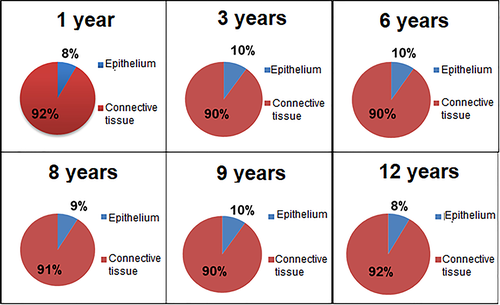
The percentages of the epithelial tissue and CT of the conjunctiva were separately calculated for one-year-old (n = 3), three-year-old (n = 3), six-year-old (n = 1), eight-year-old (n = 1), nine-year-old (n = 1) and twelve-year-old (n = 1) animals.
2.3 Data analysis
Statistical analysis was performed for the qualitative cytological evaluation using two-way analysis of variance (ANOVA). Where α < 0.05, a Sidak's multiple comparisons post-test was performed. ROUT’s and Pearson's correlation tests were performed with α < 0.1 and α < 0.05, respectively, to eliminate outliers. Quantitative cytological evaluation results were evaluated using classification scores and the percentages of different scores.
No statistical analysis was used for qualitative histological evaluation. Two-way ANOVA was used for quantitative histological evaluation, comparing values from both epithelial and CT samples.
3 RESULTS
3.1 Quantitative cytological evaluation
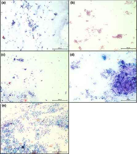
The results were grouped according to the stain quality, difficulty of evaluation, and scores (Figure 1). Fourteen samples (70%) were scored I (Figure 1a) and six (30%) were scored II (Figure 1b), suggesting that most of the microscopic fields were easily observed and evaluated. Nine samples (45%) were scored I (Figure 1c), six (30%) were scored II (Figure 1d) and five (25%) were scored III (Figure 1e) for cellularity and cell distribution per microscopic field.
3.2 Qualitative cytological evaluation
The cells showed preserved morphology; the edges were easily visualized as structures such as cytoplasm and nucleus (Figure 2). Cytoplasm varied from light pink to medium blue, depending on the staining intensity. Nuclei varied from darker pink to light purple and were always well distinguished. All cells had nuclei, with the obvious exception of anucleated superficial cells. Small numbers of leucocytes (lymphocytes, neutrophils, and monocytes) and goblet cells were dispersed in the analysed fields (Table 2). The shapes of each cell type were typical, except for those of the intermediate cells, which varied from oval to polygonal.
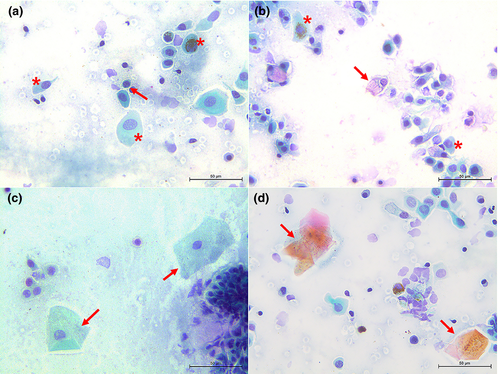
Basal cells of the conjunctival epithelium were rounded with centralized nuclei and relatively little cytoplasm (Figure 2a). Intermediate cells presented with centralized or eccentric round nuclei, and overall shape varied from rounded to polygonal and prismatic. The quantity of cytoplasm varied from small to large amounts, depending on the cell shape. Moreover, a pigment was observed in the cytoplasm of several cells (Figure 2a,b).
The goblet cells showed the presence of eccentric nuclei and a highly vacuolized cytoplasm. The cell sizes varied from elongated to rounded (Figure 2b). Nucleated and anucleated superficial cells were polygon-shaped and were the largest among those in the microscopic fields; some showed pigment granules in the cytoplasm. The nuclei of nucleated cells were centralized and occupied a small fraction of cytoplasmic area. Eventually, the cell edges folded up. (Figure 2c,d). Several regions with cell conglomerates could not be evaluated.
A high percentage of leucocytes was found in the right eyes of animals 1 and 2. Two-way ANOVA test was performed along with the multiple comparisons. Sidak's test was used to examine differences between percentages of cell types, and no significant differences were found between the left and right eyes.
The values obtained from the qualitative cytological evaluation, along with the mean and standard deviations, are shown in Table 2. Results from the Pearson's test showed a strong negative correlation of −0.69 (p < .001) between leucocytes and intermediate cell percentages (Table 3).
| % NSC | % ASC | % IC | % BC | % GC | % L | |
|---|---|---|---|---|---|---|
| % NSC | −0.19 | −0.32 | 0.22 | 0.20 | −0.16 | |
| % ASC | −0.19 | −0.06 | 0.05 | −0.41 | −0.30 | |
| % IC | −0.32 | −0.06 | −0.34 | −0.32 | −0.69* | |
| % BC | 0.22 | 0.05 | −0.34 | −0.14 | −0.15 | |
| % GC | 0.20 | −0.41 | −0.32 | −0.14 | 0.32 | |
| % L | −0.16 | −0.30 | −0.69* | −0.15 | 0.32 |
Note
- The values with asterisk represent the strong negative correlation
- Abbreviations: ACS, anucleated superficial cells; BC, basal cells; GC, Goblet cells; IC, intermediate cells; L, leucocytes; NSC, nucleated superficial cells.
3.3 Quantitative histological evaluation
Percentages of the epithelial and CTs observed using the conjunctiva of animals of different ages are shown in Figure 3. No statistical differences (p > .9) were observed among these animals.
3.4 Qualitative histological evaluation
Tissues examined displayed the epithelial and CTs (Figure 4). The epithelium was found to be stratified squamous and showed a number of layers, varying from three to seven. Deeper cells had rounded or prismatic shapes with well-stained rounded or oval nuclei. Cells in the intermediate layer were rounded and showed the same nuclei characteristics. Superficial cells were squamous and were stained more intensely than those in other layers that had flat and well-stained nuclei. A few melanocytes were noted in the basal layer of the epithelium (Figure 4a). Goblet cells were visualized using the PAS stain (Figure 4b).
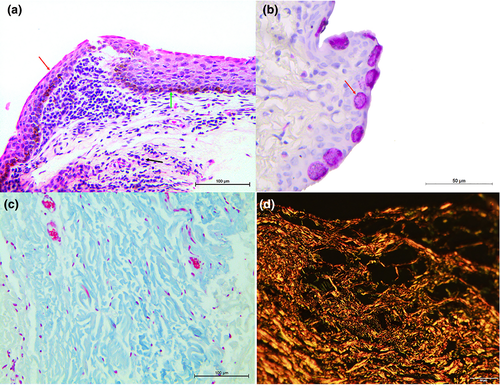
The CT was classified as loose and presented with fibroblasts, many blood vessels, few macrophages, plasmacytes and lymphocytes, generally in the perivascular region. MT staining revealed collagen fibres in the CT (Figure 4c) and picrosirius red stain showed shade differences in interference colours, varying among green, yellow and red under polarized light (Figure 4d). Tissues of animals 1 and 7 displayed bilateral polymorphonuclear and mononuclear cells infiltrates, respectively (Figure 4a).
4 DISCUSSION
The conjunctiva is the most exposed mucous membrane of an organism, and its morphofunctional design is essential for the maintenance of ocular surface health (Maggs et al., 2013). Thus, this study evaluated cells and tissues of bulbar conjunctiva of the maned wolves, aiming at obtaining relevant information on these structures (Samuelson, 2014).
There are various procedures for procuring cell samples for conducting cytological examination. Most procedures are based on exfoliative and impression cytology that differ in the quality of the obtained cells. Exfoliative cytology usually allows the collection of more intermediate cells in addition to those that occupy most of the superficial layers of the epithelium (Maggs et al., 2013; Perazzi et al., 2017). In this study, cells were collected using a cytobrush and were well-preserved; their cytoplasm and nuclei were easily observed. Abundant cellularity was observed in microscopic fields, which was consistent with previous studies (Borges et al., 2012; Perazzi et al., 2017). The exfoliative cytology of domestic carnivores (dogs and cats) usually presents good cellularity (40% of dog samples and 70% of cat samples) (Perazzi et al., 2017). In this study, this percentage was 55%. Cell types in this study did not differ from those recorded in exfoliative or impression cytological preparations from healthy dogs (Bolzan et al., 2005; Borges et al., 2012).
Different epithelial cell types could be observed in cytological preparations, from basal to superficial. Superficial cells presented in polygonal shapes, and eventually their edges are folded, as previously verified in dogs (Bolzan et al., 2005). Furthermore, they presented large amounts of cytoplasm and some cells contained melanin granules. The nuclei were small and centralized (Bolzan et al., 2005). Basal cells had rounded/oval shapes with centralized nuclei (Bolzan et al., 2005; Perazzi et al., 2017). Variation was observed in intermediate cell shape—oval to polygonal and nuclei-positioned in the centre or edge of cytoplasm (Bolzan et al., 2005). Eccentric nuclei and cytoplasm have also been corroborated by Bolzan et al. (2005) and Perazzi et al. (2017).
The results of this study are consistent with those of Oriá et al. (2015) for the examination of the Sambar cervid, Rusa unicolor, where most epithelial cells were prism-shaped with certain melanin content. Furthermore, the values of intermediate cells are in accordance with those reported by Borges et al. (2012). The number of anucleated superficial cell values was also similar; 4.55% in the study by Borges et al. (2012) and 3.26% in the present study.
All samples showed the presence of leucocytes; this finding was contradictory to that of Borges et al. (2012) in healthy dogs. The presence of leucocytes is typically a sign of an inflammatory process; however, the animals did not present with clinical/macroscopic signs that could indicate inflammation of the ocular surface. The presence of leucocytes was unexpected, and animals showed no visible alteration of the ocular surface, population; moreover, the animals were free-living and did not present any visible alteration in the ocular surface. Thus, leucocytes might be typical in the conjunctiva of maned wolves. This possibility has not been assessed before (Borges et al., 2012; Honsho et al., 2012; Perazzi et al., 2017). Pearson's correlation demonstrated a strong negative correlation between intermediate cells and leucocytes. This indicates that when higher numbers of leucocytes were found, fewer intermediate cells were found. An explanation for this correlation was not elucidated.
Studies on domestic cats used the same methodology as the present study, and Honsho et al. (2012) and Venancio et al. (2012) did not report the presence of goblet cells. Conversely, Bolzan et al. (2005) and Perazzi et al. (2017) identified these cells in dogs (25% of samples contained goblet cells) (Perazzi et al., 2017). In this study, goblet cells were observed in 95% of samples, demonstrating that the conjunctival exfoliation cytology method was effective.
Goblet cells produce mucin, which is critical for the important functions of the visual system, such as protection against pathogens and acting as a mechanical barrier (Davidson & Kuonen, 2004; Gipson, 2016). Different percentages of goblet cells between this study and that of Perazzi et al. (2017) may be related to differences between species or could be attributable to the free-living conditions of maned wolves. Free-living conditions may offer greater risks of trauma than domestic life, and a higher number of goblet cells is associated with higher mucin production for ocular surface protection (Moore & Tiffany, 1979; Davidson & Kuonen, 2004; Maggs et al., 2013).
Conjunctival goblet cell distribution and density were histologically studied in dogs (Moore et al., 1987), chinchillas (Voigt et al., 2012), guinea pigs (Gasser et al., 2011), rabbits (Doughty, 2013) and cats (Sebbag et al., 2016); moreover, the density was found to be less in the bulbar conjunctiva (Moore et al., 1987; Voigt et al., 2012; Sebbag et al., 2016). In this study, we collected samples from the temporal region of the bulbar conjunctiva, and the presence of goblet cells was 100% in the histologic samples.
PAS stain facilitates the visualization of goblet cells, staining the mucin produced and secreted by these cells magenta (PAS-positive) (Gasser et al., 2011), Isola et al., 2013) and Sebbag et al., 2016). This stain distinguishes mucin from other structures; moreover, the typical oval shape already been described (Sebbag et al., 2016) was observed.
The bulbar conjunctiva is not histologically the region of major goblet cell populations (Moore et al., 1987; Sebbag et al., 2016). However, percentages of goblet cells were significantly less in dogs (0.14% in the study by Borges et al. (2012) and 2.04% in this study). These results might be explained by the methodology used (exfoliative cytology), which is not designed to accurately quantify the percentage of goblet cells. Furthermore, the higher goblet cell concentration in maned wolves seems is likely attributable to the additional protection of the ocular system, as noted above.
The samples used for histological evaluation were grouped according age to identify possible age-related differences between the epithelium and CTs. Such differences were explored in a study by Ostergaard et al. (2013) using giraffe hearts. However, no differences in the epithelial and connective tissues were identified across ages.
The palpebral and bulbar conjunctiva epithelium may vary between species and conjunctival regions (Latkovic & Nilsson, 1978; Goller & Weyrauch, 1993; Bourges-Abella et al., 2006; Hendrix, 2013; Oriá et al., 2015). In general, the conjunctiva is formed using stratified squamous epithelium (Young & Prasse, 2009; Hendrix, 2013; Maggs et al., 2013) or prismatic stratified epithelium. Peripheral cuboidal cells are observed in guinea pigs (Latkovic & Nilsson, 1978).
The conjunctival epithelium in dogs is non-keratinized (Hendrix, 2013) and squamous stratified (Goller & Weyrauch, 1993). The maned wolf is also a canid and may display the same epithelium type; the temporal bulbar conjunctiva in the maned wolf is also stratified squamous.
The CT is seen deeper in the epithelium (Maggs et al., 2013). Collagen is present in the CT, as observed using specific stains such as MT (Suvarna et al., 2013) and picrosirius (Whittaker & Kloner, 1992).
The number of leucocytes in the conjunctiva of maned wolves is greater than those in dogs (Borges et al., 2012). The presence of leucocytes in the conjunctiva is not diagnostic for local inflammation (Azevedo et al. (2009). Leucocytes are important for conjunctiva health once the lymphoid tissue aggregates occur in ocular surface defences (Knop & Knop, 2000).
The conjunctiva of the maned wolf bulbar resembles that of domestic dogs in structure, suggesting that its physiology and pathological responses may also be similar. Scientific data are scarce for the characterization of maned wolf conjunctiva. Furthermore, samples used in this study were collected from live animals who were kept in captivity. This did not allow the procurement of large numbers of samples as routinely obtained for research on conjunctiva, which frequently uses ex vivo materials. New studies are proposed using the whole conjunctiva, when available, to assess this critical structure of the ocular system.
ACKNOWLEDGMENTS
The authors gratefully acknowledge the financial support of Sao Paulo Research Foundation (FAPESP 2017/23081-7), the Higher Education Personnel Improvement Coordination-Brazil (CAPES, Finance Code 001), Dr. Ewaldo Mattos Junior and Ms. Lilian Toshiko Nishimura for the auxiliary in the anaesthetic monitoring, Dr. Ricardo Andrade Furtado for the statistics and data manipulation, Mr. Jose Luiz de Souza and Dr. Denise Crispim Tavares for the laboratory practices auxiliary and Brazilian Company of Metallurgy and Mining (CBMM).
CONFLICT OF INTEREST
The authors would like to declare no conflict of interest.
Open Research
DATA AVAILABILITY STATEMENT
The data that support the findings of this study are available on request from the corresponding author. The data are not publicly available due to privacy or ethical restrictions.


