Morphological Variations in the Canine (Canis familiaris) Right Ventricle Trabecula Septomarginalis Dextra and a Proposed Classification Scheme
Summary
The trabecula septomarginalis dextra is a structure routinely mentioned in veterinary anatomy. While there are several studies on the morphology of this structure in select animal species, there are some characteristics that are not adequately described. The purpose of this study was to describe and classify this trabecula in the domestic dog using the number of branches that insert onto the ventricular free wall. In the 39 examined hearts, 17.95% were single stranded (type I) and 2.56% were y-shaped (IA). Then, 28.21% were web-like with 3 to 4 points of insertion (IB) and 51.28% were web-like with 5 or more points of insertion (IC). While the purpose of this study was to describe the morphology of this trabecula, it also showed there is variability in this structure within the same species. This undocumented variability could present a problem to researchers who use dogs as an animal model when testing catheters for invasive cardiac monitoring or cardiac stem cell therapy. Also a web-like trabecula could create problems during pacemaker lead placement for the treatment of symptomatic bradycardia because of the potential for catheter entanglement in these branches.
Introduction
In the human, the septomarginal trabecula1 (also known as trabecula septomarginalis) is described as a fleshy trabeculation that extends from the inferior portion of the interventricular septum to the base of the anterior papillary muscle. This structure supports the base of the anterior papillary muscle and carries part of the right branch of the AV bundle across the ventricular cavity. This ensures coordinated contraction of the papillary muscle, right ventricular wall and interventricular septum (Williams, 1999; Loukas et al., 2010; Moore et al., 2010; Banderia et al., 2011; Raghavendra et al., 2013).
In the dog, examination of the right ventricle reveals a thin, band-like structure that extends from the interventricular septum or base of the largest papillary muscle (musculus papillaris magnus) to the parietal wall of the right ventricle. This band (trabecula septomarginalis dextra), similar to the trabecula septomarginalis in the human, provides a short cut for part of the right branch of the AV bundle to the ventricular wall for coordinated contraction of all parts of the ventricle (Evans, 1993; Dyce et al., 1996; International Committee on Veterinary Gross Anatomical Nomenclature, 2005). Beyond this basic description of the trabecula septomarginalis dextra, the veterinary literature indicates that in the dog, this structure may branch near its extremity as it blends with the muscular ridges (trabeculae carneae) of the parietal wall or it may consist of several anastomosing strands that form a plexus within the right ventricle (Evans, 1993).
This unique feature of the canine trabecula septomarginalis dextra is briefly mentioned in two veterinary texts, and a search of the available cardiac literature reveals only two additional studies that examine this trabecula in canine hearts (Truex and Warshaw, 1942; Nickel et al., 1981; Dudziak et al., 1984; Evans, 1993). The first study by Truex and Warshaw (1942) is a comparative analysis of the trabecula septomarginalis (referred to as the moderator band) in humans and several animal species including the dog and cat. The results of this study do not mention any branching of the trabecula septomarginalis dextra in the dog. Instead, it indicates this band is consistently present in the cats examined but absent in the canine hearts examined. The second study by Dudziak et al. (1984) is also a comparative study that uses Canis familiaris along with several other species, and in contrast to the previous work by Truex and Warshaw (1942), a trabecula septomarginalis dextra is consistently present in the right ventricle. In this study, Dudziak et al. (1984) divide the trabecula into two components referred to as septopapillary and papillomarginal. This observation by Dudziak et al. (1984) has also been recently documented in human and primate hearts, and according to these articles, the papillomarginal component in all three species (dog, human and primate) can be single stranded or have branches that insert onto the ventricular wall (Dudziak et al., 1984; Kosinski et al., 2010, 2013).
Because there are very few detailed descriptions on the morphology of the canine trabecula septomarginalis dextra, this present work describes and classifies the branching of this structure, and details the points of attachment within the right ventricular cavity.
Materials and Methods
Canine hearts
A total of 39 canine hearts were collected from pit bull mixes, beagles and tricolour hounds with no known history or symptoms of cardiac disease. All of the hearts used in this study were collected at the time of necropsy. Humane euthanasia of the canines was according to an animal protocol submitted to, reviewed and approved by IACUC. The canine euthanasia was carried out by an IV injection of commercial veterinary euthanasia solution.
The 18 pit bull mixes were 55–70 pounds and approximately 2–5 years of age and consisted of eight females and 10 males. The eight beagles were ~20–30 pounds and approximately 2 years of age and consisted of four females and four males. The 13 hounds were 60–70 pounds, approximately 1 and 1/2 years of age and consisted of nine males and four females.
Following removal of the hearts from the thoracic cavity, the right and left ventricles were flushed with water using the pulmonary trunk and aorta. As water filled these chambers, the heart was gently massaged to remove any blood clots from the chambers. The hearts were then placed in water in small plastic bags and frozen until the time of dissection at which point the organs were then thawed and subsequently placed in a 10% buffered formalin solution and kept refrigerated for the past 12 months.
Opening of the right ventricle
Beginning at the pulmonary orifice, a curvilinear incision was made through the right ventricular wall 0.5 cm to the left and parallel to the paraconal interventricular groove. A second horizontal incision was made 0.5 cm ventral to the right coronary groove and extended towards the caudal surface of the heart. This horizontal incision was connected to the curvilinear incision in the right ventricular wall at the level of the pulmonary orifice, and the cranioventral surface of the parietal wall of the right ventricle was reflected towards the apex of the heart and the ventricular cavity was opened.
Results
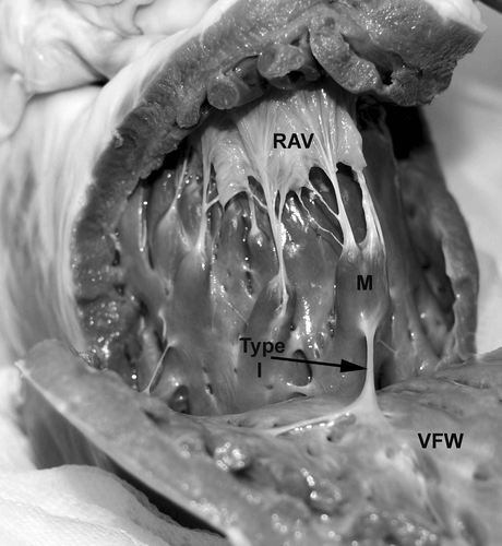
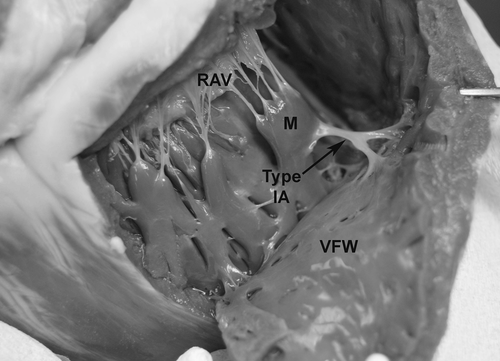
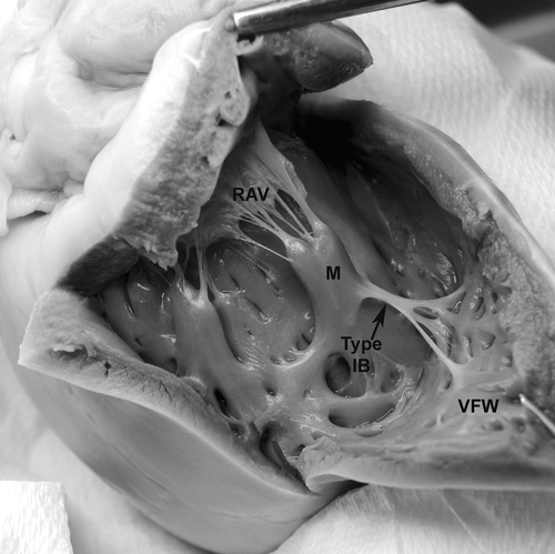
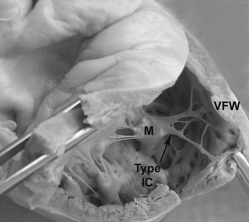
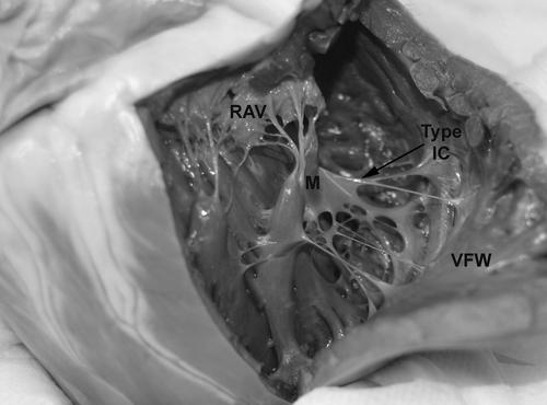
Through gross examination, a trabecula septomarginalis dextra was observed in the right ventricle of all 39 canine hearts. These trabeculae extended from the musculus papillaris magnus to the right ventricular wall and were divided into four different groups using the number of branches that inserted onto the ventricular free wall (Table 1, Figs 1-5).
| Type | Description | Number observed | Percentage |
|---|---|---|---|
| I | Single point of attachment on the Musculus papillaris magnus and ventricular free wall, single stranded | 7 | 17.95 |
| IA | Y-shaped with a single point of attachment on the Musculus papillaris magnus and 2 points of insertion on the ventricular free wall | 1 | 2.56 |
| IB | Web-like with a single point of attachment on the Musculus papillaris magnus and 3 to 4 points of insertion on the ventricular free wall | 11 | 28.21 |
| IC | Web-like with a single point of attachment on the Musculus papillaris magnus and 5 or more points of insertion on the ventricular free wall | 20 | 51.28 |
| Total number | 39 |
Discussion
In the dog, the trabecula septomarginalis dextra is described as a thin, band-like structure that extends from the interventricular septum or the base of the largest papillary muscle to the parietal wall of the right ventricle (Dyce et al., 1996; Evans, 1993; International Committee on Veterinary Gross Anatomical Nomenclature, 2005). While the general course and function of this band is similar to that of other animal species, the morphology of this structure in the dog is somewhat different and highly variable.
Previous studies on the trabecula septomarginalis in the right ventricle of human hearts describe this structure as a fleshy trabeculation that extends from the inferior portion of the interventricular septum to the base of the anterior papillary muscle. From these previous studies, Benninghoff (1942) is the first to describe two components of the trabecula septomarginalis: the septopapillary, which connects the interventricular septum to the anterior papillary muscle, and the papillomarginal, which connects the papillary muscle to the anterior ventricular wall. Using this early information, three studies describe branching of the papillomarginal component before its insertion onto the ventricular free wall (Dudziak et al., 1984; Kosinski et al., 2010, 2013). The first study by Dudziak et al. (1984) on the right ventricle of several carnivores briefly documents branching of the papillomarginal portion in C. familiaris but does not provide any description about the morphology. The two more recent studies on the trabecula septomarginalis in human and primate hearts describe branching of the papillomarginal component and document several types of trabeculae using topographic criteria and its relation to the anterior papillary muscle (Kosinski et al., 2010, 2013).
In the studies on the trabecula septomarginalis of human and primate hearts, Kosinski et al. (2010, 2013) describe four types of trabeculae. Types I, II and III are described as single trabecula that extend from the interventricular septum to the anterior ventricular wall or to the anterior papillary muscle on the ventricular wall. Types I, II and III are not divided into septopapillary or papillomarginal components. Type IV is a trabecula septomarginalis that is divided into septopapillary and papillomarginal components but is then divided into three subtypes which are labelled as A, B and C because of the morphological variability of the papillomarginal component.
In all three subtypes of type IV, the septopapillary component is a single well-developed structure, but the papillomarginal component is characterized by morphological variability. In these human and primate studies, type IVA is a trabecula in which the anterior papillary muscle is centrally located and the septopapillary and papillomarginal are equal in length. In type IVB, the anterior papillary muscle divides the trabecula into its two components, but the papillary muscle is positioned closer to the anterior ventricular wall, thus making the septopapillary portion longer than the papillomarginal portion. In type IVC, the anterior papillary muscle originates from the anterior ventricular wall and unequally divides the trabecula into a septopapillary and papillomarginal component. The unique feature of this subtype (IVC) is that in humans and primates, the papillomarginal component is a non-uniform structure that is heterogeneous in its size, course and the number of branches from it to the anterior ventricular wall (Kosinski et al., 2010, 2013).
Examination of the trabecula septomarginalis dextra in the right ventricle of the dog reveals a distinct band-like structure extending from the musculus papillaris magnus (i.e. anterior papillary muscle) to the ventricular free wall. In these hearts, it is not possible to consistently identify the septopapillary component of this trabecula and only two canine hearts of the 39 used for this study have a very small band-like structure extending from the interventricular septum to the musculus papillaris magnus. Previous work on this trabecula in select carnivores (including dogs), primates, humans and goats indicates this structure is divided into septal, septopapillary and papillomarginal components (Dudziak et al., 1984; Kosinski et al., 2010, 2013; Leao et al., 2010). In the study on select carnivores, Dudziak et al. (1984) indicates that in addition to the septopapillary and papillomarginal components described by Benninghoff (1942), there is also a ‘hidden part’ of the trabecula septomarginalis within the interventricular septum. This observation seems to correspond to the work of Banderia et al. (2011) on the human trabecula septomarginalis which describes a septal component before the septopapillary component. Banderia et al. (2011) describe the septal component as a fleshy column of tissue along the interventricular septum that is only visible by dissection. Then in a study on goats, Leao et al. (2010) describe the septal component as a flattened cord attached to the interventricular septum that detaches to go towards the papillary muscle as the septopapillary component. However, this study also indicates the septal and septopapillary components are not consistently visible in the goat hearts examined. Based on these previous descriptions, it is believed that the septopapillary component of the canine trabecula septomarginalis dextra is present but not well developed or easy to visualize when compared to the papillomarginal component.
In contrast to the septopapillary component, the papillomarginal component of the canine trabecula septomarginalis dextra is consistently present in all canine hearts examined. This structure, similar to the papillomarginal component described in type IVC by Kosinski et al. (2010, 2013), exhibits much variability in size and degree of branching as it extends towards the ventricular free wall. Using the observed branching pattern, the papillomarginal component of the canine trabecula septomarginalis dextra is divided into four different types. The most prevalent one is type IC (web-like with 5 or more points of insertion), followed by type IB (web-like with 3–4 points of insertion) then type I (single stranded) and type IA (y-shaped).
While the main purpose of this study is to provide additional information about the morphology and variability of the canine trabecula septomarginalis dextra, this study may also help with selection of this animal in biomedical research. This study shows that the canine trabecula septomarginalis dextra is highly variable in its morphology. This variability if not documented could present a problem to researchers designing and testing catheters for invasive cardiac monitoring, cardiac stem cell therapy, and techniques or devices for ventricular septal defect repair. Intuitively, the potential for catheter entanglement in a web-like trabecula or even damage to this trabecula would seem to be greater in canine hearts with a branched trabecula septomarginalis dextra than those with a single or y-shaped trabecula.
In addition to the challenges this variability in the canine trabecula septomarginalis dextra can present to researchers, there can also be challenges for veterinary cardiologists. Right heart cardiac catheterization is occasionally used for monitoring and diagnosing pulmonary hypertension or following the pharmacologic performance of vasodilators in order to identify reversible vasoconstriction (Tilley et al., 2008; Iaizzo, 2009). Passing of this catheter through the right heart and into a pulmonary artery may be difficult if it gets entangled in a web-like trabecula that is unknown to the veterinarian. More often though is the therapeutic use of pacemaker therapy for symptomatic bradycardia in dogs. Placement of permanent pacemaker leads requires fluoroscopic advancement through the external jugular vein into the right ventricle. At this point, the pacemaker lead tip is positioned into trabeculae carneae in the most apical portion of the right ventricle or into the endomyocardium (Oyama et al., 2001; Johnson et al., 2007; Tilley et al., 2008; Hilderbrandt et al., 2009).
At this time, a review of the literature on transvenous canine pacemaker placement and possible complications did not document lead entanglement in the trabecula septomarginalis dextra. Previous studies by Johnson et al. (2007), Oyama et al. (2001), Petrie (2005), Sisson et al. (1991) and Wess et al. (2006) indicate the most common complication in canine patients with transvenous pacemakers is lead dislodgement.
The only study that has been found that indicates entanglement of pacemaker leads in ‘ventricular strands’ is a human study by Loukas et al. (2008). This study describes single or multiple thin fibrous strands that traverse the right ventricular cavity of the human heart. These strands, which have no connection with valvular cusps, are not considered chordae tendineae or trabecula septomarginalis. Instead, these strands are routinely referred to as false tendons and may extend from the interventricular septum to a papillary muscle, from a papillary muscle to the ventricular free wall or from the anterior leaflet of the tricuspid valve to the ventricular free wall.
In this study, Loukas et al. (2008) clearly indicate these strands (aka false tendons) can serve as an additional obstacle, thus increasing the possibility of cardiac catheter entanglement. Loukas et al. (2008) state it is important for the interventional cardiologist to recognize the existence of these strands, not only as a possible cause of catheter entanglement, but also in order to develop techniques for dislodgement.
Given the incidence of lead entanglement in these false tendons in human hearts, it seems logical to assume that entanglement can occur in the canine trabecula septomarginalis dextra. Therefore, if a dog requires permanent pacemaker lead placement, it is important that clinicians be aware of the morphology of this ventricular structure in order to avoid lead entanglement or damage to the trabecula and its branches.
Acknowledgements
The author thanks Maura Farley Pipkins, MS Preclinical Sciences for her conscientious help with this research project.


