Environmental factors and host sex influence the skin microbiota structure of Hong Kong newt (Paramesotriton hongkongensis) in a coldspot of chytridiomycosis in subtropical East Asia
Bowen Wan and Guoling Chen contributed equally to this work.
Abstract
Chytridiomycosis, an infectious skin disease caused by the chytrid fungi, Batrachochytrium dendrobatidis and B. salamandrivorans, poses a significant threat to amphibian biodiversity worldwide. Antifungal bacteria found on the skin of chytrid-resistant amphibians could potentially provide defense against chytridiomycosis and lower mortality rates among resistant individuals. The Hong Kong newt (Paramesotriton hongkongensis) is native to East Asia, a region suspected to be the origin of chytrids, and has exhibited asymptomatic infection, suggesting a long-term coexistence with the chytrids. Therefore, the skin microbiota of this resistant species warrant investigation, along with other factors that can affect the microbiota. Among the 149 newts sampled in their natural habitats in Hong Kong, China, putative antifungal bacteria were found in all individuals. There were 314 amplicon sequence variants distributed over 25 genera of putative antifungal bacteria; abundant ones included Acinetobacter, Flavobacterium, and Novosphingobium spp. The skin microbiota compositions were strongly influenced by the inter-site geographical distances. Despite inter-site differences, we identified some core skin microbes across sites that could be vital to P. hongkongensis. The dominant cores included the family Comamonadaceae, family Chitinophagaceae, and class Betaproteobacteria. Moreover, habitat elevation and host sex also exhibited significant effects on skin microbiota compositions. The antifungal bacteria found on these newts offer an important resource for conservation against chytridiomycosis, such as developing probiotic treatments for susceptible species.
INTRODUCTION
Microbiota, which are communities of microorganisms, have been found to reside on or within animals (Hoffmann 2017). These microbiota can include symbionts that enhance the innate immunity of the host by educating the immune system (Chen & Tsao 2013; Naik et al. 2015). Additionally, they aid in combating infections through competition with pathogenic microbes for resources and space, as well as by producing antimicrobial compounds against invasive pathogens (Grice & Segre 2011; Chen & Tsao 2013). Research conducted on germ-free laboratory mice has demonstrated that the absence of skin microbiota compromises immunity, leading to abnormal populations of epidermal T cells. However, this immune deficiency can be alleviated by introducing commensal skin bacteria, such as Staphylococcus epidermidis (Chen & Tsao 2013; Naik et al. 2015). In humans, fecal microbiota transplant has emerged as a treatment for patients with recurrent Clostridioides difficile infections. This procedure involves using a fecal preparation from a healthy stool donor to repopulate the patient's guts with a balanced microbiota. This new microbial community competes with C. difficile for resources and space, effectively combating the infection (Cammarota et al. 2014). These cases highlight the crucial role that resident body microbes play in maintaining the overall health of the host, indicating a coevolutionary relationship between these organisms and their hosts (Naik et al. 2015; Colston & Jackson 2016). This hypothesis of coevolution is supported by the observation of phylosymbiosis, which reveals a significant cophylogenetic signal between the microbiota and their corresponding hosts (Brooks et al. 2016; Colston & Jackson 2016; Ross et al. 2019).
Amphibians, among vertebrates, represent the most endangered group, with 41% of all species facing the threat of extinction (IUCN 2021). The causes of amphibian population decline are numerous and often complex. Chytridiomycosis, in particular, has caused significant biodiversity loss, contributing to the decline of at least 501 amphibian species, with 90 already extinct and 124 experiencing a decline of over 90% in abundance (Scheele et al. 2019; Fisher & Garner 2020). An outbreak of chytridiomycosis in Panama resulted in the loss of 30 amphibian species, including three caudate species Bolitoglossa colonnea, B. schizodactyla, and Oedipina parvipes (Crawford et al. 2010). Before the identification of Batrachochytrium salamandrivorans (Bsal) in 2013, chytridiomycosis was believed to be caused solely by the pathogenic chytrid fungus, Batrachochytrium dendrobatidis (Bd), leaving some dramatic amphibian population declines unexplained, including the local extinction of the fire salamander (Salamandra salamandra) in the Netherlands (Martel et al. 2013; Spitzen-van der Sluijs et al. 2013). It has been subsequently discovered that Bsal specifically targets salamanders and newts, while Bd can infect a wider range of amphibian species, including frogs, salamanders, and caecilians (Martel et al. 2013, 2014; Fisher & Garner 2020).
Both Bd and Bsal infect the amphibian skin, which is the organ responsible for gaseous exchange and osmoregulation, exhibiting specific adaptations to aquatic and terrestrial habitats (Varga et al. 2019; Fisher & Garner 2020). The amphibian skin, being mucosal, water-permeable, and in direct contact with a diverse and microbially rich environment, is susceptible to pathogen invasion (Varga et al. 2019). Similar to other vertebrates (Fredricks 2001; Martín-Platero et al. 2006; Lai et al. 2010; Martín-Vivaldi et al. 2010; Pérez-Sánchez et al. 2011; Naik et al. 2015), the microbiota is one of the vital innate immune defenses of the amphibian skin (Varga et al. 2019). Coevolution might have shaped a skin microbiota that confers higher resistance to infection in amphibians (Colston & Jackson 2016; Foster et al. 2017; Bates et al. 2018). In the context of conservation, research has primarily focused on the amphibian skin microbiota, particularly its implications for understanding chytrid pathogenesis and treatment. Previous studies have demonstrated interactions between the skin microbiota and Bd infection. For example, in the Sierra Nevada yellow-legged frog (Rana sierrae), Bd was found to disturb the skin bacterial community both in the wild populations and in controlled experiments (Jani & Briggs 2014). In the red-backed salamander (Plethodon cinereus), Bd changed their microbiota and caused high host mortality across different temperatures (Muletz-Wolz et al. 2019). Bd infection history also had a significant impact on the skin microbiota of the common midwife toad (Alytes obstetricans), resulting in reduced bacterial diversity in epizootic populations (Bates et al. 2018). On the other hand, the inoculation of anti-Bd bacterial isolates identified from resistant species has been shown to reduce morbidity, mortality, and symptom severity in the mountain yellow-legged frog (Rana muscosa) (Harris et al. 2009a), the western toad (Anaxyrus boreas) (Kueneman et al. 2016), and P. cinereus (Harris et al. 2009b) when infected with Bd.
Contrary to the intensive research associated with Bd, far less is known about the emerging fungal pathogen Bsal, including its genetic and phenotypic variations, virulence, and threats to global salamander and newt populations (Fisher & Garner 2020). Genetic evidence suggests that Bd originates in East Asia (O'hanlon et al. 2018; Fisher & Garner 2020). It is worth noting that Bsal has also only been found in East and Southeast Asia, apart from the epidemic in Europe (Martel et al. 2014; Laking et al. 2017; Yuan et al. 2018). Asia, often referred to as a “coldspot” of chytridiomycosis, exhibits a low prevalence of chytrids and lacks chytridiomycosis outbreaks, suggesting that certain Asiatic amphibian species may have evolved natural resistance to Bsal or possess immune responses that limit the impact of the pathogen (Swei et al. 2011; Laking et al. 2017; Yuan et al. 2018). However, studies on amphibian skin microbiota in Asia are relatively scarce compared to Europe and North America. In European newt species, including the smooth newt (Lissotriton vulgaris), the great-crested newt (Triturus cristatus), and S. salamandra, Bsal infection has been associated with alterations in skin microbial composition (Bletz et al. 2018; Bates et al. 2019). Bsal-inhibiting bacteria have been identified from the skin microbiota of S. salamandra, suggesting that increasing their abundance could potentially slow down the progression of chytridiomycosis (Bletz et al. 2018).
In addition to chytrid fungal infection, the amphibian microbial community is affected by various host and environmental factors. The species identity of amphibians has been identified as the strongest predictor of the composition of a skin microbial community (Kueneman et al. 2014). This finding is supported by studies such as the one documenting distinct skin bacterial communities among different Panamanian frog species (Belden et al. 2015). Moreover, the effect of life history traits on skin microbial compositions has been observed in A. boreas (Prest et al. 2018) and the Cascades frog (Rana cascadae) (Kueneman et al. 2014, 2017) as they undergo significant changes from tadpole to adult stages. While the effect of host sex on the skin microbiota is not well documented in amphibian studies, its influence has been found in a plethodontid salamander, the yellow-eyed ensatina (Ensatina eschscholtzii xanthoptica) (Prado-Irwin et al. 2017). However, in other plethodontid species, skin microbiota structures are more influenced by geographical parameters such as elevation and habitat type rather than phylogenetic relatedness or life stages (Prado-Irwin et al. 2017; Bird et al. 2018; Muletz Wolz et al. 2018), as environmental factors often co-vary along the elevation gradient of a habitat (Hughey et al. 2017; Muletz Wolz et al. 2018). Similar findings have been reported in the cane toad (Rhinella marina), in which geographical location dictated the structure of the skin microbial community (Abarca et al. 2018). Additionally, co-evolution with predators might drive the recruitment and proliferation of toxin-producing bacteria in the skin microbiota of amphibians (Vaelli et al. 2020). A recent study reported that bacteria from the genera Aeromonas, Pseudomonas, Shewanella, and Sphingopyxis on the rough-skinned newt (Taricha granulosa) produce a deadly neurotoxin, tetrodotoxin (TTX), which could serve as a defense against predation (Vaelli et al. 2020). Nonetheless, the relative importance of different factors in shaping amphibian skin microbiota remains unclear and likely varies depending on the context.
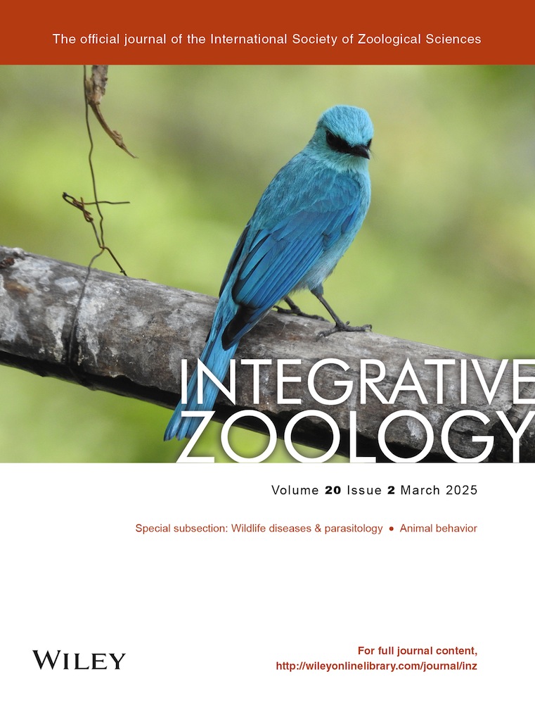
The Hong Kong newt (Paramesotriton hongkongensis) (Fig. 1a) is a subtropical salamander listed as Near Threatened by the International Union for Conservation of Nature. It has a highly restricted distribution in southern China (Fu et al. 2013; Lau 2017; Lau et al. 2017; IUCN 2021). A recent survey conducted in 2018 found that P. hongkongensis had the highest incidence of Bsal presence on the skin, with over 50% of individuals testing positive for the pathogen. This was observed in a single population located in Wutongshan near Hong Kong, China. (Yuan et al. 2018). Given the low number of studies on the skin microbiota of amphibians, specifically salamanders, in Asia and its connection to the origins of Bd and Bsal, the findings from this study can offer important insights into the host–pathogen–microbiota coevolution within the region. Furthermore, the anti-chytrid bacterial communities found on the salamander skin in this chytrid-coldspot region can be a useful resource for amphibian conservation, such as providing targets for developing probiotic treatments against chytridiomycosis. The high Bsal prevalence and absence of host symptoms reported in the previous survey make P. hongkongensis a suitable system for these purposes (Yuan et al. 2018). Here we aimed to: (1) characterize the skin microbiota compositions and the core microbiota of P. hongkongensis populations, (2) investigate whether P. hongkongensis harbors putative antifungal bacteria, and (3) determine the host and environmental factors associated with the skin microbial community structure.

MATERIALS AND METHODS
Field sampling
We collected P. hongkongensis skin swabs (n = 149) and water samples (n = 12) from 15 locations in Hong Kong, China (Fig. 1b) between October 2019 and February 2020 (Table S1, Supporting Information) during their breeding season when the newts gathered in streams to reproduce. We captured each newt with gloved hands and kept it in an individual plastic bag. A sterile swab (FLOQSwabs, Copan, Italy) was used to stroke each individual in a single direction 30 times each on the ventral and dorsal sides and five times on each leg. We recorded the snout-to-vent length (SVL), total length (TL), weight, and sex, and calculated the body condition index (BCI, weight/SVL) for each individual. One milliliter of stream water was taken from each of the 12 locations. The samples were immediately kept on ice in the field and transferred to the laboratory within 3 h. They were then stored at −80°C until DNA extraction. This study was approved by the University animal ethics committee (EC052/2021) and the Department of Health ([19-103] in DH/HT&A/8/2/6 Pt. 1).
DNA extraction, DNA library preparation, and sequencing
We extracted DNA from skin swabs and water samples using the E.Z.N.A. Tissue DNA Kit (Omega Bio-tek, USA) following the manufacturer's protocol under aseptic conditions. Four negative controls were included during DNA extraction. We also prepared mock communities (MCs) by pooling equimolar genomic DNA of multiple bacteria (Table S2, Supporting Information).
For high-throughput sequencing of the V4 region of the 16S rRNA gene to characterize the skin microbiota of P. hongkongensis, all samples, MCs, and negative controls were subjected to a two-step PCR (Cruaud et al. 2017; Huang et al. 2022) for library preparation. The first-step PCR was performed in a 30-μL reaction containing 6 μL of 5X GoTaq Flexi Buffer (Promega, USA), 0.6 μL of 10 mM dNTP Mix, 2.4 μL of 25 mM MgCl2, 0.15 μL of GoTaq G2 Flexi DNA Polymerase (Promega, USA), 1 μL of DNA, 1.5 μL each of 10-μM forward and reverse primers, 3 μL of 10% dimethyl sulfoxide (DMSO) (Sigma, USA), 0.15 μL of 20 mg mL−1 bovine serum albumin (BSA) (New England Biolabs, USA), and 13.7 μL of ultrapure water. The forward and reverse primers comprise 515F (5′-GTGYCAGCMGCCGCGGTAA-3′) (Parada et al. 2016) and 806R (5′-GGACTACNVGGGTWTCTAAT-3′) (Apprill et al. 2015), respectively, a heterogeneity spacer, and a linker. Thermocycling conditions were 95°C for 2 min, followed by 25 cycles of 95°C for 30 s, 50°C for 30 s, 72°C for 30 s, and a final extension of 72°C for 5 min.
Products from the first-step PCR were purified and used as the template in the second-step PCR, in a 45-μL reaction consisting of 9 μL of 5X GoTaq Flexi Buffer (Promega, USA), 0.9 μL of 10 mM dNTP Mix, 5.4 μL of 25 mM MgCl2, 0.225 μL pf GoTaq G2 Flexi DNA Polymerase (Promega, USA), 18.5 μL of purified DNA, 0.9 μL each of 10-μM forward and 10-μM reverse primers, 4.5 μL of 10% DMSO, and 4.675 μL of ultrapure water. The forward and reverse primers consist of the linker, an i5 or i7 index, and the adapter sequence (Cruaud et al. 2017). Each sample had a unique combination of the i5 and i7 indices. Thermocycling conditions were 95°C for 2 min, followed by 10 cycles of 95°C for 30 s, 55°C for 30 s, 72°C for 45 s, and a final extension of 72°C for 5 min. The PCR products from both PCR steps were purified with an E.Z.N.A. MicroElute Gel Extraction Kit (Omega Bio-tek, USA). The purified products from the second PCR step were quantified with the dsDNA HS Assay Kit and the Qubit 4 fluorometer (Invitrogen, USA), and pooled in an equimolar ratio to prepare a library multiplex. DNA was sequenced using NovaSeq (PE 250 bp; Illumina) by Novogene Corporation.
Sequence data processing
De-multiplexed 16S paired-end sequence reads were merged, trimmed to remove adapters, and quality-filtered. We merged the paired-end reads with -fastq_mergepairs in USEARCH v11.0.667 (Edgar 2010) and trimmed the primer sequences using CUTADAPT v2.4 (max_error_rate = 0.15) (Martin 2011). Quality assessment was performed with FastQC v0.11.8 (Andrews 2010) and -fastq_eestats2 in VSEARCH v2.15.1 (Rognes et al. 2016). Only reads with lengths between 240 and 253 bp and expected errors per read less than one were retained with the VSEARCH -fastq_filter. Pre-processed reads were then de-replicated with VSEARCH -derep_fulllength. Chimeras and singletons, representing less than 0.0001% of total reads, were removed with USEARCH -unoise3 (Edgar & Flyvbjerg 2015; Edgar 2016b). The remaining sequences were clustered at a 99% similarity level using VSEARCH -usearch_global to generate amplicon sequence variants (ASVs). Taxonomy was assigned to each ASV to the lowest identifiable taxonomic level using the SINTAX algorithm of USEARCH (Edgar 2016a) with bootstrap support higher than 80%. The EzBioCloud 16S database was used as a reference for taxonomy assignment (assessed in July 2021) (Yoon et al. 2017). The same approach was applied to identify putative antifungal ASVs, using the anti-Bd bacteria database as a reference (inhibitory ASVs only; assessed in November 2021) (Woodhams et al. 2015), with a bootstrap cutoff of 99%. Only ASVs that could be identified at the genus or species level were considered to match the sequences from putative anti-Bd taxa. ASVs with unidentified taxonomy or determined as contaminants according to negative controls were removed (Table S3, Supporting Information). ASVs represented by less than 0.018% (a threshold determined by MCs, as in Huang et al. (2021); Table S3, Supporting Information) of total reads in each sample were considered false positives and filtered out.
Data analysis
All statistical analyses were conducted using R version 4.1.0 (R Development Core Team 2021). The microbiota composition was represented by the relative read abundance (RRA) and weighted percentage of occurrence (wPOO) of each bacterial taxon in an individual, as well as frequency of occurrence (FOO) of each taxon in all swab samples. Taxa with an FOO exceeding 90% were considered as part of the core microbiota. Microbiota diversity was assessed by calculating the effective number of ASVs (hill number) for each sample (alpha diversity) and sampling site (gamma diversity) using the hilldiv package (Alberdi & Gilbert 2019). The hill number depended on the order of the diversity q (Hill 1973), where a higher q placed greater weight on abundant ASVs and resulted in a lower hill number. Differences in microbiota compositions between sites (beta diversity) were evaluated using both the pairwise Jaccard distance and Bray–Curtis dissimilarity index with the vegan package (Oksanen et al. 2022). Beta diversity was visualized using non-metric multidimensional scaling (NMDS) and principal coordinates analysis (PCoA) with the phyloseq package (McMurdie & Holmes 2013), and hierarchical clustering was performed using Ward's method with the dendextend package (Galili 2015). Pairwise permutational ANOVA using the pairwiseAdonis package (Martinez Arbizu 2020) was performed to test the differences in microbiota compositions between sites, with the Benjamini–Hochberg false discovery rate correction based on 999 permutations. The homogeneity of intra-group beta-dispersion was assessed using the phyloseq package (McMurdie & Holmes 2013). The contribution of each ASV to the microbiota composition variation was determined using the SIMPER (similarity percentage) with the Bray–Curtis dissimilarity using the vegan package (Oksanen et al. 2022). Statistical significance was checked using the Kruskal–Wallis test by ranks. Only ASVs contributing more than 0.01% were retained.
To examine the effect of environmental and host factors on skin microbiota composition (beta diversity), two statistical models were used. In the first linear model, the relationship between the geographical distance between sampling sites and microbiota similarity was tested. The pairwise Jaccard distance or Bray–Curtis dissimilarity was used as the response variable, and the geographical distance between sites was used as the explanatory variable. Pairwise distances between sites separated by the sea were excluded in the model, that is, only pairwise distances between sites in the same region (New Territories, Hong Kong Island, or Lantau Island; Fig. 1b) were included. In the second linear mixed model, principal coordinate axis 1 was extracted from the Bray–Curtis dissimilarity or Jaccard distance matrix and used as the response variable. Elevation, BCI, and sex were included as fixed effects, and the sampling site and sampling month were included as random effects.
RESULTS
Total, putative anti-Bd, and core skin microbiota of P. hongkongensis
Using high-throughput sequencing of the V4 region of the 16S rRNA gene, we identified 9433 amplicon sequence variants (ASVs; Table S4, Supporting Information), belonging to 2605 bacterial taxa classified at the lowest possible taxonomic level. In total, 42 phyla, 128 classes, and 274 orders of bacteria were identified. Among the ASVs, 314 were found in the putative anti-Bd bacteria database (Woodhams et al. 2015) and were assigned to 25 genera (Table S5, Supporting Information), accounting for 9.7% of the total reads. On average, each P. hongkongensis individual harbored 21 putative anti-Bd ASVs, and all individuals had at least three putative anti-Bd ASVs. The most prevalent putative anti-Bd ASV was present in 86.6% of all individuals, while 114 putative anti-Bd ASVs were exclusively found in single individuals. Among the ASVs, two from Acinetobacter, three from Flavobacterium, and three from Novosphingobium exhibited the highest RRA and occurrence. Rarefaction analysis indicated sufficient sequencing depth for ASV identification (Fig. S1, Supporting Information).
The most abundant phyla were Proteobacteria (59.2%), Bacteroidetes (21.2%), and Verrucomicrobia (12%), collectively accounting for over 92% of the total reads. No other phyla had an RRA >1% (Fig. 2). Within Proteobacteria, the most abundant classes were Betaproteobacteria (44.8%), Gammaproteobacteria (6.5%), Alphaproteobacteria (6.4%), Cytophagia (1.2%), and Deltaproteobacteria (1%). Among Bacteroidetes, Sphingobacteriia (5.9%) and Flavobacteria (3.5%) were the most abundant classes, while Opitutae (6.2 %) and Verrucomicrobiae (5.7%) dominated within Verrucomicrobia. The most abundant taxon was Comamonadaceae, a family in Betaproteobacteria, representing 33.3% of the total reads and present in every individual skin microbiota (Fig. 2). Five out of 16 ASVs, each accounting for >1% reads, were assigned to Comamonadaceae (Table S6, Supporting Information). Among the 14 taxa with RRA >1%, three were putatively anti-Bd (Acinetobacter, Flavobacterium, and Novosphingobium; Table S5, Supporting Information). The core microbiota comprised 13 bacterial taxa, representing 7.4% of the total reads. These taxa were primarily composed of species belonging to the Betaproteobacteria class (e.g. Undibacterium in order Burkholderiales) and the Alphaproteobacteria class (e.g. Novosphingobium in order Sphingomonadales) within the phylum Pseudomonadota. Additionally, the core microbiota included taxa from the Chitinophagia class (e.g. family Chitinophagaceae) and the Flavobacteriales class (e.g. Flavobacterium in Flavobacteriales) within the phylum Bacteroidota (Table S7, Supporting Information), two of which were putatively anti-Bd (Flavobacterium and Novosphingobium; Table S5, Supporting Information). All taxa that accounted for >1% of the total reads or were within the core microbiota belonged to one of the three most abundant phyla.

The six Bd/Bsal-positive samples showed a comparable number of putative anti-Bd ASVs (mean = 21.3, SD = 8.4) to the negative samples (mean = 21.4, SD = 11.8; Fig. 2). Among the positive samples, the Bd-positive sample had 36 putative anti-Bd ASVs, while the five Bsal-positive samples had an average of 18.4 (SD = 5) putative anti-Bd ASVs, respectively. In addition, we identified several ASVs from bacteria that are potentially capable of producing TTX in our dataset, including 1 ASV for Aeromonas, 20 ASVs for Pseudomonas, 2 ASVs for Shewanella, and 2 ASVs for Sphingopyxis. However, only ASV21 (Aeromonas sp.) was present in a majority of newt individuals (60%) and water samples (75%), and its RRA was highly variable across samples (mean = 0.4%, SD = 1.68%, range = 0–13.9%; Tables S8,S9, Supporting Information).
Microbiota diversity within and between sites
Microbiota composition and diversity differed among sites and between skin and water samples (Figs 2, 3; Fig. S2 and Tables S10–S12, Supporting Information). Individual microbiota from Hok Tau (HT), Kowloon Peak (KP), and Wu Kau Tang (WKT) exhibited the highest ASV richness (at q = 0), while those from Tai Mo Shan (TMS) exhibited the lowest (Figs 1, 3; Tables S13,S14, Supporting Information). HT and KP also showed the highest gamma diversity in microbial communities (Fig. 3b). As the value of q increased, both the alpha diversity (within each sample) and gamma diversity (within each site) of the newt skin microbiota from all sites decreased rapidly, reflecting highly uneven relative abundances of bacterial taxa in the skin microbiota.

Microbiota composition analyses showed that the skin microbiota demonstrated site differences and were differentiated from water microbiota (hierarchical clustering, Fig. 4; NMDS and PCoA, Fig. S3, Supporting Information). Skin microbiota were well differentiated by site in the hierarchical clustering analysis based on both the Bray–Curtis dissimilarity (Fig. 4; Table S15, Supporting Information) and Jaccard distance (Fig. S4 and Table S16, Supporting Information), indicating site-specific bacterial compositions. The Bray–Curtis dissimilarity analyses revealed that skin microbiota from Sunset Peak (SP) and Wong Lung Hang (WLH) were highly similar to each other (Figs 1, 4). TMS had a relatively distinct skin microbiota composition from most sites according to the results from NMDS (Fig. S3a, Supporting Information) and PCoA (Fig. S3c and Table S17, Supporting Information). The Jaccard distance analyses presented similar patterns to the Bray–Curtis dissimilarity results in NMDS (Fig. S3b, Supporting Information) and PCoA (Fig. S3d and Table S18, Supporting Information). In Bray–Curtis dissimilarity analyses, bacterial communities in water samples were clustered as a distinct group, except for the one from SP, which clustered with SP skin microbiota samples (Fig. 4; Figs S3,S4, Supporting Information).

The skin microbiota composition was significantly different between all pairwise site comparisons (adjusted P ≤ 0.002; Tables S19,S20, Supporting Information). The mean number of ASVs contributing to the top 10% dissimilarity between any two sites was 30.1 (SD = 8.7). Within the top 10% dissimilarity, seven ASVs were present in more than 50% of the 105 pairwise comparisons (Table S21, Supporting Information), while 90 ASVs contributed to the dissimilarity of just one or two site pairs. Site-specific patterns were observed in the composition of newt skin microbiota (Figs 2, 4; Fig. S4, Supporting Information). While the bacterial communities from most sites were dominated by Betaproteobacteria, those from Mui Tsz Lam (MTL) had a large proportion of Opitutae (39.3% RRA; Fig. 2). Among the samples from KP, Verrucomicrobiae was the most abundant class (25.8% RRA; Fig. 2). In half of the samples from Pak Ngau Shek (PNS), Gammaproteobacteria were present in the highest abundance (58% RRA; Fig. 2). Skin microbiota from SP and WLH, the two sites on Lantau Island, as well as TMS, the highest peak in Hong Kong, China (Fig. 1), formed relatively well-separated groups in NMDS (Fig. S3a,b,Supporting Information). Compared to other sites, bacterial communities from MTL, SP, and WLH had a particularly low abundance of Bacteroidetes (Fig. 2). Microbiota in water samples appeared to be more evenly distributed across taxa compared to skin microbiota (Fig. 3) and shower greater similarity to each other than to skin microbiota (Fig. 4; Figs S3,S4, Supporting Information).
Effects of environmental and host factors on skin microbiota
The pairwise distance between microbiota was positively correlated to the geographical distance between sites in terms of Bray–Curtis dissimilarity (P < 0.001, R2 = 0.21; Fig. 5a) and Jaccard distance (P < 0.0001, R2 = 0.23; Fig. 5b). The linear mixed model (Table 1) revealed that the skin microbiota composition changed with site elevation (P = 0.02; Fig. 5c) and showed significant differences between host sexes (P < 0.01; Fig. 5d).

| Fixed effects | Effect | SE | t | P | |
|---|---|---|---|---|---|
| (Intercept) | 9.59 × 10⁻² | 9.36 × 10⁻² | 1.02 | 0.31 | |
| Elevation | −4.40 × 10⁻⁴ | 1.82 × 10⁻⁴ | −2.42 | 0.02 | |
| BCI | −1.26 × 10⁻² | 4.25 × 10⁻² | −0.30 | 0.77 | |
| Sex | 6.52 × 10⁻² | 2.38 × 10⁻² | 2.74 | 0.01 | |
| Random effects | SD | ||||
|---|---|---|---|---|---|
| Location | 0.12 | ||||
| Month | 0.03 | ||||
| Residual | 0.16 | ||||
- Site elevation, body condition index (BCI), and sex were included as fixed effects. Sampling location and month were included as random effects. Significant effects are bolded.
DISCUSSION
Skin microbiota diversity and composition in P. hongkongensis
As the pandemic caused by chytrid fungi continues to threaten global amphibian diversity, Asia has gained increasing attention from the scientific community, as it has been suggested as the origin of these pathogens (O'hanlon et al. 2018; Fisher & Garner 2020). This study is one of the few to investigate the skin microbiota in wild amphibian populations in Asia (Sabino-Pinto et al. 2016; Bletz et al. 2017b; Bataille et al. 2018; Yang et al. 2020; Mutnale et al. 2021). Our findings reveal that the diverse bacterial communities on the skin of P. hongkongensis were predominantly composed of Proteobacteria, Bacteroidetes, and Verrucomicrobia. The skin microbiota of amphibians, as is likely in the case with newts, may produce antimicrobial compounds that protect against harmful pathogens (Rebollar et al. 2020), which will be discussed in a later section. Additionally, amphibian microbiota may actively participate in nutrient cycling by breaking down the complex organic matter present on the amphibian's skin (Brunetti et al. 2023). Moreover, these microbes may interact with the host's immune system, potentially influencing the immune response of the amphibians (Küng et al. 2014). Their presence in the skin microbiota plays a crucial role in enhancing the overall microbial diversity, thereby significantly contributing to survival and well-being (Harrison et al. 2019). The compositions of the skin microbiota were significantly different across sampling sites, and most of them were highly differentiated from the streamwater microbiota present in the habitats of P. hongkongensis. The dissimilarity between skin microbial communities was found to be positively correlated with the geographical distance between sites. Moreover, among other environmental and host factors tested, elevation and host sex were identified as significant factors in determining the skin microbiota structure of the newts.
In congruence with many existing skin microbiota studies in amphibians, Proteobacteria was the most abundant bacterial phylum on P. hongkongensis, followed by Bacteroidetes (e.g. Bletz et al. 2017c; Muletz Wolz et al. 2018; Douglas et al. 2021). Comamonadaceae, a family in Pseudomonadota, was also commonly identified as an abundant family (McKenzie et al. 2012; Belden et al. 2015; Prado-Irwin et al. 2017; Bletz et al. 2017a, 2018; Jiménez et al. 2019; Walker et al. 2020; Walke et al. 2021). While Verrucomicrobia was occasionally found among the dominant phyla, its relative abundance rarely exceeded 3% (Belden et al. 2015; Bletz et al. 2017b, 2018; Walke et al. 2021), except in the case of the Japanese giant salamander (Andrias japonicus, 9%) and the larvae of wild Japanese fire belly newts (Cynops pyrrhogaster, 20%) in Japan (Sabino-Pinto et al. 2016). However, a few phyla that are commonly found in high abundance on amphibian skin, such as Actinobacteria, Firmicutes, Acidobacteria, and Cyanobacteria (McKenzie et al. 2012; Belden et al. 2015; Prado-Irwin et al. 2017; Bletz et al. 2017a, 2018; Jiménez et al. 2019; Walker et al. 2020; Walke et al. 2021), were scarce (RRA < 1%) on P. hongkongensis. In a study on the skin microbial community structures of 89 frog species in Madagascar, Actinobacteria and Acidobacteria were found to occur in significantly higher relative abundance on terrestrial species compared to aquatic or semiaquatic species, which might explain their low abundance on P. hongkongensis, as it is primarily aquatic during the sampling period (Bletz et al. 2017a).
The core microbiota of P. hongkongensis: Potential for protection against chytrid fungal pathogens
Although there were large intraspecific variations in the skin microbiota across different sites, a core group of microbiota members was shared by most P. hongkongensis individuals, albeit with generally low relative abundances. The presence of a core microbiota, which maintains a relatively stable composition despite variations in environmental conditions experienced by the hosts, has been observed in many amphibian species, suggesting its functional importance. For example, the core microbiota structures on coqui frogs (Eleutherodactylus coqui) did not vary along the elevation gradient or with changes in forest integrity (Hughey et al. 2017). The core microbiota of P. hongkongensis comprised bacterial species that were likely well adapted to reside on the host skin and perform vital functions for the host, thus establishing a symbiotic relationship (Prado-Irwin et al. 2017; Muletz Wolz et al. 2018). However, since our sampling was limited to the breeding season of the newts, the abundance of these core bacterial species may fluctuate temporally as the newts move from an aquatic to a terrestrial environment after the breeding season, which can result in changes in various relevant host traits, for example, the levels of skin moisture or reproductive hormones (Lau 2017; Lau et al. 2017; Bletz et al. 2017b). Seasonal changes in the abundance of core microbes, such as Chitinophagaceae, which was also detected in P. hongkongensis, have been reported in the North American wood frog (Rana sylvatica) (Douglas et al. 2021). Noteworthily, P. hongkongensis produces TTX (Yotsu et al. 1990), which is believed to act as a defense against predators, although the synthesis mechanism of this deadly neurotoxin is unknown. Further investigation of the TTX-producing ability of Aeromonas sp. found on many newt individuals can help us understand whether P. hongkongensis has developed a symbiotic relationship with certain toxin-producing bacteria to increase its fitness, as observed in another poisonous newt (Vaelli et al. 2020).
Furthermore, the core microbiota might play a functional role in enhancing host resistance against Bd infection. For example, based on bioinformatic and culture tests, it was predicted that P. cinereus has maintained a core microbiota in which bacterial members provided anti-Bd functions (Loudon et al. 2014). Multiple species of Novosphingobium and Flavobacterium were detected in the core microbes of P. hongkongensis. These genera have already been shown to exhibit anti-Bd properties (Harris et al. 2006), with the latter genus being widely distributed among amphibian species, such as R. cascadae (Roth et al. 2013), the green-eyed frog (Lithobates vibicarius) (Jiménez et al. 2019), and the four-toed salamander (Hemidactylium scutatum) (Lauer et al. 2008). Comamonadaceae, Methylotenera, and Undibacterium were also identified as core taxa on the wild Oriental fire-bellied toad (Bombina orientalis) in Japan (Sabino-Pinto et al. 2016), and the abundance of Methylotenera and Rhizobiales on wild B. orientalis was found to vary with individual Bd infection status in South Korea (Bataille et al. 2016, 2018). Although the functions of core microbiota on amphibian skin are not well understood, the overlapping skin microbiota composition between P. hongkongensis and other Asian amphibians studied suggests a potential protective role of their core microbes against chytrid fungal pathogens.
It may be reasonable to assume the putative anti-Bd bacterial taxa identified here, using the anti-Bd isolates database, could potentially possess anti-Bsal properties as well. This assumption is based on the fact that many of these bacteria can produce broad antifungal compounds (Woodhams et al. 2015; Muletz Wolz et al. 2018). Putative antifungal bacteria were found on P. hongkongensis in great diversity, with high prevalence but low abundance. Notably, eight ASVs from Acinetobacter, Flavobacterium, and Novosphingobium were particularly abundant and prevalent, indicating a putative symbiotic relationship with P. hongkongensis (Muletz-Wolz et al. 2017). Most of the putative anti-Bd microbiota had restricted distributions among newt individuals and showed dissimilar taxon identities. This pattern might have resulted from the colonization by putative anti-Bd bacteria acquired from distinct pools of microbes present in different locations (Muletz-Wolz et al. 2017). The estimated relative abundances of the putative anti-Bd bacteria in this study were generally much lower compared to those found in some other amphibian species, such as three Plethodon species in North America (Muletz Wolz et al. 2018) and six frog species in India (Mutnale et al. 2021). This observation suggests that rare taxa in a microbiota may play a functionally important role. Another possible explanation could be the failure to identify significant antifungal members present in the microbiota of P. hongkongensis using the anti-Bd bacteria database (Woodhams et al. 2015). This database was compiled by testing bacterial isolates from certain amphibian species using culture methods. As a result, it is unlikely to include rare or unculturable bacteria and host-specific microbial species on untested amphibians, especially those in Asia. However, it is worth noting that putative anti-Bd bacteria were detected in all 100% of the sampled newts. This finding indicated the potential resistance or even herd immunity against chytridiomycosis in P. hongkongensis. This resistance might be attributed to historical interactions with Bd and Bsal strains endemic to Asia, where Bd exhibited a high degree of diversity and has not been found to cause morbidity or mortality in local wild amphibian populations (Bataille et al. 2013).
Previous studies have suggested that an amphibian population can achieve herd immunity to Bd when around 80% of the individuals are protected by anti-Bd microbes, allowing the population to coexist with Bd (Woodhams et al. 2007; Muletz-Wolz et al. 2017). In the case of P. hongkongensis population in Wutongshan, all Bsal-positive individuals showed no symptoms despite the high prevalence of the pathogen (Yuan et al. 2018). For the P. hongkongensis population in Hong Kong, China, the prevalence of Bd and Bsal was very low (Chen et al. 2023), with positive individuals being asymptomatic, supporting the idea of potential herd immunity achieved through antifungal skin microbes. However, in this study, no noticeable difference was observed in the skin microbiota between Bsal-positive and Bsal-negative individuals. Noteworthily, Janthinobacterium spp. (class Betaproteobacteria, phylum Proteobacteria) were detected on P. hongkongensis. The anti-Bd property of Janthinobacterium lividum has been well verified (Brucker et al. 2008). This bacterium has been shown to produce indole-3-carboxaldehyde and violacein, which can significantly inhibit Bd growth at low concentrations (Brucker et al. 2008). Moreover, the addition of J. lividum to amphibian skin can reduce both the morbidity and mortality caused by Bd infection (Becker et al. 2009; Harris et al. 2009a). In addition, numerous ASVs belonging to a few common anti-Bd genera, such as Pedobacter and Pseudomonas, were found on P. hongkongensis.
Geographic factors influencing the skin microbiota structure
One of our main findings was that the skin microbiota structure of newts was largely influenced by site, depending on environmental or host genetic factors. Previous studies have shown that the population genetic structure of P. hongkongensis aligns with the landscape topography, which cannot be fully explained by isolation by distance (Lau 2017). Based on this population structure, there were five genetic clusters of P. hongkongensis, one on Hong Kong Island (Pok Fu Lam [PFL], Tai Tam [TT]), one on Lantau Island (SP), and three on the New Territories (North: WKT; East: HC, MTL, KP; and Central: Tai Po Kau [TPK]), while an admixture of the New Territories North and East clusters was observed at PNS (Lau 2017). Consistent with the host population structure, the skin microbiota from the two sites (WLH and SP) on Lantau Island were similar to each other and distinct from other sites (Fig. 4; Fig. S4, Supporting Information). The geographical isolation of the Lantau Island populations from the mainland not only limited the gene flow between newts but likely also restricted the exchange of individual microbiota. However, this pattern was not observed between the two Hong Kong Island newt populations (PFL and TT). Their skin microbiota were more similar to those of the New Territories populations than to each other, implying that factors other than geographical isolation alone might also influence microbiota structure. Microbiota differences in the New Territories can be partially explained by geographical and host genetic distances, for example, individuals from the New Territories East sites (i.e. HC, MTL, KP, and FSH) had mostly similar microbiota, but these factors could not account for all the observed differences. Therefore, there were instances where the microbiota from certain sites did not align with either the host population structure or geographical proximity. Overall, geographical distance appeared to have a larger influence, as microbiota within the same site strongly clustered together and did not mix with those from different sites within the same genetic population of newts (Fig. 4; Fig. S4, Supporting Information).
How amphibians acquire their skin microbes in each new generation and how their microbiota are shaped and maintained remain important questions in the field. Horizontal transmission happens when microbes are selected from the habitat to form a microbiota (Bright & Bulgheresi 2010). Members of the P. hongkongensis skin microbiota might come from the living environment, such as water, soil, and leaf litter (Muletz Wolz et al. 2018). The clear difference in the microbiota between skin and stream water implies that even though the skin of P. hongkongensis might initially contact all bacterial species present in the surroundings, only a subset of these species had managed to colonize and proliferate on the skin. Since the environmental conditions, for example, water quality (Kueneman et al. 2014; Krynak et al. 2016), soil, and pH (Fierer & Jackson 2006; Muletz Wolz et al. 2018), of various sites could be different, the composition of bacteria available from the environment for horizontal transmission could certainly vary in response to the site-specific conditions. Moreover, the environmental characteristics at each site could affect host traits, so the site-wise differences in microbiota could also reflect the influence of site-specific host traits. For instance, the production of antimicrobial peptides by the skin, a key component in amphibian immune defense, could be regulated by water surface area and conductivity, thus affecting the skin microbiota composition (Krynak et al. 2016).
Influence of elevation and host sex on the skin microbiota structure
In addition to geographical distance, elevation also had an effect on the skin microbial communities of newts, similar to previous observations that skin microbiota structures varied along elevational gradients from 0 to 875 m and 700 to 1000 m above sea level (Hughey et al. 2017; Muletz Wolz et al. 2018). The influence of elevation is complicated, and many environmental factors co-vary along the gradient, which might, in turn, affect the host traits as well as the growth and interactions of different bacteria on the host (Muletz Wolz et al. 2018). Furthermore, Bd prevalence was found to positively correlate with elevation, which might have led to a stronger selection for anti-Bd bacteria on surviving hosts at higher elevations if there have been recurring infections (Bresciano et al. 2015). Considering the impact of elevation, we recommend that future studies investigate the potential influence of environmental temperatures on the skin microbiota of newts.
We also found that the variations in the microbiota structures of P. hongkongensis were associated with host sex. Although it has been shown in Blanchard's cricket frog (Acris blanchardi) that sex interacted with latitude and water surface area in affecting the skin microbiota (Krynak et al. 2016), it has no effect on E. eschscholtzii (Prado-Irwin et al. 2017) or R. sylvatica (Douglas et al. 2021). In P. hongkongensis, females exhibited cannibalism on eggs and larvae, which caused a difference in the diets between the sexes (Fu et al. 2013). As diet can influence the skin microbiota (Antwis et al. 2014), the effect of sex might be an indirect indicator of the effect of diet. Alternatively, hormonal differences between the sexes might explain their microbiota divergence. A relationship between sexual differences in host-associated microbiota and sex hormone levels has been documented in birds and primates (Escallon Herkrath 2015; Mallott et al. 2020), but whether such a relationship exists in amphibian skin microbiota has not been investigated.
CONCLUSION
The skin microbiota of P. hongkongensis was diverse, with a group of core skin microbes potentially coevolving with, and providing essential functions for, the hosts. The bacterial community structure of P. hongkongensis was highly site-specific, which was likely attributed to geographical isolation and environmental characteristics instead of host population genetics, as demonstrated by the effect of elevation. Host sex also influenced the newt skin microbiota, possibly because of the differences in diet or hormonal levels between the two sexes. The high prevalence and low abundance of putative antifungal bacteria found on the skin, together with the low prevalence and dispersed distribution of chytrid infection in the newt population, suggest that P. hongkongensis has had a history of exposure to and coexistence with the fungal pathogens in the region.
Using P. hongkongensis as a reference species, interrogation of the compositions and patterns of the skin microbiota structures of other amphibians in East Asia, where chytrids are proposed to be native, will enhance our understanding of host–pathogen–microbiota coevolution. Such knowledge can be applied to the conservation of P. hongkongensis as well as amphibian populations naïve to pathogenic chytrids. Given that the extant P. hongkongensis skin microbiota has likely been shaped by the coevolution with chytrids, it is a promising source for identifying and characterizing antifungal bacteria not yet known to us, and thereby developing novel probiotic treatments against chytridiomycosis. Further investigations into the factors governing the differences in the skin microbiota between sites and sexes could improve our understanding and our ability to develop effective probiotic treatments.
ACKNOWLEDGMENTS
We would like to thank Yik-Hei Sung and Wing Ho Lee for their assistance during sample collection, Pei-Yu Huang for her advice on analysis, and Charis May Ngor Chan for her technical assistance. The computations were performed using the research computing facilities offered by the Information Technology Services at the University of Hong Kong. This work was supported by the Start-Up Fund granted to S.Y.W.S. by the University of Hong Kong. This work was approved by the Agriculture, Fisheries and Conservation Department [Permit no.: (29) in AF GR CON 09/51Pt.7] of the HKSAR Government.
CONFLICT OF INTEREST STATEMENT
The authors declare no conflict of interest.
Open Research
DATA AVAILABILITY STATEMENT
The sequencing data have been archived in the National Center for Biotechnology Information (NCBI) under the BioProject accession number PRJNA1113385.