Endoscopic Ultrasound for Diagnosis and Management of Pediatric Gastrointestinal Stromal Tumor
The authors report no conflicts of interest.
Written parental and patient consent and assent were obtained for publication of the details of these cases.
Abstract
Gastrointestinal (GI) stromal tumors arise from the interstitial cells of Cajal and are rare in the pediatric population. The most common clinical manifestation is anemia secondary to GI bleeding. Endoscopy is commonly used for diagnostic and therapeutic interventions of an obstructing mass or gastrointestinal bleed, while experience with endoscopic ultrasound (EUS) and EUS fine needle aspiration (EUS-FNA) for pediatric patients with suspected gastric tumors is limited. We report 2 cases, a 14-year-old male and an 11-year-old female, who presented with symptomatic anemia. Both patients were diagnosed with GI stromal tumors of the stomach using EUS and EUS-FNA. This report shows that EUS and EUS-FNA are safe and effective diagnostic tools for pediatric patients.
INTRODUCTION
Gastrointestinal stromal tumors (GISTs) are neoplasms of mesenchymal origin, specifically the interstitial cells of Cajal (1). GISTs are most commonly diagnosed in a patient's sixth and seventh decade of life (1). Therefore, the diagnosis of GIST in a child may be delayed due to clinicians’ lack of familiarity with this tumor (1). Presenting signs and symptoms are variable depending on the size and anatomic location of the tumor. Over 80% of GISTs in children arise from the stomach, and the most common presenting symptoms are secondary to anemia (2). In the pediatric population, females develop GIST at a higher rate than males, while the adult population has equal gender distribution (2).
The role of endoscopic ultrasound (EUS) in adult patients is well established; however, in pediatric patients, experience with this technology is limited (3). We present two cases of pediatric patients who presented with symptomatic anemia and were found to have GIST in the stomach, diagnosed using EUS and EUS fine needle aspiration (EUS-FNA).
CASE REPORTS
Case 1
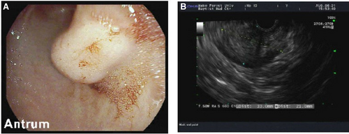
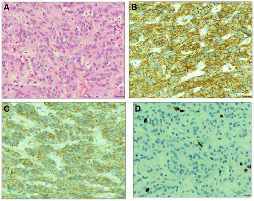
A 14-year-old male presented with episodes of dizziness and dark stools for 4 days. Physical examination was notable for pallor, tachycardia (124/bpm), and tachypnea (24/bpm). A complete blood count showed hemoglobin of 7 g/dL, MCV 83.1 FL, reticulocyte count 3.8%, and ferritin 10 ng/mL. Stool was positive for occult blood. The patient was transfused with two units of packed red blood cells. Computed tomography (CT) did not initially reveal a mass, but on later review [after esophagogastroduodenoscopy (EGD)] showed a possible mass in the gastric antrum which did not demonstrate enhancement characteristics beyond what would be expected from normal gastric folds or ingested food. CT revealed no evidence of metastatic disease. (Fig. 1) Initial EGD showed a submucosal mass in the gastric antrum with overlying ulceration (Fig. 2A). Biopsies of the stomach and duodenum showed no evidence of malignancy. The patient underwent EUS the following day when EUS-FNA was performed (Fig. 2B). EUS demonstrated a hypoechoic, subepithelial mass in the distal stomach that appeared to originate from the fourth hypoechoic layer. Cytology revealed a spindle and epithelioid neoplasm with uniform cellularity and bland nuclear features, most consistent with GIST. The patient underwent exploratory laparotomy and partial gastrectomy with successful removal of the tumor. Pathology revealed a 3 cm × 2.5 cm × 2 cm firm submucosal mass, completely resected (0.7 cm from each margin). Lesional cells were positive for DOG-1 and C-kit with negative staining for CK, S100, MSA, SMA, synapthophysin, and desmin, confirming the diagnosis of GIST. MIB-1 showed a low proliferation rate (mitotic rate of <5/5 mm2; Fig. 3). No adjuvant therapy was administered. There have been no complications or signs of recurrence in his short postoperative period.

CT scan of gastric antrum lesion in patient 1. CT = computed tomography.

Appearance of submucosal mass on EGD and EUS in patient 1. A) Appearance of submucosal mass on EGD. B) Appearance of submucosal mass on EUS. EGD = esophagogastroduodenoscopy; EUS = endoscopic ultrasound.

Histological sections of GIST and immunostaining in patient 1. A) Histological section showing spindle and epithelioid neoplasm. B) Lesional cells positive for DOG-1. C) Lesional cells positive for C-kit. D) MIB-1 showing a low proliferation rate. GIST = gastrointestinal stromal tumor.
Case 2
An 11-year-old female presented with a short history of fatigue, pallor, episodes of dizziness, and abdominal pain. Physical exam was notable for pallor and a II/VI systolic ejection murmur. Laboratory tests revealed a hemoglobin of 6.9 g/dL, MCV 70 FL, reticulocyte count 3.8%, and ferritin 3 ng/mL. Stool was heme positive. EGD revealed bleeding ulcers associated with tumors in her stomach. EUS-FNA cytology findings were consistent with multifocal GIST. Since the patient was diagnosed prior to the use of the electronic medical records, the details of the EUS findings are not available. She underwent distal gastrectomy with Billroth I gastroduodenostomy. Pathology confirmed GIST, with five nodules clustered together, the largest with a 5 cm diameter. Microscopy revealed mixed epithelioid and spindle cell morphology. C-kit was positive, and CD34 was negative. Mitotic count was low at 30 mitoses per 50 high-powered fields. The surgical margins were free of tumor, and there was no evidence of nodal metastases. Follow-up CT showed no evidence of local recurrence or metastatic disease. No adjuvant therapy was administered. At routine follow-up visit 1 year later, she was again found to have iron deficiency anemia. Stool hemoccult was negative on several samples. EUS-FNA revealed gastric polyps, but no evidence of GIST. The anemia resolved with oral iron supplementation and birth control pills. EUS was subsequently utilized multiple times to scan the patient for GIST recurrence or other GI pathologies. She is doing well 14 years from original diagnosis with no sign of disease recurrence.
DISCUSSION
GIST have a reported incidence of 10–15 cases per million overall; however, due to the rarity of GIST in pediatric patients, the incidence in this population is difficult to quantify (4). Previous reports suggest 0.4% of all GIST patients are <20 years old and that patients aged 8–20 make up approximately 1.64% of all GIST cases (4). The immunohistochemical features of pediatric GISTs are similar to adult GIST, usually having a strong expression of kit protein (CD117) (2). Spindle cell morphology is most common, and epithelioid is the second most common (2). However, pediatric GISTs have some important pathologic differences compared with adult GIST, which typically harbors a mutation of a receptor tyrosine kinase, typically KIT or PDGFRA (5). Pediatric wild-type GIST with KIT or PDGFRA mutations occur in 11% of the pediatric population, while it occurs in 80%–85% of the adult population (5).
EGD is commonly used in pediatric patients for diagnosis of an obstructing mass or gastrointestinal bleed. However, a few studies report EUS and EUS-FNA for pediatric patients with suspected gastric tumors. In adult patients, EUS and EUS-FNA are a well-established techniques for an accurate diagnosis of a GIST (6). Several reports show that EUS and EUS-FNA in pediatric patients are safe, feasible and have a significant impact on the management of various disorders of the gastrointestinal tract (6-8). Additionally, EUS can be used for periodic surveillance for recurrent disease (3). As seen in one of our patients, EUS can be used safely to scan for recurrent disease and to diagnose additional gastrointestinal pathology, especially if symptoms are concerning for recurrence at follow-up visits. Our cases add to the limited body of knowledge, demonstrating that EUS and EUS-FNA are feasible and can be accurate for the diagnosis of GIST in pediatric patients.
ACKNOWLEDGMENTS
B.C.K. and P.C. designed the study, collected data, and drafted the manuscript. H.R., N.W., J.P., M.L., and T.W.M. collected data, interpreted data, and critically revised the manuscript for important intellectual content. All of the authors read and approved the final manuscript, and agree to be accountable for all aspects of the work in ensuring that questions related to the accuracy or integrity of any part of the work are appropriately investigated and resolved.


