Influence of κ-rhythm when assessing drowsiness
Abstract
Abstract κ-Rhythm appears at the highest amplitude on a bipolar T3–T4 derivation and its frequency range is 7–10 Hz. When a contra-lateral earlobe is used as a reference, the κ-rhythm causes serious problems in assessing drowsiness. Assessment of drowsiness using contra- and ipsi-lateral earlobes as references in 129 subjects who showed κ-rhythm was compared. Drowsiness could be assessed properly in only 26% of subjects using the contra-lateral earlobe and in 90% of subjects using the ipsi-lateral earlobe. These results suggest that the ear on the ipsi-lateral earlobe should be used as a reference in subjects who show κ-rhythm.
INTRODUCTION
An earlobe reference electrode is sometimes activated by spontaneous electrical potential occurring in the temporal lobe. The most frequent cause of activation is κ-rhythm, which appears at the highest amplitude on a bipolar T3–T4 derivation, and its frequency range is 6–12 Hz (predominantly 7–10 Hz), which is similar to α-waves. It does not disappear during sleep or upon waking,1,2 and its polarity is opposite between the right and left hemispheres. The κ-rhythm causes serious problems when assessing drowsiness because it obscures the disappearance of α-waves, ripple waves, etc., which are the characteristic features of drowsiness. The present study examined the influence of κ-rhythm when assessing drowsiness, using both earlobes as references.
METHOD
A total of 129 inpatients and outpatients (mean ± SD 57.7 ± 10.1 years; range 38–75 years; 80 men and 49 women) who showed κ-rhythm on a clinical electroencephalogram (EEG) examination and then fell asleep were included in the study. The EEG was recorded by a digital EEG recorder (2514; NEC Medical Systems, Tokyo, Japan), and the data displayed on a PC monitor with a SynaViewer ES5003 (NEC Medical Systems). Sleep stages were scored manually with a unipolar C3–A2 or C4–A1 derivation, and with a C3–A1 or C4–A2, separately. Bipolar T3–T4 and A1–A2 derivations were also displayed simultaneously on a PC monitor for checking the presence of κ-rhythm. When the earlobe reference was activated, source derivation (SD) methods, in which the potentials of four active electrodes around an active electrode were averaged and used as a reference, were used. An O1–SD or O2–SD derivation was displayed for checking α-waves. For those subjects who showed a strong κ-rhythm, a C3–SD or C4–SD derivation was used for sleep scoring.
RESULTS
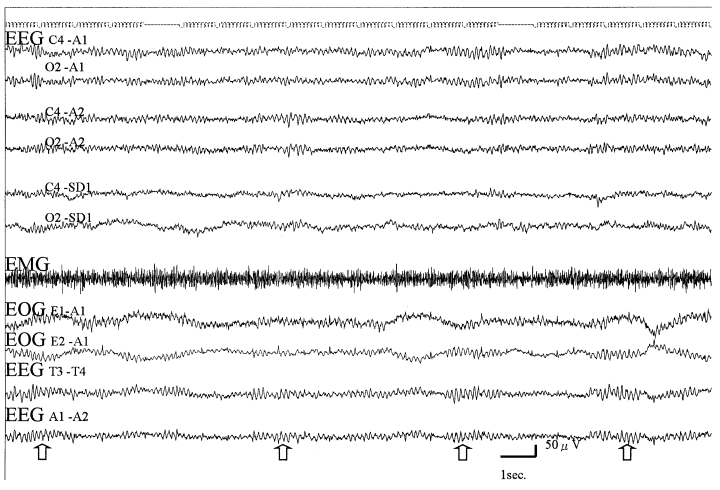
Figure 1 shows the polysomnogram (PSG) of a representative subject. A prominent κ-rhythm appeared on T3–T4 and A1–A2 derivations. On a C4–A1 derivation, α-like waves appeared continuously, but on C4–A2 and C4–SD1 derivations α-like waves sometimes disappeared, indicating drowsiness. We judged drowsiness using both earlobes as references in 129 subjects who showed a κ-rhythm on the T3–T4 derivation. When the contra-lateral earlobe was used as a reference, drowsiness could be judged properly in 26.4% of subjects. Conversely, drowsiness could be judged properly in 90.0% of subjects using the ipsi-lateral earlobe. It was impossible to assess drowsiness in 10.1% of subjects using either earlobe as a reference. Using the ipsi-lateral earlobe was significantly better than the contra-lateral earlobe (P < 0.0001, χ2 test) for assessment of drowsiness. When we used the SD method, drowsiness could be judged in all subjects (Fig. 1), but at least 19 active electrodes were required.

. Polysomnogram of a representative subject. κ-Rhythms (arrows) appeared on an A1–A2 derivation and activated an earlobe electrode.
DISCUSSION
In the present experiment, assessment of drowsiness was improved significantly by using an ipsi-lateral earlobe. When the amount of κ-rhythm increases, it is necessary to counterbalance the rhythm, which activates not only an earlobe but also active electrodes, by using the ipsi-lateral earlobe. We have reported previously that was observed in 21.6% of 440 subjects (mean age 48.4 years).4 Furthermore, the presence rates of κ-rhythm were 9.8% for subjects aged in their 20s, 6.6% in their 30s, 15.1% in their 40s, 24.2% in in their 50s, 39.5% in their 60s, and 43.8% in their 70s. Kennedy et al. have reported that κ-rhythm was observed in 30% of healthy adults.3
The present study’s results suggest that the bipolar T3–T4 derivation should be monitored and checked for the presence of κ-rhythm, and that the ipsi-lateral earlobe or the SD method should be used as a reference, especially in the elderly. Using these methods, amplitude was reduced by approximately 5–15% and has very little influence on judging drowsiness.


