Portal biliopathy
Abstract
Abstract In patients with portal hypertension, particularly with extrahepatic portal vein obstruction, portal biliopathy producing biliary ductal and gallbladder wall abnormalities are common. Portal cavernoma formation, choledochal varices and ischemic injury of the bile duct have been implicated as causes of these morphological alterations. While a majority of the patients are asymptomatic, some present with a raised alkaline phosphatase level, abdominal pain, fever and cholangitis. Choledocholithiasis may develop as a complication and manifest as obstructive jaundice with or without cholangitis. Endoscopic sphincterotomy and stone extraction can effectively treat cholangitis when jaundice is associated with common bile duct stone(s). Definitive decompressive shunt surgery is sometimes required when biliary obstruction is recurrent and progressive.
INTRODUCTION
The changes of portal biliopathy have been reported to be more common in patients with portal vein thrombosis than in patients with non-cirrhotic portal fibrosis or cirrhosis of the liver. Unless the clinical and radiological profile of this entity is well appreciated, the condition could be overlooked.
Portal biliopathy is a recent terminology used to describe biliary ductal and gallbladder wall abnormalities seen in patients with portal hypertension.1–3 These changes are predominantly seen in patients with extrahepatic portal vein obstruction (EHPVO) and include abnormalities (stricture and dilatation) of both extra- and intrahepatic bile ducts and varices of the gallbladder. Occasionally, these changes become significant to give rise to overt obstructive jaundice and possibly contribute to the development of choledocholithiasis.4
Biliary changes suggestive of biliopathy are reported to be quite common in patients with EHPVO. While some investigators have reported the ductal anomalies in all patients with EHPVO,5 we had observed these changes in 80% of our patients.1 The present review deals with the pathogenesis, natural history, diagnosis and management of patients with portal biliopathy.
Vascular supply of the bile duct
The arterial supply of the bile duct is through branches of gastroduodenal artery, but it is the venous drainage that is of particular importance in the development of portal biliopathy.
The venous drainage of the common bile duct (CBD) is mostly by veins that ascend along its course. They form epicholedochal venous plexus (of Saint) and paracholedochal venous plexus (of Petren) (Fig. 1). The former forms a fine reticular venous plexus on the common bile duct and hepatic ducts, and is in intimate contact with their outer surface.6–8 The veins of this plexus vary in size, but normally are not larger than 1 mm. The paracholedochal veins course parallel to the CBD and are connected to the gastric veins, the pancreaticoduodenal vein, the portal vein and to the liver directly.7,8 Varices of the plexus alter the normal smooth intraluminal surface of CBD and produce fine irregular mural changes.9
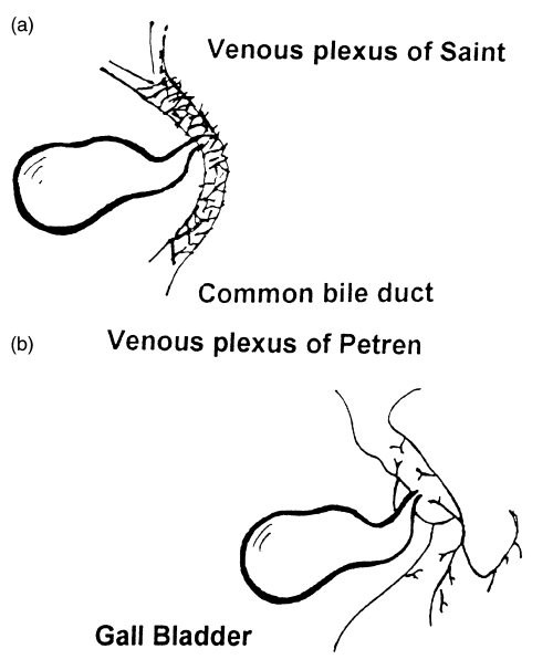
(a) Epicholedochal venous plexus. (b) Paracholedochal venous plexus.
Pathogenesis
Bile duct varices
The earliest evidence of choledochal varices was noted by Hunt in 1965.10 Later on, Meredith et al.11 and others12,13 reported a few cases of common bile duct obstruction caused by extensive collateral venous circulation at the porta hepatis. These patients had varied etiologies of portal hypertension like EHPVO, alcoholic cirrhosis, congenital hepatic fibrosis, and few of them were jaundiced also. The cholangiographic changes caused by choledochal varices were first demonstrated by William et al.14
The development of portal hypertension leads to the opening up of numerous venous collaterals that decompress into the systemic circulation.15 The common collaterals include esophageal and gastric varices. Varices have also been noted in the duodenum, colon, rectum, abdominal wall, retroperitoneum and at surgical anastomotic sites.
Portal biliopathy has been observed mostly in EHPVO patients, although such changes have been reported in a milder form in patients with cirrhosis, non-cirrhotic portal fibrosis,1 and congenital hepatic fibrosis. A precise explanation for a higher frequency in EHPVO patients is not clear. In the idiopathic or primary variety of EHPVO, which is possibly due to omphalitis or umbilical sepsis at birth, is an early age of onset of portal vein thrombosis and a long duration of portal hypertension are likely to be contributory. Following thrombosis of the portal vein, several new collaterals develop, which bypass the obstruction and produce ‘cavernomatous transformation’ of the portal vein or formation of a ‘portal cavernoma’.11 The same process leads to formation of collaterals around the gallbladder and the bile ducts.
The bile duct wall is thin and pliable, and allows protrusion of the varicose paracholedochal veins into the lumen, resulting in an appearance resembling that of esophageal varices.11 These luminal features can be seen better at surgery than at cholangiography.
The biliary strictures that develop in patients with biliopathy could form because of ischemia and vascular bile duct injury, or prolonged compression of the biliary tree from the portal cavernoma. Khuroo et al. have speculated that vascular injuries at the time of portal vein thrombosis are responsible for these CBD abnormalities.2 An extension of the thrombotic process may cause ischemic necrosis of bile duct(s) and subsequent stricture formation, irregularities in the caliber of the ducts and cholangiectases. Webb and Sherlock reviewed the presentation and natural history of EHPVO patients and noted that out of 97 patients, 13 had an elevated bilirubin level.16 They postulated that partial obstruction of the CBD because of mechanical compression by engorged venous collaterals could be responsible for this. Besides ischemia and compression, some investigators have speculated an infective basis for these biliary anomalies. Dilwari and Chawla who had observed biliary changes in all 20 EHPVO patients studied (Table 1), called the radiological abnormalities ‘pseudosclerosing cholangitis’.5 This term, however, is misleading because cholangitis is not a common presentation of these patients, and is rather a late event.
Clinical features
Portal biliopathy produces symptoms that are caused by partial or rarely, complete bile duct obstruction. Abdominal pain, recurrent fever with chills and jaundice, alone or in combination, suggests the possibility of biliary obstruction. Majority of the patients are asymptomatic and demonstrate characteristic changes on endoscopic retrograde cholangiopancreatography (ERCP) only (Table 2). Only 14% of patients were symptomatic in one study2 and 5% in another.5 In our initial study, choledocholithiasis was observed in five of 23 (17%) patients.1 However, none of these patients had biliary symptoms at onset. Only during follow up, three of the four patients developed cholangitis, and had to be treated with endoscopic stone extraction.
| ERCP changes | EHPVO(n = 47) | NCPF(n = 10) | Cirrhosis(n = 12) |
|---|---|---|---|
| Abnormal cholangiogram | 41 (87.3) | 4 (40) | 4 (33.3) |
| Extrahepatic duct | 41 (87.3) | 4 (40) | 3 (24.9) |
| Left hepatic duct | 34 (72.3) | 2 (20) | 2 (16.6) |
| Right hepatic duct | 26 (55.3) | 1 (10) | 1 (8.3) |
| IHBR | 6 (12.8) | 1 (10) | 2 (16.6) |
| Dilated | Pruned | Pruned | |
| CBD stone(s) | 8 (17) | 0 | 0 |
- ERCP, endoscopic retrograde cholangiopancreatography; IHBR, intrahepatic biliary radicle; EHPVO, extrahepatic portal vein obstruction; NCPF, non-cirrhotic portal fibrosis; CBD, common bile duct. All percentages in parentheses.
Gibson et al. reviewed 28 cases of EHPVO and observed jaundice in five of them.17 These authors did not specifically study the biliary abnormalities. Ductal anomalies are likely to contribute to a large extent to jaundice because EHPVO patients generally have near normal liver functions. While jaundice in a patient with portal biliopathy could be caused by the development of a narrowing or stricture in the CBD or because of choledocholithiasis, prolonged obstruction could lead to the development of secondary biliary cirrhosis. In the majority (80%) of patients with EHPVO and portal biliopathy, serum alkaline phosphatase levels were elevated.2 However, the exact significance of raised alkaline phosphatase is often hard to interpret if the EHPVO patient is a child or an adolescent. The other tests of liver function are also not helpful. However, late in the course of the illness, the serum albumin may decrease with derangement of coagulation functions because of the development of secondary biliary cirrhosis. An abdominal ultrasound is very helpful in establishing the diagnosis of portal cavernoma and in the detection of venous collaterals compressing the bile ducts and the gallbladder.18
Endoscopic retrograde cholangiopancreatography abnormalities
Portal biliopathy changes on ERCP are protean. The biliary abnormalities are most prominent in the common bile duct (Table 1). The observed abnormalities include: irregularities in the CBD and hepatic ducts (i.e., smooth tapering strictures that may be single or multiple, short or long); localized saccular dilation and filling defects suggestive of CBD calculi etc.1,2,5 Some of these changes are similar to those described in sclerosing cholangitis.2 As in recurrent pyogenic cholangitis, the changes in the left hepatic duct are seen more often in portal biliopathy. The common differential diagnoses of this condition include CBD calculi with associated stricture formation, biliary ascariasis and sclerosing cholangitis. In the former, the presence of concomitant gallstones and occurrence of CBD stones proximal to the distal stricture provides a clue to diagnosis. In biliary ascariasis, linear or curved filling defects on ERCP and similar echogenic shadowing on an ultrasound point to the diagnosis. Biliary anomalies can be seen in portal hypertension caused by varied etiologies.
In patients with cirrhosis, pruning of intrahepatic biliary radicles is often seen. However, in a small proportion (25%) of patients with cirrhosis, cholangiographic anomalies of the extrahepatic bile ducts may be evident.19 Similarly, in approximately 40% of patients with non-cirrhotic portal fibrosis (NCPF), cholangiographic abnormalities are detectable.19 These biliary anomalies are, however, mild.
We have proposed a simple classification for portal biliopathy based on the location and extent of cholangiographic abnormalities. The diagrammatic representation of these changes is depicted in Fig. 2 and cholangiographic abnormalities have been shown in Fig. 3.
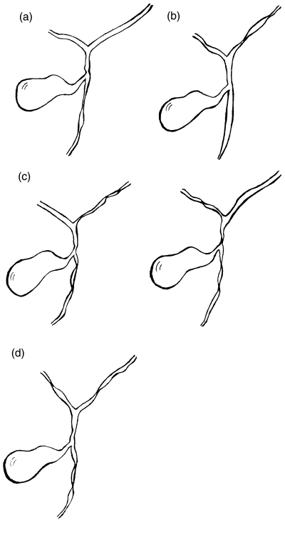
Schematic representation of endoscopic retrograde cholangiopancreatography changes in portal biliopathy. (a) Type I, involvement of extrahepatic bile duct; (b) type II, involvement of intrahepatic bile ducts only; (c) type IIIa, involvement of extrahepatic bile duct and unilateral intrahepatic bile duct (left or right); (d) type IIIb, involvement of extrahepatic bile duct and bilateral intrahepatic ducts.
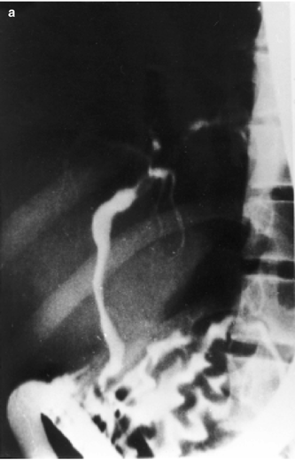

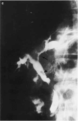
Endoscopic retrograde cholangiopancreatography changes in portal biliopathy. (a) Type I cholangiographic changes, (b) type IIIa cholangiographic changes; (c) type IIIb cholangiographic changes with common bile duct stone (arrowhead) and left hepatic duct stricture (arrow). Continued on next page.
Cholangiographic studies are essential for the diagnosis of portal biliopathy. However, ERCP is a somewhat invasive procedure. Discretion needs to be used, especially if the patient is asymptomatic and has no radiological or biochemical evidence of biliary obstruction. Magnetic resonance cholangiography (MRC) could be helpful in defining the bile duct changes. Magnetic resonance cholangiography has been shown to be useful in several biliary tract disorders. It would be worthwhile to compare the two modalities in diagnosing the condition in patients with suspected portal biliopathy. There is little available information on the use of radionuclide scanning in this situation.
Ultrasound abnormalities
Ultrasound studies are complimentary to ERCP findings. While ERCP studies delineate the biliary ductal changes, ultrasound provides additional information regarding the presence of gallbladder varices, and is thus helpful in providing the complete spectrum of portal biliopathy changes.20 Gallbladder varices were observed by us in 34% of patients with EHPVO, 24% of patients with NCPF and 13% of patients with cirrhosis.21 Gallbladder varices appear as tortuous dilated vessels in or around the wall of the gallbladder, or in the bed of the gallbladder fossa (Fig. 4). To investigate whether these collaterals impede the contractility of the gallbladder, motor functions of the gallbladder were studied by us. No significant difference was noted in the ejection fraction of the gallbladder in patients having gallbladder varices. However, the fasting gallbladder volume was found to be increased in such EHPVO patients. The clinical significance of the latter observation remains to be determined.21
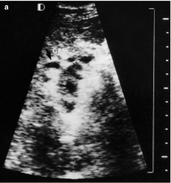

(a) Gallbladder varices (GBV); (b) a Doppler study showing the flow in gallbladder varices.
Magnetic resonance cholangiography
Because ERCP is an invasive procedure and an ultrasound of the abdomen may not give a definite outline of the biliary ductal anomalies, there is a need to have a good non-invasive modality to study biliopathy changes. Magnetic resonance cholangiography has been found to be helpful in the diagnosis of several biliary tract anomalies. Magnetic resonance cholangiography needs to be evaluated in patients with suspected portal biliopathy and could be compared with the results of ERCP studies.
TREATMENT
At present, strategies for the management of portal biliopathy are selective and directed to symptomatic patients only (Fig. 5). Asymptomatic patients do not need any treatment, especially if the tests of liver function are normal. If there is a persistent elevation of alkaline phosphatase in a given patient with portal hypertension, especially an EHPVO patient, then an ultrasound, MRCP or, if needed, an ERCP could be considered for use to detect portal biliopathy.

An algorithmic approach to diagnosis and management of portal biliopathy. CBD, common bile duct; IHBR, intrahepatic biliary radicles; USG, ultrasonogram; ALP, alkaline phosphatase; ERCP, endoscopic retrograde cholangiopathy; ⋆; rule out other causes of increased ALP.
Choledocholithiasis is a known complication of portal biliopathy. It was encountered in 17% of patients in the present study. Once stones produce obstruction or are detected otherwise, they can be safely removed by the use of endoscopic sphincterotomy.22,23 However, care should be taken while performing a sphincterotomy as large venous collaterals in the ampullary and juxta-ampullary area could be present and might carry a risk of bleeding associated with this procedure. However, we have not encountered any complications in the seven patients who had received sphincterotomy at our center (GB Pant Hospital). It is important to differentiate duodenal folds around the ampulla from collaterals before cutting the ampulla. There is no rationale of giving octreotide as a preventive therapy before sphincterotomy. The chances of bile duct stone recurrence are high because the primary obstructive bile duct lesions often persist.
Sometimes, obstructive jaundice could be caused by a dominant stricture or significant abnormalities of the bile duct without any evidence of CBD stones. In such patients, the placement of a biliary stent with balloon dilation of the strictured segment of CBD is recommended. The stent often needs to be exchanged and sometimes, two or more stents need to be placed to keep the lumen patent.
In the presence of symptomatic biliary obstruction not amenable to endoscopic therapy, a portosystemic shunt is indicated.24–27 This may relieve the symptoms of biliary obstruction through portal decompression. Biliary tract surgery in the presence of portal hypertension is fraught with the danger of massive blood loss because of unavoidable damage to collaterals that are present in abundance in EHPVO patients.24 Hence, biliary surgery should always be preceded by a portocaval shunt. Following shunt surgery, changes of portal biliopathy have been shown to regress in most, but not all, patients. In a proportion of patients, bile duct changes persist25 and produce symptoms. If these changes get well established, surgery is probably of little consequence in reverting them. This necessitates a second stage surgery in the form of hepaticojejunostomy to relieve the biliary obstruction.28 This lends support to the hypothesis that ischemic changes of the bile duct are, in part, responsible for this condition.
In conclusion, patients with portal hypertension who have features of biliary obstruction (clinical, sonographic and or cholangiographic), portal biliopathy should be suspected as a possible diagnosis. The natural history of portal biliopathy is ill-defined and, as mentioned already, varies from an asymptomatic state to the development of various sequelae-like choledocholithiasis, cholangitis and secondary biliary cirrhosis. In a single institution (GB Pant Hospital) experience of a follow up of more than 6 years, two of 55 patients (4%) with EHPVO and portal biliopathy have succumbed to these sequelae. A high index of suspicion, an early diagnosis and clear treatment protocols would help in the better management of this often overlooked entity.


