Identification and characterization of a germination operon on the virulence plasmid pXOl of Bacillus anthracis
Abstract
The spores of Bacillus anthracis, the agent of anthrax disease, germinate within professional phagocytes, such as murine macrophage-like RAW264.7 cells and alveolar macrophages. We identified a cluster of germination genes extending for 3608 nucleotides between the pag and atxA genes on the B. anthracis virulence plasmid pXOl. The three predicted proteins (40, 55 and 37 kDa in size) have significant sequence similarities to B. subtilis, B. cereus and B. megaterium germination proteins. Northern blot analysis of total RNA from sporulating cells indicated that the gerX locus was organized as a tricistronic operon (gerXB, gerXA and gerXC). Primer extension analysis identified a major potential transcriptional start site 31 bp upstream from the translation initiation codon of gerXB. Expression of the gerX operon was studied using a gerXB–lacZ transcriptional fusion. Expression began 2.5–3 h after the initiation of sporulation and was detected exclusively in the forespore compartment. A gerX null mutant was constructed. It was less virulent than the parental strain and did not germinate efficiently in vivo or in vitro within phagocytic cells. These data strongly suggest that gerX-encoded proteins are involved in the virulence of B. anthracis.
Introduction
Bacillus anthracis, the causative agent of anthrax, is a Gram-positive, endospore-forming, aerobic, rod-shaped bacterium. Anthrax infection starts after the intradermal inoculation, ingestion or inhalation of spores (Laforce et al., 1969; Friedlander et al., 1993). Therefore, spore germination is required for the establishment of anthrax disease. We showed recently, in a murine inhalation infection model, that the alveolar macrophage is the primary site of B. anthracis spore germination (Guidi-Rontani et al., 1999). B. anthracis infection causes death via multiplication of the bacilli and the expression of virulence factors. The major known virulence factors of B. anthracis are an antiphagocytic poly-γ-d-glutamic acid capsule (Green et al., 1985) and the lethal and oedema toxins produced during exponential growth (Beall et al., 1962; Friedlander, 1986). The genes necessary for synthesis of the capsule, capB, capC, capA and dep, are located on pXO2 (95 kb) (Uchida et al., 1993a). pXO1 (185 kb) harbours the structural genes, pag, lef, cya and atxA, encoding the three toxin components (Leppla, 1991) and their transcriptional activator respectively (Uchida et al., 1993b; Koehler et al., 1994). These genes are located in a 40 kb region of pXO1 flanked by two inverted repeat elements (J. M. Hornung and C. B. Thorne; EMBL accession U30715; U30713). The development of B. anthracis strains cured of pXO1 in the host is impaired, whereas that of strains deficient in the production of toxin components only is not (Pezard et al., 1995). Therefore, other virulence factors, encoded by pXO1, unrelated to the toxins are probably involved.
In the search for such factors, we identified and characterized a tricistronic germination operon within the 40 kb toxin-encoding region of pXO1. We also investigated the contribution of the products of this operon to the pathogenesis of B. anthracis.
Results
Sequence analysis of gerXB, gerXA and gerXC
The nucleotide sequence of the pag-atxA region of B. anthracis pXOl was obtained by oligonucleotide walking and analysed using the GCG software package. Three open reading frames (ORFs) were identified (GenBank accession no. AF108144), on the same strand, 7975 bp from the atxA transcription on the opposite strand (EMBL accession no. l13841). These ORFs consisted of 359, 492 and 318 codons, respectively, and had a high AT content (CG% 33.8). Each ORF started with an ATG initiation codon, and together they spanned 3608 bases of the pag-atxA region.
A tfasta search of the EMBL database (Pearson and Lipman, 1988) using the deduced amino acid sequences for the three ORF-encoded proteins showed significant similarity to the products of the gerB (Corfe et al., 1994; EMBL accession no. L16960) and gerK (Irie et al., 1996; EMBL accession no. D78187) operons of B. subtilis, the gerA operon (K. Tani, unpublished; EMBL accession no. U61380) of B. megaterium and the gerI operon (Clements and Moir, 1998; EMBL accession no. af067645) of B. cereus. The three genes were therefore designated gerXB, gerXA and gerXC (for germination pXOl related), and the encoded putative proteins with calculated molecular sizes of 40.839, 55.106 and 37.084 kDa were designated GerXB, GerXA and GerXC respectively. The greatest similarity was observed with the gerA operon products of B. megaterium using the algorithm of Needleman and Wunsch (1970): between GerXB and GerAB (20.0% identity and 36.8% similarity); between GerXA and GerAA (30.1% entity and 44.1% similarity); between GerXC and GerAC (20.9% identity and 30.4% similarity). Kyte–Doolittle hydrophobicity plots (Kyte and Doolittle, 1982) demonstrated that GerXB, GerXA and GerXC had patterns of hydropathy (Fig. 1) typical of germination proteins of the gerA operon of B. subtilis. Therefore, GerXB, GerXA and GerXC are homologues of GerAB, GerAA and GerAC respectively (Feavers et al., 1985; Irie et al., 1993; 1996). Hydrophobic cluster analysis showed that GerXB (X35-Ycter) (Fig. 1A) and GerXA (V250-F466) (Fig. 1B) contained 10 and seven membrane-spanning sequences, respectively, which predicted transmembrane domains (Kyte and Doolittle, 1982). Further analysis with plotsimilarity (data not shown) showed that the most conserved region between GerXA and GerAA of B. megaterium corresponds to (V250–F466; 37.3% identity and 50% similarity). The deduced sequence of GerXC predicted a hydrophilic protein (Fig. 1C) with a putative lipoprotein signal (L12LMFLFLLTGC22; Hayashi and Wu, 1990) as observed in the GerAC homologues (Irie et al., 1993).
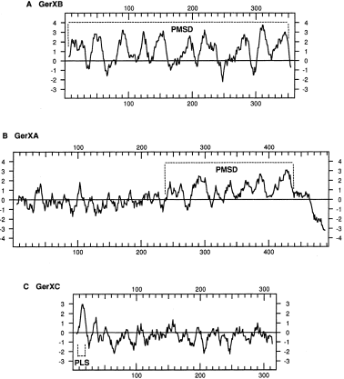
. Hydropathy profiles of the predicted gene products of gerX. The hydropathy values of 11-amino-acid windows centred on each residue along the GerXB (A), GerXA (B) and GerXC (C) sequences are plotted. The y-axis corresponds to hydrophobicity. The horizontal line represents the average hydropathy of a large number of sequenced proteins according to the algorithm of Kyte and Doolittle (1982). The putative membrane-spanning domains (PMSD) and the putative lipoprotein signal (Ll2LMFLFLLTGC22) (PLS) are indicated.
Regulation of gerX expression
The ATG initiation codons of the three gerX genes were preceded by potential ribosome binding sites with homology to the 3′ end of the B. anthracis 16S ribosomal RNA molecule (Ash et al., 1991), with ‘spacers’ of 3, 10 and 7 nucleotides, respectively, the optimum ‘spacer’ in Bacillus being 7 nucleotides (Vellanoweth and Rabinowitz, 1992). The initiation codon of gerXB was preceded by one potential sigma-G (σG)-dependent promoter sequence 5′-TGAATA-n18-TATAATA-3′ (Nicholson et al., 1989) (Fig. 2). GerXC overlapped with gerXA by 14 bp, and neither promoter-like nor terminator-like sequences were found around gerXA and gerXC. Therefore, these three genes may be co-transcribed and expressed during sporulation. We determined the size of the gerXB, gerXA and gerXC transcripts by carrying out Northern blot analysis with total RNA isolated from B. anthracis 7702 during the exponential phase and at t3 (tn is the number of hours after the end of the exponential phase) (Fig. 3). No mRNA from these genes was detected in total mRNA extracted during the exponential phase (Fig. 3, lanes Al and Bl). In contrast, an mRNA species of ≈ 3.5 kb in size was detected at t3, with both specific gerXB (Fig. 3, lane A2) and gerXC probes (Fig. 3, lane B2). The estimated size, 3.5 kb, was close to the predicted minimum size (3569 bases) for a polycistronic mRNA covering the three cistrons gerXB, gerXA and gerXC.

. Schematic representation of the gerX locus. The structural genes gerXB, gerXA and gerXC, located on pXOl between the atxA and pag genes, are shown as open boxes. The nucleotide sequence shows the −35 and −10 box sequences in bold, consensus sequences to the sigma-G (σG)-dependent promoter. The transcriptional start site (Pl), determined by primer extension (Fig. 4), is indicated by an arrow. The potential ribosome binding site of gerXB is underlined. Recombinant B. anthracis strains GX11 and GX12 were constructed by deletion between nucleotides 572 of gerXB and 186 of gerXC (indicated above the schematic representation) and insertion of a spectinomycin resistance cassette or the lacZ-spectinomycin cassette respectively.
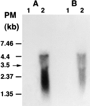
. Transcriptional analysis of the gerX locus. Hybridizations with the gerXB (A) and gerXC (B) probes are shown. Total RNA (20 μg) was extracted from B. anthracis 7702 during the exponential growth phase (lanes l) and 3 h after the end of the exponential growth phase (lanes 2). The arrow on the left marks the position and size of the transcript.
The 5′ end of the transcript was then determined by primer extension analysis (Fig. 4) using a synthetic end-labelled primer extending from nucleotide position 53 to 75 bp downstream from the translational start site of gerXB. Total RNA was isolated at the end of the exponential phase and into the stationary phase. At t3, two adjacent bases were identified as start sites C and A, which are 31 bp and 30 bp upstream from the putative translational start site of the gerXB cistron respectively.
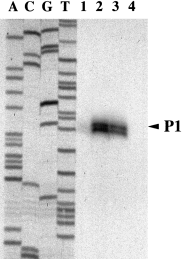
. Mapping of the gerX transcriptional start sites. Primer extension analysis of the 5′ terminus of gerX mRNA is shown. Total RNA (20 μg), extracted from B. anthracis 7702 at t2.5, t3 and t3.5 (lanes 1–3 respectively), was subjected to primer extension with reverse transcriptase and a synthetic 21-mer as described in Experimental procedures. The transcription start site, P1, is indicated by an arrow.
We analysed the expression of the gerX locus in space and time by constructing a gerXB–lacZ transcriptional fusion, which was introduced into pXOl at nucleotide 572 downstream from the putative translational start site of gerXB (Fig. 2). Direct evidence that gerX is expressed exclusively in the forespore compartment was obtained by growing the recombinant cells (GX12) carrying the gerX–lacZ fusion in the presence of the fluorescent lipophilic β-d-galactosidase substrate, C12FDG (Zhang et al., 1991). No fluorescence was detected in the mother cell compartment; the fluorescence was limited exclusively to the forespore compartment (Fig. 5A), thus demonstrating that gerX was expressed in this compartment after septation. The expression of gerX was also studied in a time course experiment. β-d-Galactosidase activity was barely detectable during the exponential phase (Fig. 5B), but it increased rapidly during the stationary phase, reaching a peak 3 h after the end of the exponential phase (2014 U mg−1 protein).
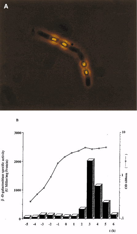
. Growth phase-dependent expression of gerXB–lacZ. The β-d-galactosidase activity of B. anthracis GX12, expressing a gerXB–lacZ gene fusion, was assessed (A) using ImaGene green fluorescein isothiocyanate (FITC) or (B) as described by Pardee et al. (1959). β-d-Galactosidase specific activity is expressed in Miller units (U) mg−1 protein. Time zero is the end of the exponential growth phase and tn is time, with n being the number of hours before (−) or after time zero.
Thus, gerXB, gerXA and gerXC are organized into a single operon (gerX ) expressed during the sporulation phase.
Germination of the gerX null mutant within macrophage
We showed recently that B. anthracis germinates within professional phagocytes (Guidi-Rontani et al., 1999). We investigated the possible involvement of gerX in this process by producing a gerX null mutant, GX11 (Fig. 2). The fate of the 7702 and GX11 spores within murine macrophages such as RAW264.7 was investigated. The germination of spores associated with macrophages was assessed by testing for a loss of heat resistance (30 min at 65°C) and by indirect immunofluorescence 3 h after infection. In heat resistance tests, we observed that the macrophage-7702 fraction had significantly higher levels of germination (26%) than the macrophage-GX11 fraction (8%). Immunofluorescence analysis showed that antibodies against bacilli recognized a large proportion of germinated spores in the 7702-macrophage population (Fig. 6A and B). In contrast, very few germinated spores were detected in the macrophage-GX11 fraction (Fig. 6C and D).
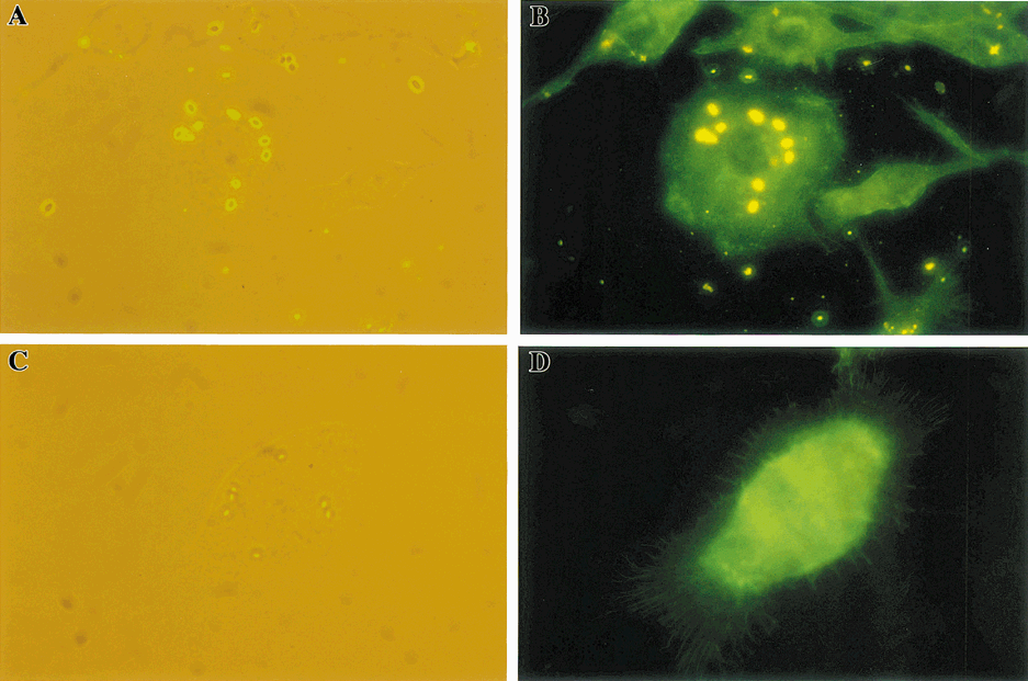
. Fluorescence microscopy of RAW264.7 macrophages 3 h after infection with B. anthracis gerX null mutant. Germinated spores from the 7702 (A and B) or GX11 (C and D) strains were detected in RAW264.7 cells with the antibacillus serum and a rhodamine-conjugated secondary antibody (see Experimental procedures). RAW264.7 cells were stained with Oregon green–488 phalloidin (green) (B and D). The total spore population was detected by light microscopy (A and C). Germinated spores were detected by immunofluorescence (yellow) (B and D). Images acquired during each of these recordings are shown in pairs. (A) and (B) are from the same field, as are (C) and (D).
In vivo contribution of gerX in B. anthracis virulence
We investigated the involvement in vivo of the factors encoded by the gerX operon in anthrax pathogenesis by studying the virulence properties of GX11. The cumulative mortality of mice was assessed after subcutaneous inoculation with 5 × 10⁴ spores of 7702 or GX11, a dose equivalent to one LD50 for the parental strain 7702 (Fig. 7). Whereas 50% of mice died after inoculation with 7702, only 10% of mice died after GX11 inoculation. We followed the germination of GX11 and 7702 spores over time within the host after the injection of 1.7 × 10⁵ spores. Tissue was collected from the inoculation site. Large subcutaneous oedema developed in tissues adjacent to the site of inoculation after 12 h with 7702 and after 24 h with GX11. This suggested that oedema toxin is synthesized soon after the germination of 7702 and GX11 spores. The germination of spores was assessed by testing for a loss of heat resistance (Fig. 8). A decrease in the number of 7702 dormant spores was observed 12 h after inoculation. This decrease became large over time until only 2.5 × 10³ cfu remained. Bacteria then grew rapidly, and large numbers were present 18 h and 24 h after inoculation. In contrast, there was no significant reduction in the number of dormant GX11 spores until 24 h after inoculation (2.5 × 10⁴ cfu). However, at 24 h and in the absence of heat treatment, there was a significant increase in cfu for GX11. Thus, the multiplication of some germinated forms had already been initiated. Consequently, GX11 germination was less efficient than that of 7702, and this was associated with a significant delay in the onset of germination.
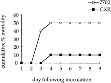
. Virulence of B. anthracis GX11 and 7702 strains. Swiss mice were inoculated subcutaneously with 5 × 10⁴ spores per mouse (groups of 10 mice). Mortality was recorded daily and plotted as the cumulative number of deaths.
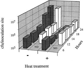
. In vivo germination of B. anthracis GX11. Swiss mice inoculated subcutaneously with 1.7 × 10⁵ spores (groups of three mice) were killed at intervals after infection (6 h to 24 h). The inoculation sites were removed and tested for the germination and survival of spores with (+) or without (−) heat treatment. The data are expressed as cfu (bars) for 7702 (dark bars) and for the gerX null mutant (white bars).
Discussion
This paper describes the first germination operon (gerX) to be identified in B. anthracis. We demonstrated, using the lacZ reporter gene and primer extension analysis, that gerX transcription was restricted to the sporulation phase, as reported for other germination operons (Stragier and Losick, 1990; Errington, 1993). There were significant levels of gerX expression 3 h after the initiation of sporulation, and this expression was exclusively associated with the developing forespore after septation. In the promoter sequence, a consensus sequence for a B. subtilis sigma-G-like factor was identified (Karmazyn-Campbelli et al., 1989; Sun et al., 1989). Transcription of the gerX operon may therefore be mediated by a σG-like, forespore-specific sigma factor. Sequence similarity search and hydrophobic cluster analysis suggested that GerXB, GerXA and GerXC are germinant-specific components and are homologous with GerAB, GerAA and GerAC (Feavers et al., 1985; Zuberi et al., 1987; Irie et al., 1996), respectively, as defined in B. subtilis. The gerX locus is the only germination operon known that is located on a plasmid (pXOl). The gerX operon is part of the large toxin-encoding fragment (40 kb) of pXOl, which is flanked by two inverted repeat elements (J. M. Hornung and C. B. Thorne; EMBL accession nos U30715 and U30713). This fragment has virulence-associated functions and some features typical of the pathogenicity islands described by Galan (1996). It may therefore be a pathogenicity island in B. anthracis, which is the only highly pathogenic agent among the Bacilli.
We have shown that B. anthracis germinates efficiently within murine alveolar macrophages and within macrophage-like RAW264.7 (Guidi-Rontani et al., 1999). Germination was significantly less efficient for the gerX null mutant, GX11, than for the wild-type strain. Therefore, the gerX-encoded proteins may be involved in the detection of specific germinants within the macrophage. These proteins may act as an ‘environment–sensor complex’ transducing the environmental signal into the B. anthracis spore. Such a model would require membrane-associated proteins, such as the putative integral transmembrane protein, GerXB, and a highly hydrophobic protein with potential membrane-spanning helices, such as GerXA. GerXC, a putative lipoprotein, would probably be attached to the membrane of the forespore and would expose its hydrophilic domains to the environment. B. anthracis spores may possess a unique system for detecting specific germinants in the host. However, B. anthracis strains lacking pXO1 and pXO2 are able to germinate in vitro (Cataldi et al., 1990). There must, therefore, be at least one other germination operon, located on the chromosome. B. anthracis, like B. subtilis, probably possesses several germinant-specific receptors, enabling the spore to respond to different stimuli. Major germinant systems have been described: (i) the gerA operon (Zuberi et al., 1985) required for the l-alanine response and the gerB (Corfe et al. 1994) and gerK (Irie et al., 1996) operons required for the l-asparagine, glucose, fructose and potassium ion responses in B. subtilis; and (ii) the gerI locus (Clements and Moir, 1998) required for inosine germination in B. cereus.
Deletion of the gerX operon affected the in vivo virulence of B. anthracis, providing evidence for the involvement of the gerX-encoded proteins in the pathogenesis of B. anthracis. Greater insight into the mechanisms involved in the attenuation of GX11 virulence was obtained by analysing the time course of spore germination and multiplication of GX11 and its parental strain. The onset of germination was delayed in GX11. However, after 24 h, the germinated GX11 cells were able to multiply and invade the host. Therefore, the rate of germination may have a strong effect on the fate of the infection. The establishment of anthrax disease may require the rapid emergence of the vegetative form in a favourable niche, and this may occur in the macrophage, a key cell in the pathogenesis of B. anthracis. Whether or not the macrophage is the only site of germination remains to be investigated.
Experimental procedures
Bacterial strains, plasmids and culture media
B. anthracis Sterne strain 7702 (pXOl+) (Pasteur Collection) was used. Escherichia coli harbouring the Gram-positive suicide vector, pAT113 (Trieu-Cuot et al., 1991), was used for transferring DNA into B. anthracis. E. coli was grown in L broth (Miller, 1972), and B. anthracis was grown in brain–heart infusion (BHI; Difco Laboratories), HCT medium (Lecadet et al., 1980) or NBY medium for the preparation of spores (Green et al., 1985). Antibiotic concentrations in the media were as follows: spectinomycin, 100 μg ml−1 for B. anthracis; kanamycin, 50 μg ml−1 and 20 μg ml−1 for E. coli and B. anthracis respectively; erythromycin, 180 μg ml−1 and 100 μg ml−1 for E. coli and B. anthracis respectively. Stocks of B. anthracis spores were prepared as described previously (Pezard et al., 1991).
Antiserum preparation
Antiserum was raised against vegetative forms as described previously (Guidi-Rontani et al., 1999).
DNA isolation, sequencing and plasmid construction
pXOl was isolated from B. anthracis strain 7702 as described by Green et al. (1985). Methods for recombinant DNA manipulation were as described by Maniatis et al. (1982). DNA was sequenced by Genome Express, and the sequence strategy applied to both strands was ‘oligonucleotide walking’.
Construction of gerX null strains and mating procedure
Strains GX11 and GX12 were obtained by deletion of the fragment between nucleotides 572 of gerXB and 186 of gerXC (Fig. 2) and insertion of a spectinomycin resistance cassette, spc (Etienne-Toumelin et al., 1995), or of a lacZ-spectinomycin cassette, as described previously by Sirard et al. (1995). The constructs were integrated into the gerX locus of pXO1 by homologous recombination using the suicide plasmid, pAT113 (Trieu-Cuot et al., 1991). Conjugative transfer from E. coli to B. anthracis 7702 was as described by Pezard et al. (1991) and Sirard et al. (1995).
β-d-Galactosidase assays
β-d-Galactosidase (EC 3.2.1.23) was assayed as described by Pardee et al. (1959). Briefly, B. anthracis GX12 was cultured in HCT medium (Lecadet et al., 1980) at 34°C with shaking. Bacterial suspensions were then treated with SDS (0.2%) and chloroform (2%) at 28°C. Specific activities are expressed in Miller units (U) mg−1 protein. The enzyme was detected within GX12 bacilli using the fluorescent lipophilic, β-d-galactosidase substrate, 5-dodecanoylaminofluorescein di-β-d-galactopyranoside (C12FDG; 330 μM; Molecular Probes) in BHI medium containing 0.4% agar.
RNA extraction, Northern analysis and primer extension
RNA extraction and primer extension were performed as described previously (De-Souza et al., 1993). Electrophoresis of 20 μg of formaldehyde-denatured RNA and transfer to nitrocellulose membranes (Hybond-C; Amersham) were performed as described elsewhere (Sambrook et al., 1989). The specific gerXB and gerXC probes were a 544 kb internal fragment (+ 28 to + 572) and a 545 kb internal fragment (+186 to + 731) of the respective genes. Probes were labelled with [32P]-dATP (nick translation, Amersham). Hybridizations and washes were performed at 65°C in standard solutions (Amersham). For primer extension experiments, a synthetic 21-mer oligonucleotide was used, 5′-CGACCAGAATGATCTAATAGC-3′, which was complementary to the 5′ end of the gerXB gene (+ 101 to + 121). It was 5′ end-labelled with [32P]-ATP (4500 Ci mmol−1) using T4 polynucleotide kinase.
Cell culture and macrophage infection
The murine macrophage-like cell line RAW264.7 was grown routinely as described previously (Guidi-Rontani et al., 1999). RAW264.7 cells in PBS were infected at a multiplicity of infection (MOI) of 10 spores per macrophage. After 30 min of incubation, the cells were washed thoroughly and incubated with RPMI-1640 medium containing gentamicin (2.5 μg ml−l). The germination of spores within cells was assessed by testing for a loss of heat resistance (30 min at 65°C). Colony counts (cfu) without heat treatment correspond to total spore population (dormant and germinated spores). Viability after heat treatment was taken as a measure of the population of dormant spores. All experiments were carried out at least twice, on at least two independent spore preparations. One representative experiment is shown.
Immunofluorescence microscopy
Germinated spores were detected by indirect immunofluorescence with rabbit polyclonal antiserum specific for germinated spore (Guidi-Rontani et al., 1999) (1:1000) and rhodamine-conjugated goat anti-rabbit IgG (H + l; 1:50; Kirkegaard and Perry Laboratories). F-actin was detected by incubating fixed cells suspended in PBS with Oregon green 488–phalloidin (1:50; Molecular Probes). Labelling was carried out as described by Guidi-Rontani et al. (1999).
Infection of mice
Seven-week-old female Swiss mice were supplied by IFFA-CREDO. Mortality was monitored by subcutaneously inoculating groups of 10 mice with 5 × 10⁴ spores from 7702 or GX11. Bacterial germination in host tissues was monitored by infecting mice subcutaneously above the groin with 1.7 × 10⁵ spores in 0.1 ml of PBS. Groups of three infected mice were killed at intervals. The entire site of inoculation, including subcutaneous oedema, was removed, homogenized in PBS (pH 7.2) and tested for the presence of spores or bacilli as described above.
Data handling and computer analysis
DNA sequences were compiled and analysed using the programs frames, translate, fasta, motfs and plotsimilarity from the GCG Sequence Analysis Software Package.
Nucleotide sequence accession number
The nucleotide sequence of the gerXB, gerXA and gerXC genes of B. anthracis strain 7702 have been submitted to GenBank and have been assigned the accession number AF108144.
Acknowledgements
We would like to thank F. Brossier and R. Okinaka for valuable discussions, A. Moir for the communication of gerI sequence before publication, H. Agaisse for advice about Northern analysis and primer extension, and E. Duflot for technical assistance.


