Gata-1 transcription factor is mutated in cmk megakaryoblastic cell line
The zinc finger transcription factor GATA-1 is the founding member of the GATA family that binds to the WGATAR consensus sequence (Shivdasani, 2001). GATA-1 is expressed in erythroid cells, megakaryocytes, mast cells and eosinophils, and has been shown to be essential for normal erythropoiesis and megakaryocyte differentiation. Defective GATA-1 function due to missense mutations in the GATA-1 gene has been described in human X-linked macrothrombocytopenia (Shivdasani, 2001). Lineage-specific disruption of GATA-1 in mice has demonstrated that megakaryocytes from these mice proliferate profusely in vitro as a result of a unique megakaryocyte differentiation arrest (Shivdasani et al, 1997; Vyas et al, 1999). However, there are no reports on the involvement of this transcription factor in malignancy or leukaemia. In this study, we investigated possible mutations of the GATA-1 gene in megakaryoblastic leukaemia cell lines.
Four human megakaryoblastic leukaemia cell lines, CMK (Institute for Fermentation, Osaka, Japan), MEGA2, MEGO1 and UT7/TPO (a gift from Dr Komatsu, Jichi Medical School, Tochigi, Japan), were examined (reviewed in Saito, 1997). Total RNA was extracted using the Catrimox reagent (Takara Shuzo, Otsu, Japan), and first-strand cDNA was synthesized using ThermoScript reverse transcriptase and oligo-dT primer (GibcoBRL, Life Technologies, Rockville, MD, USA). Polymerase chain reaction (PCR) amplification was performed on the cDNA with three primer pairs as described by Mehaffey et al (2001). The target sequences were amplified with AmpliTaq Gold DNA polymerase (Applied Biosystems, Foster City, CA, USA) in a total volume of 20 µl for 35 cycles of 30 s at 95°C, 30 s at 60°C and 45 s at 72°C in a thermal cycler (GeneAmp PCR System 9600; Perkin-Elmer Cetus, Norwalk, CT, USA). Amplified DNA fragments were purified and subjected to direct cycle sequence analysis on an ABI 373A DNA sequencer.
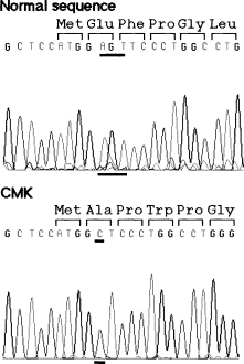
In each cell line, the amplified cDNA fragments were of predicted size compared with the normal sequences. In CMK cells, there was a complex deletion/insertion mutation in which 3 base pairs (bp), AGT, were deleted at nucleotide 5–7 and 1 bp, C, was inserted at the same position (5–7delAGTinsC) (Fig 1). This mutation would result in a frameshift that produces only the translation initiation methionine and additional 36 altered amino acids before a premature termination codon. Sequence analysis of the genomic DNA showed the presence of the same mutation as in cDNA. In addition, there was a synonymous 154C→T substitution at Leu52 (CTG→TTG) in MEGO1 cells. Direct sequencing analysis showed normal GATA-1 cDNA sequences in other cell lines.

DNA sequence analysis of the GATA-1 gene in the CMK cell line. PCR-amplified cDNA fragments were purified and directly sequenced. In CMK cells, there was a complex deletion/insertion mutation, 5–7delAGTinsC, resulting in a premature termination. The position of the substitution is underlined.
CMK was established from a boy with Down's syndrome and acute megakaryoblastic leukaemia (Sato et al, 1989). This cell line has megakaryoblastic features, and has been used extensively for studies of megakaryocyte growth and differentiation as well as platelet development. Because the GATA-1 gene is located on chromosome X and CMK was derived from a male patient, this cell possesses only the mutated GATA-1 gene. Furthermore, the 5–7delAGTinsC is essentially a loss-of-function mutation. Our results suggest that inactivation of the GATA-1 gene may be a possible feature in megakaryoblastic cell lines and may play a role in lineage-committed leukaemogenic processes or the establishment of these cell lines. The CMK cell line may be useful for further studies of this transcription factor such as complementation analysis. It remains to be determined whether the alteration of the GATA-1 gene occurred during in vitro culture or was present in the original myeloid leukaemia cells.
Disclosures
There is no conflict of interest regarding the submitted article.


