Visceral Adiposity and Subclinical Coronary Artery Disease in Elderly Adults: Rancho Bernardo Study
Abstract
Objective: Despite growing attention to central obesity as a predictor of clinical coronary heart disease (CHD), there are few reports about the association between directly measured visceral obesity and subclinical coronary atherosclerosis in elderly adults. We examined this association in elderly, community-dwelling adults without clinically recognized CHD.
Methods and Procedures: Elderly adults (190 men, BMI 27.2 ± 3.6 kg/m2; 220 women, BMI 25.8 ± 4.6) aged 55–88 years (median 69 years) with no history of CHD or coronary revascularization had an electron beam computed tomography (EBCT) to measure coronary artery calcification score (CACS), an estimate of coronary plaque burden. Visceral and subcutaneous adiposity were assessed by a triple-slice EBCT scan at the lumbar 4–5 disc level and height, weight, and waist and hip circumferences were measured.
Results: In sex-specific ordinal logistic regression analyses, no measure of obesity or body fat distribution, including BMI, waist-to-hip ratio, waist girth, and visceral and subcutaneous fat by EBCT, was significantly associated with CACS before or after adjusting for multiple covariates of CACS (age, smoking, alcohol intake, exercise, pulse pressure, low-density lipoprotein (LDL)/high-density lipoprotein (HDL)-cholesterol ratio, and fasting plasma glucose).
Discussion: In elderly adults without clinically recognized CHD, body weight and fat distribution do not predict coronary artery plaque burden. These results raise questions about the value of weight reduction diets for preventing heart disease in elderly survivors without clinical heart disease.
Introduction
Obesity is a powerful risk factor for cardiovascular disease (1,2,3,4,5,6), predicting coronary heart disease (CHD) independent of other risk factors in long-term follow-up studies (7,8,9). Growing attention has been paid to particular patterns of fat distribution, especially central (presumably visceral) obesity, as a predictor of both clinical coronary events (10) and angiographically documented CHD, independent of BMI (11,12).
However, most studies of the association between central obesity and CHD have estimated visceral obesity using waist circumference or waist-to-hip ratio. Directly measured visceral fat mass yields a more precise estimate of visceral obesity. The volume of visceral adipose tissue (VAT) and subcutaneous adipose tissue (SAT) can be measured directly as well as simultaneously by electron beam computed tomography (EBCT). Most studies of visceral adiposity and heart disease have been performed in patients who had clinical coronary events or coronary angiography performed to validate a suspected diagnosis. Body weight and fat distribution could have changed after diagnosis in these patients.
Age influences the pathogenesis of atherosclerosis both by inducing physiological vascular changes and by increasing the duration of exposure to cardiovascular risk factors (13,14). Several studies have shown that the association of CHD risk factors with CHD is attenuated in old age (15,16,17). The association between visceral obesity and CHD could be different in elderly adults, especially those without clinical CHD, but there has been no published study about this in a community-dwelling, elderly, middle to upper-middle class, white population.
Ultrafast high-intensity EBCT is a noninvasive method to measure coronary artery calcification (CAC), which reflects the amount of coronary artery plaque as assessed by pathology (18) and predicts subsequent cardiac events in both symptomatic and asymptomatic adults (18,19). EBCT can also be used for assessing abdominal body fat distribution to the visceral and subcutaneous compartments (20). The objective of this study was to examine the association of several parameters of obesity, including VAT and SAT directly measured by EBCT, with subclinical coronary atherosclerosis, also measured by EBCT, in relatively healthy, elderly adults who had a low prevalence of hyperglycemia and hypertension and were without clinically manifest CHD.
Methods and procedures
Study population
Participants were community-dwelling adults from the Rancho Bernardo Study (21), an ongoing population-based study of healthy aging begun in 1972–1974 when 82% of adult residents of a middle-class, white community in southern California were enrolled. Between 1997 and 1999, ∼70% of surviving local, noninstitutionalized, ambulatory members of this cohort participated in a research evaluation for chronic diseases, when lifestyle, medical history, and CHD risk factor data were ascertained. Between 2001 and 2002, participants who were aged 55–88 years and had no history of myocardial infarction, angina pectoris, or coronary artery revascularization were invited to have an EBCT scan for CAC. Among the eligible participants, 82% joined the EBCT study.
Data collection and clinical evaluation
Participants were evaluated at the University of California, San Diego Rancho Bernardo Research Clinic. All gave written informed consent, and the study was approved by the Institutional Review Board of the University of California, San Diego. Structured questionnaires were used for determining demographic characteristics; behaviors including cigarette smoking (current/past/never), alcohol use (≥3 alcohol drinks per week), and physical activity (exercise ≥3 times per week); physician-diagnosed diseases; hospitalizations; and surgeries. Participants who were using prescription or nonprescription medications (in the month before the clinic visit) brought pills and prescriptions to the clinic to be verified and recorded by a nurse.
Two morning blood pressure readings were recorded in seated subjects using a mercury sphygmomanometer and the Hypertension Detection and Follow-up Program protocol (22). Pulse pressure was calculated as the difference between systolic and diastolic pressure.
Plasma glucose was measured using the glucose-oxidase method, and insulin was measured by radioimmunoassay in a diabetes research laboratory. Fasting plasma total cholesterol, high-density lipoprotein (HDL) cholesterol, and triglyceride levels were measured using enzymatic methods in a laboratory certified by Center for Disease Control (23). Low-density lipoprotein (LDL) cholesterol was calculated according to the Friedewald formula (24). The LDL/HDL cholesterol ratio was computed as a marker of the atherogenic dyslipidemic state.
Measurement of obesity parameters. Height and weight were measured with participants wearing lightweight clothing without shoes. BMI (kg/m2) was computed as an estimate of obesity. Waist circumference was measured at the bending point (the natural indentation when bending sideways) and the minimum circumference; these measurements were correlated by 98%, and the bending-point measurement was used for these analyses. Hip circumference was measured as the largest girth below the waist.
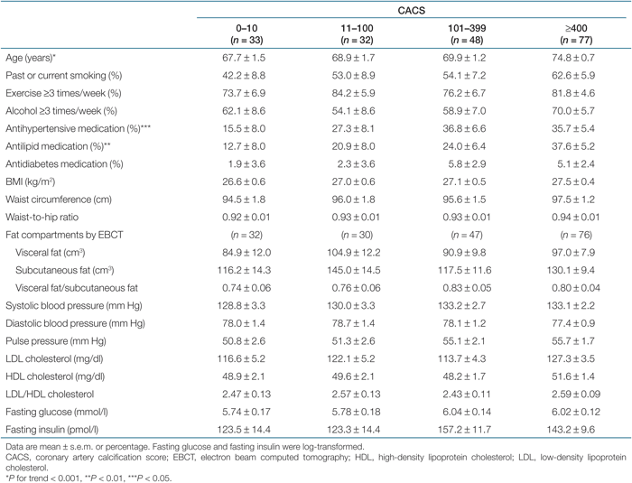
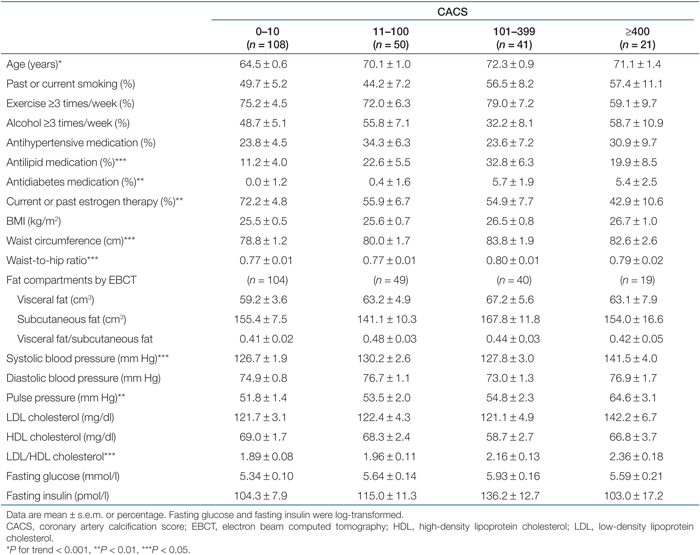
Measurement of CAC score and visceral and subcutaneous fat mass by EBCT. An Imatron C-150 ultrafast CT scanner (Imatron, San Francisco, CA) was used for assessing CAC and visceral adiposity. Images were obtained with 100-ms scan time at Dr Michael Wright's LifeScore Clinic in San Diego, California. Approximately 40–45 slices were obtained for each subject's heart, using 3-mm slices starting at the level of the carina and proceeding to the level of the diaphragm. Tomographic imaging was electrocardiographically triggered at 40 or 65% of the relative risk interval, depending on the subject's heart rate. CAC was defined as ≥2 pixels (area 0.67 mm2) with a density ≥130 Hounsfield units. Quantitative calcium scores were calculated using the method described by Agatston et al. (25), which is the product of the area of calcification per coronary tomographic segment and a factor rated 1–4 based on the calcium X-ray density. The CAC score (CACS) was the sum of all coronary artery calcium, and was classified according to the severity: minimal, 0–10; mild, 11–100; moderate, 101–399; and severe plaque, ≥400. Concurrent with the EBCT scans, a triple 6-mm slice at the lumbar 4–5 disc level was obtained to determine VAT and SAT expressed in cubic centimeters.
Statistical analysis
Data were analyzed using SPSS (version 11) and SAS (version 9.1). The association of CACS with related variables was evaluated using ANOVA for continuous variables and χ2 for categorical variables. Fasting glucose and fasting insulin levels had skewed distributions; analyses were performed using log-transformed values. Sex-specific standardized values were used for body size and fat distribution variables as well as all other continuous covariates.
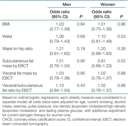
The association of various obesity parameters with CACS was examined by ordinal logistic regression analysis with minimal CACS (0–10) as the reference. Each odds ratio for each parameter of obesity and fat distribution was obtained separately after controlling for age, current smoking, alcohol (3+ drinks per week), regular exercise (3+ times per week), pulse pressure, LDL/HDL cholesterol, and fasting plasma glucose; analyses for women were also adjusted for current estrogen use. Variables used in multivariate analyses were chosen on the basis of known associations in the literature; for consistency, the same variables were used in the multivariate models for both sexes. In ordinal logistic regression, the outcome variable is interpreted as a set of cumulative logits based on an increasingly severe CACS, i.e., the odds of greater severity compared to lesser severity; χ2-tests of the proportional odds assumption were P ≥ 0.10 for all ordinal logistic regressions, indicating the proportional odds assumption was met.
A post-hoc power analysis for logistic regression models for coronary atherosclerosis based on CACS>10, a prevalence of 50%, and sample sizes of N = 220 for women and 190 for men, indicate there is 77% power for women and 72% for men to detect an odds ratio as small as 1.5 for each body size measurement. To our knowledge, no formulas exist for power calculations using ordinal regression analyses (as done in this study), however, power with ordinal regressions should be greater than that with logistic regressions as there is less partitioning of the data.
Results
There were 410 participants (190 men and 220 women) with no history of heart disease who had measures of CACS, VAT, and SAT by EBCT. Men were older (71.0 ± 8.7 years) on average than women (67.9 ± 7.2 years). The average BMI was 27.2 ± 3.6 kg/m2 in men and 25.8 ± 4.6 kg/m2 in women; 15.5% of men and 17.7% of women were obese (BMI ≥ 30 kg/m2). Most of them reported a healthy lifestyle, with current smoking in only 3.6% of men and 5.9% of women, and regular physical activity in 79.8% of men and 73.6% of women. Current use of postmenopausal estrogen was reported by 63% of women.
Men showed higher CACS than women. In sex-specific analyses, divided into four groups according to CACS (minimal, 0–10; mild, 11–100; moderate, 101–399; and severe plaque, ≥400), men showed a higher proportion of severe and moderate plaque than women (40% vs. 10% in severe; 18% vs. 12% in moderate) and a lower proportion of minimal and mild plaque than women (18% vs. 50% in minimal; 24% vs. 30% in mild). Table 1 (men) and Table 2 (women) show age and age-adjusted CHD risk factors and body size and composition measures by CACS category. Of these variables, age and current use of blood pressure and lipid medication differed by CACS category in men. In women, age, current use of lipid and diabetes medication, systolic blood pressure, pulse pressure, LDL-cholesterol/HDL-cholesterol, waist girth, and waist-to-hip ratio increased significantly with increasing CACS, whereas current or past use of estrogen therapy decreased; other variables did not differ.
 |
 |
As shown in Table 3, no parameter of overall or central obesity was significantly associated with CACS in men or women in multivariate ordinal logistic regression models adjusted for age, smoking, alcohol intake, exercise, pulse pressure, LDL/HDL-cholesterol ratio, fasting plasma glucose, and current estrogen therapy (women only). Of these variables, only age was independently associated with CACS in men (5 years increase of age, odds ratio (95% confidence interval), 1.41 (1.17–1.70), P < 0.001); whereas age, pulse pressure, and current estrogen therapy were each independently associated with CACS in women (5-year increase of age, odds ratio (95% confidence interval), 2.03 (1.60–2.56), P < 0.001; 1 s.d. (15.4 mm Hg) increase of pulse pressure, odds ratio (95% confidence interval), 1.61 (1.19–2.19), P = 0.002; current estrogen therapy, odds ratio (95% confidence interval), 0.35 (0.19–0.67)). Additional adjustment for the use of diabetes medications (8 men; 2 women) or lipid-lowering drugs (51 men; 40 women) did not materially alter the results (data not shown).
 |
Discussion
The objective of this study was to examine the association of obesity including visceral adiposity with subclinical coronary atherosclerosis measured by EBCT-generated CACS in older adults. As expected, male sex and age were independently associated with CACS, but no parameter of obesity was significantly associated with the degree of CAC in men or women before or after adjusting for multiple covariates.
The atherosclerotic process begins with fatty streaks and plaques in young adulthood (26). By the time clinical symptoms appear decades later, the opportunity for primary prevention has been lost (27). In order for primary prevention efforts to be effective, they must focus on risk factors that contribute to the earlier development of disease. Calcification of atherosclerotic plaques begins as early as the second decade of life and continues to progress as the plaque matures (28). Others have reported that age and sex are the strongest clinical correlates of CAC (29,30,31,32), compatible with results shown here. After controlling for body size and other cardiovascular risk factors, men in this cohort had a nearly fivefold increased prevalence of moderate-to-severe coronary artery plaque burden compared to women.
We did not observe a significant association of CACS with any obesity parameter. In contrast, Snell-Bergeon et al. (33) reported that BMI, waist circumference, and visceral fat by CT were significantly associated with CACS in 383 men and 379 women; VAT was not superior to BMI or waist circumference. This cohort was much younger (aged 20–58 years) than the Rancho Bernardo cohort (aged 55–88). In the large Rotterdam Coronary Calcification Study, elderly adults with or without a history of CHD had less favorable CHD risk factors than our study, and CACS was associated with BMI. The median values of CACS in the Rotterdam study were higher than those in Rancho Bernardo (men, median (interquartile range), 312 (62–969) in the Rotterdam study vs. 201 (41–720) in our study; women, 56 (5–261) vs. 13 (0–119)) (34). In a study of 443 asymptomatic younger adults (aged 20–58 years), waist circumference, waist-to-hip ratio, and BMI were positively related to 9-year progression of CAC only in those at relatively low risk of CHD (10-year risk <10% according to the Framingham risk equation) (35). These studies suggest that the association between CACS and overall, central, and visceral obesity may differ in younger vs. elderly populations.
The association of obesity with CACS may be more difficult to identify in elderly persons, against a background of lifetime exposure to cardiovascular risk factors. Newman and colleagues reported that the extent of CAC was strongly associated with age through the ninth decade, but risk factor levels were only weakly associated with CACS after age 65 years, concordant with our results (36). This and other studies show that the association of risk factors with CHD is attenuated in old age (15,16,17). Stevens and colleagues reported that greater body weight increased the 12-year risk of death from cardiovascular disease, but the relative risk was the lowest among older persons (14). Overall, these studies support the hypothesis that the risk factors associated with obesity may have worked their spell years before, that is, that weight and fat distribution measures are more important at younger ages, and that high-risk individuals have clinical CHD by age 65. Alternatively, weight loss in elderly adults may have obscured an obesity–CHD association.
The Rancho Bernardo Study cohort was elderly, middle to upper-middle class, white, and had a healthy lifestyle and access to health care. Almost all saw a physician at least once a year. Most importantly, those with a history of coronary artery disease were excluded from the EBCT test because the goal of our EBCT study was to examine the association between obesity and subclinical coronary atherosclerosis. This design may have lessened the statistical power to show obesity–CACS associations because individuals with clinically manifest CHD were excluded. Further, all studies of the elderly are subject to survivor bias. However, there was wide range of CACSs (58% of men and 21% of women had severe or moderate plaque), and ∼15% were obese individuals by the BMI criteria of ≥30 kg/m2. Finally, low power may have limited identification of obesity–CAC associations with odds ratios <1.5.
This is a cross-sectional study, and therefore cannot establish causality. It seems unlikely, however, that absence of subclinical heart disease caused a more favorable weight, waist, or VAT pattern. Despite the limitations of survival bias and cross-sectional design, this study is concordant with other reports showing clinically measured obesity is not a strong risk factor for coronary atherosclerosis in older adults.
In conclusion, neither overall obesity nor directly measured visceral obesity significantly predicted atherosclerosis in older survivors without clinical heart disease. The importance of weight control to prevent CHD in healthy elderly survivors is uncertain.
Disclosure
The authors declared no conflict of interest.
Acknowledgments
This research was supported by the National Institute of Aging grant NIA 5R01 AG07181 and the National Institute of Diabetes and Digestive and Kidney Diseases grant NIDDK 5R01 DKK31801.