Brain magnetic resonance imaging examination in a patient with non-magnetic resonance conditional pacemaker
Abstract
Clinical dilemmas arise when patients with a non-magnetic resonance (MR) conditional pacemaker are required to undergo magnetic resonance imaging (MRI). We encountered a pacemaker patient with debilitating non-motor symptoms of Parkinson׳s disease, who required an MRI prior to deep brain stimulation (DBS) surgery. MRI was performed safely without adverse events despite the presence of a conventional pacemaker.
1 Case Report
A 62-year-old man who visited Nihon University Hospital for regular pacemaker examination was suffering from the debilitating effects of parkinsonian tremors and was thus considered a candidate for deep brain stimulation (DBS) by the department of neurosurgery. The conventional single-chamber pacemaker (Nexus I Plus, Boston Scientific, Natick, MA, USA) had been implanted to treat sick sinus syndrome, diagnosed several years earlier. DBS lead implantation requires that brain imaging [magnetic resonance imaging (MRI) in this case] be performed to identify the proper targets. Thus, we were consulted to determine whether the patient׳s pacemaker should first be replaced with a magnetic resonance (MR) conditional device. However, replacement would have required extraction of the two existing leads, and although the patient desired it, the procedure would have posed significant risks because one of the two leads had been in place for over 20 years. Interestingly, one of the leads was found to be non-functional. We consulted a team of doctors, including those at Kyorin University Hospital, where the lead extraction would have been performed. The fact that the patient was not dependent on the pacemaker was encouraging. He had an intact intrinsic conduction, and therefore, it was decided that MRI would be performed with the existing device in place. The provision was that if subsequent examination of the pacemaker after DBS surgery revealed pacing malfunction, the pacemaker could be safely turned off and a new pacemaker implanted soon after. The case was submitted to and discussed with the ethical review board of Nihon University Itabashi Hospital, which approved the MRI examination.
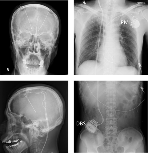
Before MRI, the pacemaker was examined, and all operating parameters, such as pacing threshold, ventricular sensing, and lead impedance were checked. The remaining battery life was 1.5 years at most. The pacemaker was set to VVI mode and programmed at 60 beats per minute (bpm). Lead impedance was 500 Ω, ventricular pacing threshold was 0.75 V at 0.35 ms, and ventricular sensing was 7.8 mV. Telemetry recordings revealed 80% ventricular sensing and 20% ventricular pacing over the past 6 months; hence, we reset the pacemaker to VVI mode at 40 bpm for the MRI examination. During the examination, the pulse oximeter displayed a heart rate of 100 bpm, which was also the magnet rate. We inquired of the patient throughout the procedure and confirmed if he was feeling any discomfort. The examination was completed without incident, and subsequently, we re-examined the pacemaker for any dysfunction or arrhythmic events. The examination report showed the expected magnet response but no arrhythmic events. All parameters were rechecked, and the pacemaker was reprogrammed to the previous pacing mode. The DBS surgery was performed on the same day, and the patient was discharged without complications (Fig. 1).

Head, chest, and abdominal radiographs performed 1 day after implantation of the deep brain stimulation (DBS) device showing the pacemaker and the DBS device. The pacemaker is seen on the left side of the chest, and the DBS device on the right side of the abdomen.
The patient visited the Nihon University Hospital pacemaker clinic 1 and 6 months after the MRI examination, and no pacemaker dysfunction was detected. Programmed settings such as lead impedance, pacing threshold, sensing signal amplitude, and battery voltage remained stable during the chronic period (Table 1). Additionally, DBS had no effect on the pacemaker function. The patient׳s intrinsic sinus rhythm was generally maintained with a very low percentage of pacing.
| Parameter | Before MRI | After MRI | 1 week later | 1 month later | 6 months later |
|---|---|---|---|---|---|
| Magnet rate | 100 bpm | 100 bpm | 100 bpm | 100 bpm | 100 bpm |
| Battery life | 1.5 years | 1.5 years | 1.5 years | 1.5 years | 1.0 years |
| Lead impedance | 530 Ω | 500 Ω | 520 Ω | 520 Ω | 500 Ω |
| Pacing threshold | 0.7 V/0.35 ms | 1.0 V/0.35 ms | 0.7 V/0.35 ms | 0.7 V/0.35 ms | 0.7 V/0.35 ms |
| Ventricular sensing | 8.3 mV | 8.8 mV | 8.1 mV | 8.7 mV | 8.2 mV |
- bpm=beats per minute, the omega symbol=ohms, V=volts.
2 Discussion
MRI has become almost indispensable in the diagnosis and treatment of diseases of the central nervous system and the heart muscle. Since MR conditional pacemakers have been developed, MRI has been performed widely in patients with implantable cardiac devices. This case is the first case in which we performed MRI in a patient with a conventional (non-MR conditional) pacemaker. There have been reports outlining the safe performance of MRI in patients with a conventional pacemaker [1], [2]. However, serious adverse events can occur during MR scanning [3].
Our patient׳s motor symptoms had severely affected the quality of life, and DBS was considered the only avenue for improvement. MRI examination was necessary before the DBS surgery. We could have performed lead extraction under the current guidelines [4]; however, on weighing the benefits against the risks, we decided to perform the MRI examination without extracting the pacemaker leads. Our decision was based on three concerns. First, one of the two pacemaker leads had been in place for over 20 years, and the complication rate associated with older leads is relatively higher [5]. Second, the remaining battery life was less than 1.5 years; thus, we were not concerned with the need for pacemaker replacement, should the device itself have been damaged by the MRI examination. Third, the replacement of the old lead with a new lead after the DBS surgery was viewed as a good option, in case any lead-related dysfunction was discovered subsequent to the MRI examination. Our rationale did not adhere strictly to the current guidelines. However, in all aspects of medical care, it is sometimes necessary to intervene beyond the clinical guidelines for the patient׳s sake. Reports of MR scanning in patients with a conventional pacemaker from Europe and the United States [1]-[3] were very useful to us while handling this case.
It should be noted that the particulars of the patient made the MRI examination possible in this case. There are many patients for whom such an examination would not be safe. For example, if a patient׳s spontaneous sinus rate is high, MRI cannot be performed safely. Tachycardia, whether sinus tachycardia or atrial fibrillation, retrograde ventricular atrial conduction during VOO pacing, competition between the paced rhythm and the patient׳s spontaneous sinus rhythm, and simultaneous arrival of the pacing signal and T wave can occur, which may result in a life-threatening ventricular arrhythmia. Although occasional ventricular pacing had been documented in our patient, there had been no retrograde conduction; hence, we considered the risk of pacemaker-induced tachycardia as very low. On careful consideration, we believed that the patient׳s intrinsic heart rate, which was neither too fast nor too slow, did not pose an undue risk. Alternatively, if there had been no spontaneous beats and the pacemaker had stopped pacing due to oversensing of the magnetic field, the situation could have been dangerous. Thus, in patients with a non-MRI conditional device, MRI should be performed with a careful consideration of all associated risks. It is important to confirm the presence of an intrinsic heartbeat, the rate of the intrinsic beat, and ensure whether atrioventricular conduction remains intact when the pacemaker is set to VOO mode.
Our case stands as an example of both complex decision-making for patients with a conventional pacemaker who require MRI and safe performance of MRI that does not fall strictly within the pacemaker guidelines. In any patient with a conventional pacemaker for whom MRI is being considered, the benefits and risks associated with MRI as well as lead extraction should be carefully evaluated. Once the decision to perform MRI has been made and the examination has begun, careful observation is necessary. We believe that, in addition to the fact that our patient was not pacemaker dependent, we were able to perform the examination safely because we had prior experience with MR scanning among patients with an MR conditional device. Such experiences advanced our understanding of the potential effects of MRI on the pacemaker and helped us prepare for several possible scenarios.
Conflict of Interest
None.


