Asymptomatic sonographic abnormalities of the hindfoot region in Division I collegiate gymnasts
Abstract
Background
The hindfoot region is commonly injured in gymnasts, and musculoskeletal ultrasound can be used to identify structural abnormalities in this region. Although prior studies have shown that sonographic abnormalities may not correlate with symptomatic pathology, the presence of asymptomatic sonographic abnormalities of the hindfoot in Division I collegiate gymnasts has not been evaluated.
Objective
To identify and describe commonly seen asymptomatic sonographic abnormalities of the hindfoot region in Division I collegiate gymnasts.
Design
Cross-sectional study.
Setting
Tertiary care academic medical center.
Participants
39 Division I NCAA men's and women's collegiate gymnasts without current hindfoot pain or history of hindfoot injury.
Interventions
Diagnostic musculoskeletal ultrasound of the hindfoot region.
Main Outcome Measures
Sonographic appearance of the hindfoot region, specifically the plantar fascia, plantar fad pad, and Achilles tendon.
Results
A total of 37 of 39 gymnasts included in the study were found to have at least one asymptomatic sonographic abnormality of the hindfoot region. A total of 28.2% of athletes were found to have sonographic abnormalities within the Achilles tendon, with Doppler flow being the most common finding, and 35.8% of athletes were found to have a Haglund's deformity. However, only 7% of athletes with a Haglund's deformity demonstrated abnormal sonographic findings within the tendon. Sonographic abnormalities of the plantar fascia and plantar fat pad were seen in 30.7% and 69.2% of athletes, respectively.
Conclusions
Asymptomatic sonographic abnormalities of the hindfoot region are common in collegiate gymnasts. Clinicians should use clinical judgment when interpreting these findings as they may not represent symptomatic pathology.
INTRODUCTION
The Olympic sport of artistic gymnastics consists of several events for both men and women that involve repetitive running, jumping, and landing. These events result in significant forces placed through the lower limb, in particular the hindfoot region, which includes structures such as the plantar fascia, plantar fat pad, and Achilles tendon.1 As a result of these forces, the hindfoot region is the most commonly injured anatomic region in National Collegiate Athletic Association (NCAA) women's gymnasts at a rate of 3.64 per 1000 athletic exposures and the second most commonly injured region injured in NCAA men's gymnasts at a rate of 1.54 per 1000 athletic exposures.2-5
Accurate and early diagnosis of hindfoot injuries is critical to prevent prolonged time lost from sport and activity. Although radiographs are useful for various bone pathologies, their utility for soft tissue injuries is limited. Magnetic resonance imaging (MRI) has commonly been used for the evaluation of hindfoot injuries; however, it comes at a high cost, may have limited availability, and has multiple contraindications.6
Musculoskeletal ultrasound (US) is a high-resolution, dynamic imaging modality commonly used for evaluation of many soft tissue structures, including those of the hindfoot region.7, 8 The resolution of US is greater than MRI, producing real-time images with submillimetric resolution.9 This allows US to evaluate anatomic structures in much finer detail and potentially identify more subtle pathology. It is important to note, however, that not all sonographic pathology is symptomatic pathology.
Sonographic abnormalities, both symptomatic and asymptomatic, have been described in athletes from various sports,10-18 including collegiate and recreational runners,10, 12 soccer players,13 fencers,14 and ballet dancers.15 However, common sonographic abnormalities in gymnasts have been minimally described in the literature.19, 20 Further, the clinical relevance of these sonographic abnormalities has not been elucidated. Therefore, we present a descriptive study aiming to describe the sonographic appearance of the hindfoot region in a cohort of asymptomatic, Division I collegiate gymnasts. We hypothesized that asymptomatic sonographic abnormalities of the hindfoot would be frequently seen in these athletes.
METHODS
Participants
After the study was approved by the University of Iowa Institutional Review Board, a convenience sample of 40 NCAA Division I men's and women's gymnasts at the home institution of the primary author were approached on a voluntary basis to participate in this descriptive study. Athletes were eligible to participate in the study if they were an active member of the varsity gymnastic team at the home institution of the primary author and available on campus at the time of the sonographic evaluation. The athletes verbally confirmed that they were asymptomatic in respect to their hindfoot at rest, during training, and during competition the week prior to and at the time of the sonographic evaluation. Athletes were excluded if they reported symptoms in the hindfoot, had a history of injury or surgery to the hindfoot region, or did not wish to participate. Only one male athlete was excluded due to a history of an Achilles tendon repair. Thirty-nine athletes who met inclusion criteria and voluntarily went through the informed consent process were included in the study.
Equipment
All sonographic evaluations were performed in the athletic training room with a portable Phillips Lumify US unit (Philips Healthcare, Bothwell, WA), utilizing a 12–4 MHz linear array transducer. All ultrasound examinations were performed by either the primary author or the sports medicine fellow physician at the primary author's institution, and the examinations were performed on a single day in the athletes' offseason.
Procedure specifics
Participants underwent a diagnostic US of their bilateral hindfoot regions with a specific focus on the plantar fascia, plantar fat pad, and Achilles tendon.
Each US exam was performed using a systematic, consistent protocol. All US images were reviewed and interpreted by the primary author who at the time had more than 3 years of experience independently performing and interpreting musculoskeletal US. Each US image was deidentified and reviewed by the primary author at two separate time points, 2 weeks apart. Interpretation of each image was consistent at both time points without any discrepancies.
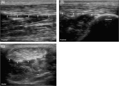
All examinations were performed with participants laying prone on the exam table with their feet hanging off the edge of the bed. The Achilles tendon was evaluated in both in long axis as well as short axis (Figure 1). Achilles tendon thickness in short axis was measured at its thickest portion. Abnormalities of the Achilles tendon were defined as the loss of normal echotexture (hypoechogenicity, heterogeneity, increased thickness) as well as the presence of hyperemia or Doppler flow.10, 16
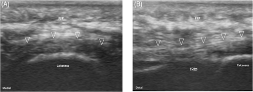
Next, the plantar fascia was evaluated in both long and short axis (Figure 2). The thickest portion of the plantar fascia was measured in short axis. Plantar fascia abnormalities were defined as loss of normal fascial echotexture, the presence of Doppler flow, as well as plantar fascia thickness of >4 mm.10, 16
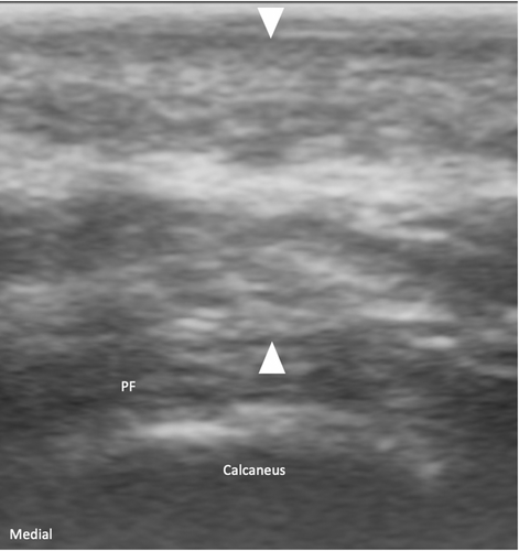
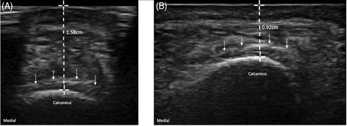
The plantar fat pad was then evaluated with the participant in the same position. The plantar fat pad was evaluated in a transverse, anatomic coronal plane (Figure 3). Abnormalities of the plantar fat pad were defined as the loss of normal echotexture, presence of hyperemia, and an abnormal compressibility index. Compressibility index was calculated by measuring the distance from the transducer to the calcaneus at baseline with no transducer pressure, and then again with maximal transducer compression. A compressibility index of 0.44–0.62 was considered normal.21
Data analysis
Baseline demographics were analyzed using descriptive statistics. Sonographic abnormalities of the Achilles tendon, plantar fascia, and plantar fat pad were also analyzed with descriptive statistics. Statistical analysis was performed using JMP statistical analysis software.
RESULTS
Demographics
Thirty-nine asymptomatic Division I collegiate gymnasts (24 males and 15 females) without a history of hindfoot injury participated in the study after completing the voluntary consent process. Baseline demographics are shown in Table 1, and the number of athletes that participated in each event is shown in Table 2. Thirty-seven of 39 athletes (23 males and 14 females) were found to have at least one sonographic abnormality of the hindfoot region, and the sonographic findings in these athletes are presented in Table 3.
| Gender (n) | Age, years | Height, inches | Weight, lbs | BMI, kg/m2 |
|---|---|---|---|---|
| Male (24) | 21.2 (18–23) | 68.1 (65–71) | 156.8 (132–186) | 23.8 (21.2–29.5) |
| Female (15) | 19.4 (18–23) | 65.3 (58–70) | 143.5 (103–167) | 23.6 (20.2–27.1) |
- Note: Age, height, weight, and BMI are presented as mean (range).
- Abbreviation: BMI, body mass index.
| Gender | AA | R | PB | V | HB | FX | B | UB | PH |
|---|---|---|---|---|---|---|---|---|---|
| Male | 17 | 8 | 4 | 9 | 7 | 8 | NA | NA | 2 |
| Female | 9 | NA | NA | 6 | NA | 7 | 7 | 3 | NA |
- Note: All events are presented as total number of athletes.
- Abbreviations: AA, all around; B, beam; FX, floor exercise; HB, horizontal bar; NA, event not performed by this gender; PB, parallel bars; PH, pommel horse; R, rings; V, vault; UB, uneven bars.
| Structure | Sonographic findings | Male (N = 24) | Female (N = 15) |
|---|---|---|---|
| Hindfoot region | Any structural abnormality | 23 (95.8%) | 14 (93.3%) |
| Achilles tendon | Any tendon abnormality | 6 (25%) | 5 (33.3%) |
| Doppler flow | 5 (20.8%) | 2 (13.3%) | |
| Focal thickening | 3 (12.5%) | 2 (13.3%) | |
| Hypoechogenicity | 4 (16.6%) | 2 (13.3%) | |
| Haglund's deformity | 9 (37.5%) | 5 (33.3%) | |
| Plantar fascia | Any abnormality | 7 (29.1%) | 5 (33.3%) |
| Doppler flow | 2 (8.3%) | 1 (6.6%) | |
| Focal thickening | 0 (0%) | 0 (0%) | |
| Hypoechogenicity | 7 (29.1) | 5 (33.3%) | |
| Plantar fat pad | Any abnormality | 16 (66.6%) | 11 (73.3%) |
| Abnormal echotexture | 8 (33.3%) | 7 (46.6%) | |
| Doppler flow | 10 (41.6%) | 10 (66.6%) | |
| Abnormal compressibility index | 2 (8.3%) | 1 (6.6%) |
Achilles tendon
Of the participating athletes, 28.2% (25% of males and 33.3% of females) were found to have any abnormal sonographic findings within the Achilles tendon. The most common abnormal finding was Doppler flow, which was present in 17.9% of athletes with abnormal tendons (Figure 4). A total of 15.3% of athletes had tendons with regions of hypoechogenicity, with 10% of this group also demonstrating concomitant Doppler flow.

The average Achilles tendon thickness in short axis (measured in an anterior to posterior dimension at the region of maximal tendon thickness) was 5.43 mm (range 3.5–6.2 mm) in females and 5.82 mm (range 3.7–7.1 mm) in males. Using a upper limit of normal of 4.5 mm,22 12.5% of male and 13.3% of female athletes were found to have a thickened tendon.
A Haglund's deformity was found in 35.8% of the athletes; however, only 7% of athletes with a Haglund's demonstrated abnormal sonographic findings within the tendon
Plantar fascia
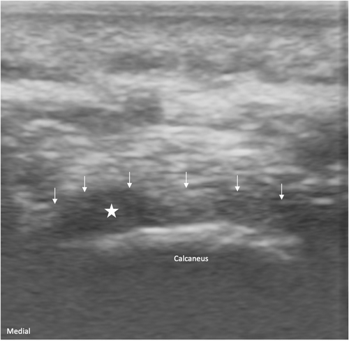
Sonographic plantar fascia abnormalities were found in 30.7% of athletes. All abnormal plantar fascias demonstrated regions of hypoechoic heterogeneity without fascial thickening23 (Figure 5). The average thickness of the plantar fascias measured 2.3 mm (range 1.7–3.8 mm) in female and 3.2 mm (range 2.1–3.9 mm) in male athletes. Plantar fascia Doppler flow was seen in only 7.6% of all athletes, and no partial or full thickness tears were seen.
Plantar fat pad
An abnormal sonographic finding of the plantar fat pad was present in 69.2% of athletes. Abnormal plantar fat pad echotexture was found in 38.4% of all athletes (46.6% female athletes, 33.3% male athletes), and 25.6% of athletes had abnormal plantar fat pad echotexture bilaterally. Most athletes demonstrated diffuse plantar fat pad echotexture changes, with only 5.1% having focal regions of abnormal echotexture. Plantar fat pad Doppler flow was seen in 51.2% of athletes.
The average plantar fat pad compressibility index was 0.59 (range 0.41–0.6) in males and 0.52 (range 0.42–0.61) in females (Figure 6). An abnormal compressibility index was seen in 7.6% of athletes, all of which were lower than the normal range.21
DISCUSSION
The primary finding in our study is that asymptomatic sonographic abnormalities of the hindfoot region are common among Division I collegiate men's and women's gymnasts, and 37 of 39 gymnasts included in the study were found to have at least one sonographic abnormality in this region. In our cohort, the plantar fat pad was the most commonly seen abnormal structure (69.2% of athletes) followed by the plantar fascia (30.7%) and Achilles tendon (28.2%).
A total of 69.2% of our athletes had sonographic abnormalities of the plantar fat pad, with Doppler flow being the most common finding. Similar abnormalities of the plantar fat pad have been seen in populations of runners and patients with systemic inflammatory conditions.10, 24-26 Although only 7.6% of athletes were found to have an abnormally low compressibility index, all athletes with an abnormal compressibility index also had abnormal findings within their plantar fascia. We hypothesize that the increased compressibility seen with a low compressibility index results in increased force transmitted to the plantar fascia, resulting in the pathologic sonographic findings.
Sonographic abnormalities of the plantar fascia were seen in 30.7% of our athletes, with all abnormal plantar fascias demonstrating regions of hypoechoic heterogeneity. This percentage is higher than prior studies evaluating sonographic abnormalities of the plantar fascia in the asymptomatic, general population.27, 28 However, the activity level and degree of repetitive running and jumping activities seen in a collegiate gymnast is likely significantly greater than that of the older, general population, and therefore the higher percentage in our athletes is not surprising.
Doppler flow was seen in only 7.6% of our athletes in the plantar fascia. Interestingly, no athletes were found to have abnormal plantar fascia thickening. This is in contrast to a prior study evaluating sonographic abnormalities of the plantar fascia in asymptomatic runners, which found that 41% of running athletes had abnormal plantar fascia thickness.10 There are likely a few reasons that explain this difference. Endurance runners experience constant, repetitive forces through the hindfoot structures, often for hours at a time. In gymnastics, the hindfoot structures endure short bursts of high impact stress and therefore a smaller cumulative volume of force. Additionally, the average age of the cohort of runners was 39.3 years (range 20–67 years), which is much older than our cohort of gymnasts with an average age of 20.3 years (range 18–23 years). Older age with larger and more frequent force through the hindfoot likely results in thickening and chronic adaptive changes within the plantar fascia, which was not seen in our group of athletes.
A total of 28.2% of our athletes were found to have sonographic abnormalities of the Achilles tendon, with the most common finding being Doppler flow. This is consistent with prior studies on the prevalence of Doppler flow in asymptomatic Achilles tendons. Risch et al. found that 46% of asymptomatic Achilles tendons in their cohort had the presence of intratendinous Doppler.29 Additionally, Doppler flow within asymptomatic Achilles tendons has also been demonstrated both runners and badminton players.30, 31
Only 12.5% of males and 13.3% of females in our study had a thickened Achilles tendon midsubstance. In a cohort of gymnasts, Emerson et al. found that 22.5% of female and 30% of male gymnasts had focal thickening of their Achilles tendons, nearly twice as high as our cohort.19 The reason for this difference is not entirely clear; however, we hypothesize that it may be the result of differences in the gymnasts' training load at the time of the US evaluations. Prior studies have shown that tendons may demonstrate a transient increase in thickness during a bout of exercise.32 The average training load in the study by Emerson et al. was 22.8 h/week. Although we did not document the training load of our athletes, we performed our US evaluations during the offseason when the expected load is minimal. This presumed difference in training load may explain these findings.
We also found that whereas 35.8% of athletes had a Haglund's deformity, only 7% of those athletes also had an abnormal sonographic appearance of the Achilles tendon. Important to note is that the Achilles tendon abnormalities seen in our athletes involved the tendon midsubstance rather than the insertion. Additionally, it has been shown that the presence of a Haglund's deformity is not necessarily symptomatic nor always correlated with abnormal sonographic findings within the Achilles tendon.33
Although the clinical relevance of these findings cannot be fully elucidated by our study, we suspect that these abnormal findings may be suggestive of “preclinical” or “presymptomatic” injury. A meta-analysis in 2016 found that individuals with asymptomatic sonographic abnormalities of their Achilles tendon had a relative risk of 7.33 of developing future symptomatic Achilles tendinopathy.11 A later study in elite soccer players showed a 45% risk of developing symptomatic Achilles tendinopathy during the season when preseason US scans showed asymptomatic sonographic abnormalities.13 The same predictive value has not been found in ballet dancers15 or fencers.14 A future, prospective observational study would allow for evaluation of whether these sonographic abnormalities predict future injury in gymnasts.
There are limitations to our study. First, we used a convenience sample of athletes with a relatively small sample size, which may limit generalizability to other athletes and other sports. Future studies should be done with a larger cohort of athletes from various sports, which may allow for better generalization. Additionally, although two sports medicine trained physicians acquired the US images, only a single physician reviewed and interpreted all images. Although the interpreting physician had extensive diagnostic US training and the images were reviewed in a protocolized manner, it is possible that other physicians may interpret the images differently. Lastly, US images were acquired using a portable, handheld US unit. These portable units generally have much lower resolution than a cart-based machine, and as such more subtle pathologic findings may not be easily visualized. However, our cohort consisted of young athletic individuals with minimal adiposity, which we feel allowed for excellent, adequate resolution to detect even subtle sonographic tissue changes.
CONCLUSION
Abnormal sonographic findings of the hindfoot region in asymptomatic Division I collegiate gymnasts are common. The most commonly seen abnormal structure in our cohort was the plantar fat pad followed by the plantar fascia. Although the clinical relevance of these abnormal findings is not yet fully understood, these findings may be indicative of future symptomatic pathology. Additional studies are needed to confirm or refute this.
DISCLOSURE
None.


