Sexual Dimorphisms in Adult Human Brown Adipose Tissue
Abstract
Objective
This study aimed to quantify and compare the amount, activity, and anatomical distribution of cold-activated brown adipose tissue (BAT) in healthy, young, lean women and men.
Methods
BAT volume and 18F-fluorodeoxyglucose uptake were measured by positron emission tomography and computerized tomography in 12 women and 12 men (BMI 18.5-25 kg/m2, aged 18-35 years) after 5 hours of exposure to their coldest temperature before overt shivering.
Results
Women had a lower detectable BAT volume than men (P = 0.03), but there was no difference after normalizing to body size. The mean BAT glucose uptake and relative distribution of BAT did not differ by sex. 18F-fluorodeoxyglucose uptake consistent with BAT was observed in superficial dorsocervical adipose tissue of 6 of 12 women but only 1 of 12 men (P = 0.02). This potential BAT depot would pose fewer biopsy risks than other depots.
Conclusions
Despite differences in adiposity and total BAT volume, we found that healthy, lean, young women and men do not differ in the relative amount, glucose uptake, and distribution of BAT. Dorsocervical 18F-fluorodeoxyglucose uptake was more prevalent in women and may be a remnant of interscapular BAT seen in human newborns. Future studies are needed to discern how BAT contributes to whole-body thermal physiology and body weight regulation in women and men.
Study Importance
What is already known?
- Brown adipose tissue (BAT) expends energy in the form of heat and is therefore a potential target for therapeutic interventions to address obesity and metabolic disease.
- Sexual dimorphisms exist in terms of general adiposity and metabolism.
- Most BAT studies are conducted in rodents; most quantitative clinical trials of human BAT have been conducted in men.
What does this study add?
- We conducted a quantitative analysis of cold-induced BAT in healthy, lean, young women and men to explore the differences in amount, activity, and anatomical distribution.
- Women have less BAT volume than men, but the relative amount, metabolic activity, and distribution of detectable BAT are similar.
- Superficial dorsocervical adipose tissue with BAT-like glucose uptake is more prevalent in women and is a probable remnant of interscapular BAT seen in rodents and human newborns.
Introduction
Obesity results from caloric intake exceeding expenditure. White adipose tissue stores surplus energy. Brown adipose tissue (BAT) expends energy as heat (1) after cold or pharmacologic stimulation (2, 3), and it is a therapeutic target for treating obesity and metabolic disease.
Most prospective studies of human BAT have been conducted on men (4), yet many adipose tissue–related sexual dimorphisms exist. Women tend to have proportionally more total body fat (5) and greater subcutaneous fat but less visceral fat (6) than men. Despite these observations, quantitative BAT differences based on sex have been largely unstudied. Here we determined whether women and men have different BAT amounts, activity, or anatomical distribution.
Methods
Protocol design
Twenty-four young (aged 18-35 years), lean (BMI 18.5-25 kg/m2), healthy female and male volunteers (12 each) were admitted to the National Institutes of Health Clinical Center for a continuous 7- to 13-day inpatient protocol approved by the Institutional Review Board (ClinicalsTrial.gov identifier: NCT01568671) and described previously (7, 8). All participants were nonsmokers, had no metabolic or psychological conditions, and took no medication except oral contraceptives for women. Women were measured in the follicular phase. Participants provided written informed consent.
Participants wore sleeveless shirts, shorts, and light socks with a combined thermal insulation of 0.34 clo. Women additionally wore a sports bra (+ 0.01 clo). Skin temperatures of the left deltoid, pectoralis major, anterior thigh, and shin were measured via wireless thermistor probes (iButton; Maxim Inc., San Jose, California), and the weighted mean skin temperature is reported (9). From 8:00 am to 1:00 pm, fasted participants were exposed to a constant ambient air temperature between 16.0°C and 31.0°C, which differed each day in randomized order. Shivering was assessed via a visual analog scale, participant report, and direct observation. On the final day, preceding BAT imaging by 18F-fluorodeoxyglucose (18F-FDG) positron emission tomography (PET) and computed tomography (CT), participants were reexposed to their coldest temperature before overt shivering. Participants were dosed with 370 MBq 18F-FDG by intravenous injection at 12:00 pm, remained in the cold until 1:00 pm, and were scanned at approximately 1:30 pm using four 5-minute bed positions on a Siemens Biograph mCT (Siemens Healthcare, Erlangen, Germany).
Body composition
Body composition was measured by dual-energy x-ray absorptiometry (Lunar iDXA; GE Healthcare, Chicago, Illinois).
PET/CT image analyses
We created regions of interest on each 3.5-mm axial section of the PET/CT images in ImageJ, as previously described (8, 10). We then applied a previously established CT Hounsfield unit threshold (−300 to −10) and an individualized PET threshold normalized to a lean body mass/total body mass ratio (8) to define active BAT voxels, and after applying this normalization, we verified that mean 18F-FDG standardized uptake values (SUVs) of three reference tissues (deltoid muscle, liver, and cerebellum) were similar between men and women (data not shown). Voxels identified as BAT in each region of interest were used to calculate total body BAT volume, SUV mean, and activity (BAT volume × SUV mean) (11).
BAT quantification
BAT distribution was categorized by six anatomical depots (cervical, supraclavicular, axillary, paraspinal, mediastinal, and abdominal), as previously described (8, 10). For this study, we used the PET/CT-based image analysis criteria to define potential BAT, in a manner consistent with the BAT-reporting consensus statement (12), in an additional depot in the subcutaneous fat on the dorsal side of the cervical spine, and we refer to it as superficial dorsocervical BAT (sdBAT).
Statistical analyses
Statistical tests were performed in SAS JMP version 14.0.0 (SAS Institute, Inc., Cary, North Carolina). Anthropometric variables and BAT were compared by sex using independent sample t tests. Normalization of BAT volume to anthropometric variables was performed by dividing BAT volume by each variable. BAT volume in each depot was compared by sex using ANCOVA and controlling for total BAT volume and multiple comparisons. Correlations between sdBAT and total BAT volume were assessed using Spearman rank correlation (ρ). Prevalence of sdBAT by sex was compared using the χ2 test. Results are median ± interquartile range unless otherwise specified. P ≤ 0.05 was considered significant.
Results
Demographics
Women were older and had a higher body fat percentage and less lean mass than men. Neither ambient temperature (20.7 ± 1.5°C vs. 21.6 ± 2.4°C) nor skin temperature (30.3 ± 0.9°C vs. 30.1 ± 1.3°C) was different on scan day (Table 1).
| Male (n = 12) | Female (n = 12) | P value | |
|---|---|---|---|
| Characteristics and body composition | |||
| Age, y | 22.5 ± 4.9 | 27.8 ± 3.6 | 0.01 |
| Weight, kg | 77.8 ± 8.8 | 61.4 ± 8.8 | < 0.001 |
| Height, cm | 183.0 ± 7.0 | 166.8 ± 7.0 | < 0.001 |
| BMI | 23.2 ± 1.9 | 22.0 ± 2.5 | 0.22 |
| Fat mass, kg | 16.2 ± 5.5 | 19.8 ± 6.2 | 0.13 |
| % fat | 20.6 ± 5.7 | 31.8 ± 6.6 | < 0.001 |
| Lean mass, kg | 57.8 ± 6.3 | 40.0 ± 4.4 | < 0.001 |
| Scan day conditions | |||
| Ambient temperature, °C | 21.2 ± 1.5 | 20.3 ± 1.3 | 0.12 |
| Weighted mean skin temperature, °C | 29.8 ± 0.8 | 30.3 ± 0.5 | 0.13 |
| Individualized BAT SUV threshold for PET image analysis | 1.6 ± 0.1 | 1.9 ± 0.2 | < 0.001 |
| BAT volume, mL | |||
| Total | 334.0 ± 187.6 | 194.3 ± 95.1 | 0.03 |
| Superficial dorsocervical | 0.5 ± 1.9 | 1.8 ± 3.4 | 0.27 |
| Cervical | 42.3 ± 30.6 | 27.7 ± 18.0 | 0.17 |
| Supraclavicular | 119.6 ± 66.8 | 60.5 ± 38.7 | 0.01 |
| Mediastinal | 15.6 ± 17.0 | 7.0 ± 9.2 | 0.14 |
| Axillary | 22.6 ± 15.7 | 20.0 ± 18.5 | 0.71 |
| Paraspinal | 93.9 ± 53.7 | 51.8 ± 25.7 | 0.02 |
| Abdominal | 45.5 ± 43.3 | 21.5 ± 14.3 | 0.08 |
| BAT SUV mean | |||
| Total | 4.91 ± 1.64 | 5.01 ± 1.51 | 0.88 |
| Superficial dorsocervical a | 2.4 | 3.0 ± 0.9 | NA |
| Cervical | 5.8 ± 2.5 | 6.0 ± 2.3 | 0.86 |
| Supraclavicular | 5.4 ± 1.8 | 5.4 ± 2.0 | 0.97 |
| Mediastinal | 3.7 ± 1.0 | 3.3 ± 1.0 | 0.40 |
| Axillary | 3.6 ± 1.0 | 3.5 ± 1.1 | 0.77 |
| Paraspinal | 4.6 ± 1.9 | 5.3 ± 1.8 | 0.35 |
| Abdominal | 4.5 ± 1.4 | 3.5 ± 0.9 | 0.06 |
| BAT activity (18F-FDG uptake) | |||
| Total | 1,890.8 ± 1,277.8 | 1,094.2 ± 765.0 | 0.08 |
| Superficial dorsocervical | 1.3 ± 4.6 | 6.6 ± 14.0 | 0.23 |
| Cervical | 291.0 ± 259.6 | 187.6 ± 170.6 | 0.26 |
| Supraclavicular | 730.4 ± 471.3 | 389.6 ± 329.8 | 0.052 |
| Mediastinal | 64.0 ± 69.7 | 29.3 ± 43.9 | 0.16 |
| Axillary | 91.2 ± 77.7 | 83.7 ± 86.9 | 0.83 |
| Paraspinal | 498.7 ± 355.8 | 292.8 ± 207.8 | 0.10 |
| Abdominal | 236.4 ± 258.3 | 83.7 ± 62.2 | 0.06 |
| BAT radiodensity from CT (HU) | |||
| Total | −45.1 ± 6.8 | −43.1 ± 10.2 | 0.79 |
| Superficial dorsocervical a | −46.5 | −57.3 ± 10.5 | NA |
| Cervical | −15.5 ± 12.9 | −40.5 ± 14.6 | 0.005 |
| Supraclavicular | −41.5 ± 14.5 | −52.1 ± 10.4 | 0.12 |
| Mediastinal | −61.4 ± 19.0 | −41.6 ± 20.4 | 0.07 |
| Axillary | −47.7 ± 20.6 | −52.5 ± 14.7 | 0.78 |
| Paraspinal | −49.2 ± 15.6 | −35.8 ± 10.0 | 0.03 |
| Abdominal | −60.9 ± 31.0 | −37.6 ± 12.0 | 0.07 |
- a Averages from one male participant and six female participants with superficial dorsocervical BAT.
- All values are mean ± SD. Values with P < 0.05 are in boldface.
- HU, Hounsfield units; NA, not applicable.
BAT volume and activity
Women had a lower detectable volume of active BAT than men (179.4 ± 230.6 mL vs. 404.8 ± 202.3 mL; P = 0.03) (Figure 1A), which persisted after normalizing to fat mass. However, there was no sex difference in BAT volume after normalizing to lean body mass, total body mass, or body surface area. The BAT SUV mean, which reflects the concentration of tracer taken up in the region of interest relative to the injection dose, was similar among women and men. Women and men also had similar total BAT 18F-FDG uptake, reflecting similar overall BAT metabolic activity (900.6 ± 1,190.0 vs. 2,147.2 ± 1,900.8; P = 0.08).

BAT distribution
The sex difference in BAT volume came primarily from the supraclavicular and paraspinal depots; men had significantly more active BAT in only these depots (Figure 1B). After adjusting for total active BAT volume, women and men had similar relative BAT distributions (Figure 1C-1D).
Superficial dorsocervical depot
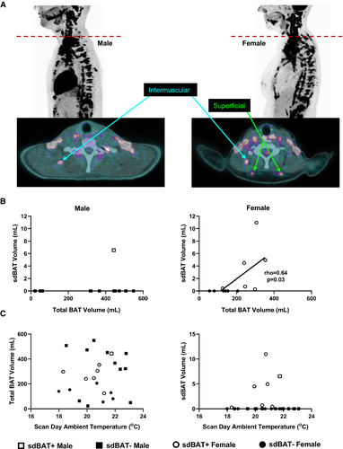
Although continuous with the classical intermuscular cervical BAT depot via a thin fascial layer, a distinct, superficial adipose depot with 18F-FDG uptake consistent with BAT was identified on the dorsal side of the cervical spine in 6 of 12 women but only in 1 of 12 men (P = 0.02; Figure 2A).

In women, sdBAT volume and 18F-FDG uptake correlated with total BAT volume and 18F-FDG uptake, respectively (both, P = 0.03; Figure 2B and not shown). However, in women who had sdBAT, it accounted for only 1% of the total BAT volume. Neither total BAT nor sdBAT volumes correlated with ambient temperature on scan day (Figure 2C) or glucose uptake in reference tissues (data not shown) in men or women.
Discussion
Sexual dimorphisms in human adiposity have been long established, yet little is known about BAT differences between men and women. In retrospective studies of thousands of patients undergoing PET/CT, BAT 18F-FDG uptake is consistently found more frequently in women than in men (11, 13). However, in controlled cohort studies, there has not been an apparent dimorphism (14). Here, a more nuanced story emerged: during cold exposure, women had a lower volume of active BAT than men, but when normalized to total body mass or body surface area, this difference did not persist. This indicates that differences in absolute BAT volume may be due to body size differences between the sexes. In terms of either mean or total metabolic activity, BAT glucose uptake was comparable between men and women.
By anatomical depot, women and men have similar BAT distributions. However, a sexual dimorphism does exist in the presence of what we have termed sdBAT. Heaton (15) first recognized histological evidence for BAT in the “subcutaneous interscapular area” in adult autopsies but did not comment on depot-specific sexual dimorphisms. In our study, half of the women, but only one man, had potential BAT in this depot, consistent with other reports (16, 17). In particular, Martinez-Tellez et al. (16) identified dorsocervical adipose tissue 18F-FDG uptake in 23 of 133 young participants, nearly all women (n = 22). Our analysis now provides quantification of this region as it relates to full-body BAT (16). Although sdBAT accounted for < 1% of total BAT in those who had it and although we did not observe a clear hierarchical cold-stimulated response pattern of BAT glucose uptake by depot, sdBAT volume and 18F-FDG uptake correlated with total BAT volume and 18F-FDG uptake, suggesting that women with a greater total BAT volume are more likely to present sdBAT.
Based on its location, we hypothesize that sdBAT is a remnant of interscapular BAT seen in human newborns (18) and a homolog of the principal functional BAT depot in rodents (1). However, because this depot may be susceptible to imaging artifacts, such as compression and partial volume effects, future studies must confirm whether sdBAT performs thermogenesis in vivo and contains BAT biomolecular markers. It is imperative to understand the functional role of sdBAT, to understand why an apparent involution of this depot occurs, and to understand why it is more prevalent in women than in men. Practically, procuring biopsy tissue from this region should involve less patient risk than other BAT depots because it has fewer proximate nerves and major blood vessels and because it is the most superficial depot identified thus far (19).
This study has several limitations. We used 18F-FDG uptake for BAT quantification, which does not account for fatty acid consumption (20). Repeated cold exposures can increase BAT 18F-FDG uptake (20). We mitigated this effect by randomizing ambient temperature order. It is unknown whether men and women have comparable adipocyte heterogeneity in fat depots. Finally, this study included only healthy, lean, young participants.
In summary, despite differences in adiposity and total BAT volume, healthy, lean, young women and men do not differ in relative amount, glucose uptake, and distribution of BAT. However, sexual dimorphisms do exist; sdBAT is more prevalent in women than in men. Based on its anatomy, we hypothesize that sdBAT is a remnant of interscapular BAT seen in human newborns. Our findings highlight the need to further investigate how BAT contributes to whole-body thermal physiology and body weight regulation in women and men.
Acknowledgments
We thank the study participants, the Metabolic Clinical Research Unit nurses and nutrition team, the National Institutes of Health PET/CT department for administering scans, Ilan Tal for software development, and Sungyoung Auh for statistical assistance. Deidentified data are available upon request.


