Genetic Markers of Brown Adipose Tissue Identity and In Vitro Brown Adipose Tissue Activity in Humans
Funding agencies: This work was supported by the Dutch Diabetes Research Foundation (2014.11.1714), the Netherlands Organization for Scientific Research (TOP 91209037), and EU FP7 project DIABAT (HEALTH-F2-2011-278373).
Disclosure: The authors declared no conflict of interest.
Clinical trial registration: metc.mumc.nl identifier METC 10-3-012; ClinicalTrials.gov identifier NCT03111719.
Abstract
Objective
Human brown adipose tissue (BAT) activity decreases with age and obesity. In addition to uncoupling protein 1 (UCP1), several genetic markers of BAT in humans have been published. However, the link between human BAT activity and genetic markers has been inadequately explored.
Methods
White adipose tissue (WAT) and BAT biopsies were obtained from 16 patients undergoing deep neck surgery. In vitro differentiated adipocytes were used to measure norepinephrine-stimulated mitochondrial uncoupling as a measure of in vitro BAT activity. Gene expression was determined in adipose tissue biopsies.
Results
Norepinephrine increased in vitro BAT activity in adipocytes derived from human BAT, and this increase was abolished by propranolol. Furthermore, in vitro BAT activity showed a negative correlation to age and BMI. UCP1 messenger RNA (mRNA) expression showed a positive correlation to in vitro BAT activity, while zinc finger protein of cerebellum 1 (ZIC1) mRNA showed a negative correlation to in vitro BAT activity. In human BAT biopsies, UCP1 mRNA showed negative correlations to age and BMI, while ZIC1 mRNA showed positive correlations to age and BMI.
Conclusions
Differentiated adipocytes derived from human BAT maintain intrinsic characteristics of the donor. High ZIC1 mRNA does not necessarily reflect high BAT activity.
Introduction
An imbalance of increased caloric intake over energy expenditure leads to the storage of excess energy in white adipose tissue (WAT), ultimately resulting in obesity. Currently, obesity is a worldwide health issue that predisposes people to develop other metabolic disorders, including type 2 diabetes mellitus, cardiovascular disease, and cancer (1). Brown adipose tissue (BAT) fulfills the opposite function of WAT by preventing hypothermia in hibernating mammals and young infants. The unique characteristic of BAT is its ability to uncouple electron transport from ATP production via uncoupling protein 1 (UCP1) in the electron transport chain, resulting in heat production while using glucose and fatty acids as substrates (2). Therefore, activating BAT might be a strategy to combat obesity and obesity-related metabolic disorders.
The interest in human BAT was renewed with the discovery of active BAT following cold exposure in adult humans (3-5). We have previously shown that in vivo human BAT activity correlates negatively with body mass index (BMI) (3), suggesting that the reactivation of BAT in people with obesity might be a strategy to promote weight loss (6, 7). Furthermore, it has been suggested that human BAT activity decreases with age (8, 9). However, in these studies, BAT activity was determined indirectly by measuring cold-induced glucose uptake in the neck/supraclavicular region by using 2-deoxy-2-(18F)fluoroglucose and positron emission tomography (PET)/computed tomography analysis. To date, it is unknown whether intrinsic mitochondrial uncoupling capacity of brown adipocytes is altered with obesity or aging. Therefore, the first aim of the present study was to examine the capacity for mitochondrial uncoupling in differentiated adipocytes derived from human BAT from volunteers with a wide range in age and BMI and to test the hypothesis that aging and obesity reduce intrinsic mitochondrial uncoupling activity in human BAT.
Studies in humans that include human BAT biopsies have mainly focused on gene expression to identify BAT. However, the field of BAT has been further complicated by the discovery of beige, brite, or recruitable brown adipocytes (10, 11). Just like brown adipocytes, these adipocytes express high levels of UCP1 in the mitochondria, enabling uncoupling. Different genetic markers have been found to demonstrate the presence of UCP1-expressing adipocytes in mouse and man. These markers have provided evidence that human BAT can be composed out of pure brown or beige adipocytes or can be a mixture of beige and brown adipocytes (12-15). However, to date, it remains unknown whether genetic markers of UCP1-expressing adipocytes have predictive value regarding BAT activity. Therefore, the second aim of the current study was to examine whether previously described genetic markers have predictive value for mitochondrial uncoupling capacity in differentiated adipocytes derived from human BAT.
Methods
Study approval
The study was reviewed and approved by the ethics committee of Maastricht University Medical Center (METC 10-3-012, NL31367.068.10). Informed consent was obtained before surgery, and participants did not receive a stipend.
Human biopsies and primary adipocyte cultures
Paired human BAT and subcutaneous WAT biopsies were collected during deep neck surgery in patients with normal thyroid function. Stromal vascular fractions were obtained from adipose tissue biopsies by using collagenase digestion. Isolated cells were grown to confluence before differentiation was initiated as previously described (16).
Mitochondrial respiration experiments
The stromal vascular fractions derived from adipose tissue biopsies were plated in XF96 cell culture microplates (Agilent Technologies, Santa Clara, California). After cells were fully differentiated, oxygen consumption and mitochondrial function were measured by using the Seahorse XF96 extracellular flux analyzer from Agilent Technologies. Cells were incubated for 1 hour at 37°C in unbuffered XF assay medium supplemented with GlutaMAX (2 mM) (Thermo Fisher Scientific, Waltham, Massachusetts), sodium pyruvate (1 mM), and glucose (25 mM). The basal oxygen consumption rate was measured, followed by an injection of 1 µM oligomycin and then an injection of 1 µM norepinephrine (NE). Oligomycin, propranolol, and NE were purchased from Sigma-Aldrich (St. Louis, Missouri). Mitochondrial uncoupling was examined as mitochondrial respiration after the inhibition of ATP synthase with oligomycin (which was set to 100%), thus reflecting mitochondrial uncoupling because of proton leak. Data were plotted as a percentage calculated from the fourth measurement after the injection with oligomycin.
RNA isolation and quantitative polymerase chain reaction analysis
RNA was isolated from human adipose tissue biopsied material and cultured adipocytes by using TRIzol extraction (Thermo Fisher Scientific) followed by purification by using the RNeasy kit from Qiagen (Hilden, Germany) according to the manufacturer's instructions. Complementary DNA was synthesized by using the High-Capacity RNA-to-cDNA Kit from Applied Biosystems (Foster City, California) according to the manufacture's instructions. Quantitative polymerase chain reactions were performed on a CFX384 Touch Real-Time PCR Detection System from BioRad Laboratories (Hercules, California). Gene expression was calculated by using the 2-ΔΔCt method. Relative gene expression was normalized to TATA box-binding protein. The following SYBR green primers were used to analyze gene expression: adiponectin (ADIPQ) (fwd: accaggaaaccacgactcaa, rev: accaataagacctggatctcctttc), leptin (fwd: gctgtgcccatccaaaaagtcc, rev cccaggaatgaagtccaaaccg), and zinc finger protein of cerebellum 1 (ZIC1) (fwd: acatgaaggtccacgaatcct, rev: cttgtggtcgggttgtctgt). Primers detecting mitochondrial tumor suppressor 1 (MTUS1) (Hs00368183_m1) and potassium channel subfamily k member 3 (KCNK3) (Hs00605529_m1) were from Applied Biosystems. Primers detecting adipocyte protein 2 (AP2) (13), UCP1 (17), epithelial v-like antigen (EVA1) (12), and TATA box-binding protein (12) have been described previously.
Statistics
For respiration experiments, differences were analyzed by using a two-way analysis of variance (ANOVA) in GraphPad Prism (GraphPad Software Inc., San Diego, California), and the correlation analyses (Spearman) were also performed in GraphPad Prism. P < 0.05 was considered significantly different.
Results
NE-stimulated mitochondrial uncoupling is specific for differentiated adipocytes derived from human BAT and is dependent on β-adrenergic receptor
Paired BAT and WAT biopsies were collected from 16 individuals with a wide range in BMI and age undergoing deep neck surgery (Table 1) and were used for culturing preadipocytes from the stromal vascular fraction. In vitro BAT activity was measured as mitochondrial uncoupling followed by stimulation with natural β-adrenergic receptor agonist NE. In accordance with the characteristic capacity of BAT, acute stimulation with NE in differentiated adipocytes derived from human BAT significantly increased mitochondrial uncoupling to 256 ± 3.3%, an effect that was largely absent in differentiated adipocytes derived from human subcutaneous WAT coming from the same region (125 ± 9.0%; Figure 1A), which is in agreement with our previously published results (16). To demonstrate that this mitochondrial uncoupling is directly due to β-adrenergic stimulation, we used β-adrenergic receptor antagonist propranolol. Indeed, NE-stimulated mitochondrial uncoupling was completely abolished by propranolol in adipocytes derived from human BAT (Figure 1A).

Mitochondrial uncoupling capacity in differentiated adipocytes derived from human BAT and the relationship to the donor's age and BMI. (A) Oxygen consumption rate (OCR) was measured in oligomycin (OG)-treated differentiated adipocytes derived from BAT (black circles) and WAT (white circles) following 1 µM NE (upward-pointing arrow). Differentiated adipocytes derived from human BAT (black triangles) and WAT (white triangles) were pretreated for 1 hour with 1 µM propranolol and, subsequently, OCR was measured in OG-treated differentiated adipocytes following 1 µM NE. Results are presented as mean ± SD measured in quadruplicate (n = 1). *P < 0.05 for brown versus brown + propranolol. #P < 0.05 for white versus white + propranolol. (B) Spearman correlation analysis (n = 16) of NE-stimulated OCR in differentiated adipocytes derived from human BAT to age (years). (C) Spearman correlation analysis (n = 16) of NE-stimulated OCR in differentiated adipocytes derived from human WAT to age (years). (D) Spearman correlation analysis (n = 16) of NE-stimulated OCR in differentiated adipocytes derived from human BAT to BMI (kilograms/meters squared). (E) Spearman correlation analysis of NE-stimulated OCR in differentiated adipocytes derived from human WAT to BMI (kilograms/meters squared).
| Subject | Sex | Age (y) | BMI (kg/m2) |
|---|---|---|---|
| 1 | F | 45 | 22.6 |
| 2 | F | 48 | 21.9 |
| 3 | F | 66 | 33.6 |
| 4 | F | 58 | 29.4 |
| 5 | F | 69 | 30.0 |
| 6 | F | 76 | 28.4 |
| 7 | F | 68 | 31.9 |
| 8 | M | 51 | 26.6 |
| 9 | F | 59 | 26.0 |
| 10 | F | 31 | 18.6 |
| 11 | F | 55 | 22.5 |
| 12 | F | 48 | 25.1 |
| 13 | F | 29 | 26.0 |
| 14 | F | 47 | 22.3 |
| 15 | F | 19 | 16.1 |
| 16 | M | 33 | 21.2 |
NE-stimulated mitochondrial uncoupling in differentiated adipocytes derived from human BAT is related to age and BMI
Differentiated adipocytes derived from human BAT in the current study originated from volunteers with a wide range in BMI and age. Although age and BMI are related to each other, this allowed us to investigate whether the intrinsic NE-stimulated mitochondrial uncoupling activity of adipocytes derived from human BAT or WAT was related to the BMI or age of the donor. NE-stimulated mitochondrial uncoupling in differentiated adipocytes derived from human BAT displayed a negative correlation to age (ρ = −0.69; P = 0.004) and BMI (ρ = −0.58; P = 0.019), while no such correlation was established in differentiated adipocytes derived from human WAT (Figure 1B-1E). These findings are consistent with previous reports demonstrating that 2-deoxy-2-(18F)fluoroglucose glucose uptake in the human neck region is negatively associated with age and BMI (3, 5, 8).
ZIC1 messenger RNA negatively correlates with in vitro BAT activity in differentiated adipocytes derived from human BAT
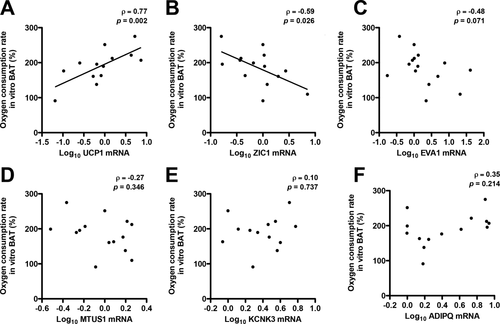
The exact nature of human BAT is currently under debate. Thus far, studies devoted to genetic markers of beige and brown adipocytes in human BAT biopsies have included ZIC1 (13, 18), EVA1 (12, 18), KCNK3 (19), and MTUS1 (19) with the aim to distinguish human brown and beige from white adipocytes. Next, we investigated how messenger RNA (mRNA) expression of these genes relates to in vitro BAT activity measured in in vitro differentiated adipocytes. UCP1 mRNA was positively associated with intrinsic mitochondrial uncoupling activity in differentiated adipocytes derived from human BAT (ρ = 0.77; P = 0.002) (Figure 2A). Interestingly, ZIC1 mRNA showed a negative correlation to in vitro BAT activity (ρ = −0.59; P = 0.026) (Figure 2B). mRNA expression of EVA1, MTUS1, and KCNK3 did not correlate with in vitro BAT activity (Figure 2C-2E). In order to monitor the effects of differentiation in these experiments, we examined ADIPQ mRNA expression and correlated that to in vitro BAT activity. ADIPQ mRNA did not show a correlation with in vitro BAT activity (Figure 2F), indicating that in vitro BAT activity was not dependent on the differentiation state of the adipocytes because ADIPQ is an important indicator of adipocyte differentiation (20, 21).

Correlation of genetic markers for human BAT to in vitro BAT activity. Spearman correlation analysis (n = 15) of (A) UCP1, (B) ZIC1, (C) EVA1, (D) MTUS1, (E) KCNK3, and (F) ADIPQ mRNA expression to NE-stimulated oxygen consumption rate (OCR) in differentiated adipocytes derived from human BAT.
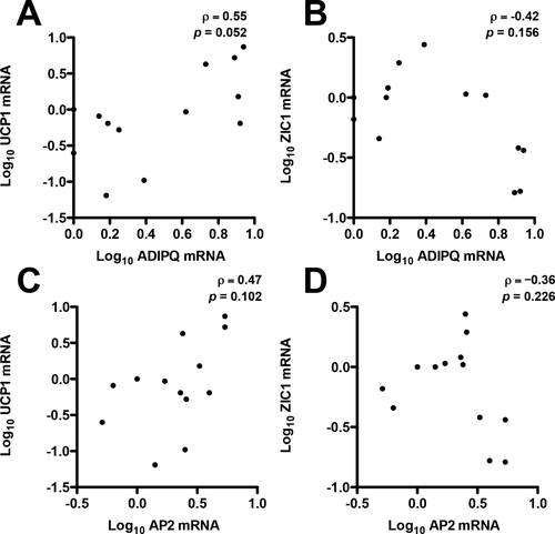
No correlations to ADIPQ or AP2 were detected for UCP1 mRNA (Figure 3A-3C). Also, no correlations to ADIPQ or AP2 were detected for ZIC1 mRNA expression (Figure 3B-3D). These results further indicate that differentiation state does not seem to be a driver for UCP1 or ZIC1 expression in adipocytes derived from human BAT.

Correlation of genetic markers of BAT in differentiated adipocytes derived from human deep neck BAT biopsies. (A,B) Spearman correlation analysis (n = 15) of UCP1 and ZIC1 mRNA expression to ADIPQ mRNA expression in differentiated adipocytes derived from human BAT. (C,D) Spearman correlation analysis (n = 15) of UCP1 and ZIC1 mRNA expression to AP2 mRNA expression in differentiated adipocytes derived from human BAT.
Expression of genetic markers in human BAT biopsies from the neck
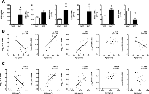
Next, the previously discussed markers were analyzed in paired BAT and WAT biopsies of adult human subjects undergoing deep neck surgery. As expected, UCP1 expression was higher in BAT biopsies compared with WAT biopsies (Figure 4A), and leptin expression was higher in WAT biopsies compared with BAT biopsies (Figure 4A). In addition, expression of MTUS1, EVA1, and KCNK3 was higher in human BAT biopsies compared with WAT biopsies (Figure 4A). Although ZIC1 mRNA was increased in BAT biopsies compared with WAT biopsies, this increase did not reach statistical significance (Figure 4A).

Genetic markers of BAT in human deep neck biopsies and the relationship to the donor's age and BMI. (A) mRNA expression of UCP1, ZIC1, EVA1, MTUS1, KCNK3, and leptin examined in human BAT biopsies (black bars) and WAT biopsies (white bars). A Student t test was used to compare WAT with BAT. Results are presented as mean ± SEM (n = 15). *P < 0.05. (B) Spearman correlation analysis (n = 15) of UCP1, ZIC1, EVA1, MTUS1, and KCNK3 mRNA to age (years). (C) Spearman correlation analysis (n = 15) of UCP1, ZIC1, EVA1, MTUS1, and KCNK3 mRNA to BMI (kilograms/meters squared).
Correlation analysis of genetic markers in human BAT biopsies from the neck to age and BMI
Because our BAT biopsies were derived from volunteers with a wide range of age and BMI, we next determined if these markers were associated with increased age and BMI. Interestingly, UCP1 and KCNK3 displayed a negative correlation to age (ρ = −0.67; P = 0.006 and ρ = −0.56; P = 0.031), while only UCP1 displayed a negative correlation to BMI (ρ = −0.53; P = 0.044) (Figure 4B-4C). In contrast, ZIC1 and EVA1 showed a positive correlation to age (ρ = 0.70; P = 0.004 and ρ = 0.70; P = 0.004) and BMI (ρ = 0.77; P = 0.001 and ρ = 0.64; P = 0.010) (Figure 4B-4C). No correlations to age or BMI were detected for MTUS1 (Figure 4B-4C).
Discussion
In the present study, we show that the intrinsic capacity of differentiated adipocytes derived from human BAT for mitochondrial uncoupling decreases with age and obesity. Although it cannot be deduced from the current study which factors are responsible for this reduction in BAT activity in humans, the strong correlation with UCP1 and not other markers may suggest that an age- or BMI-related downregulation of UCP1 may be key. It has been reported that higher human in vivo BAT activity changes metabolism through increased energy expenditure (6, 16). However, the current topic of debate questions whether we fully grasp the complete thermogenic potential of active human BAT (22). The finding that ZIC1 was positively associated with age and BMI and negatively associated with BAT activity is interesting because, to the best of our knowledge, no links have been established between BAT activity and ZIC1. ZIC1 does show specificity for human adipose tissue biopsies taken from the deep neck region in adults compared with WAT (13, 14). In the current study, we have used BAT biopsies derived from the neck area. Further research would be required to establish a similar role for ZIC1 in other human BAT depots. In human neonates, ZIC1 expression is higher in interscapular adipose tissue compared with visceral adipose tissue (23); however, no connections have been made to assess the involvement of age. ZIC1 overexpression in pluripotent murine stem cells has a negative impact on adipogenesis and expression of mitochondrial proteins (24), which fits with our findings that high ZIC1 expression is linked to low in vitro BAT activity. ZIC1 is a transcription factor with zinc finger domains primarily found in the adult cerebellum. High levels of ZIC1 have been detected in liposarcomas (25); however, hypermethylation of ZIC1 has been detected in gastric cancer (26). Future studies using differentiated adipocytes derived from human BAT could be used to unravel the mechanisms underlying the age-related decline in BAT activity with a specific interest for the contribution of ZIC1.
Conclusion
We have demonstrated that in vitro BAT activity decreases with age and BMI comparable with in vivo BAT activity in humans. Furthermore, we have provided evidence that previously reported genes found to identify beige and/or brown adipocytes may not have predictive value regarding in vitro BAT activity and perhaps also in vivo BAT activity in humans.
Acknowledgments
The authors would like to thank all participants who joined the study.


