Lumbar spondylolysis in ancient Siberian Eskimo
Abstract
Previous studies have shown that North American and Greenland Arctic groups are characterized by high incidence of spondylolysis. The hypotheses explaining the high predilection to spondylolysis in these populations range from genetic predisposition to lifestyle characteristics. To date, however, no study assessed the presence of spondylolysis in the Siberian Eskimo. Thus, the current study presents new data from the Asian side of the Beringia, the original homeland of the North American and Greenland Inuit groups. The skeletal material originates from the Ekven burial site in Chukchi Peninsula, Russia. Most burials belong to the Old Bering Sea tradition of marine mammal hunters, embracing the period from the beginning of the 1st to the beginning of the 2nd millennium AD. Totally, 71 individuals were studied. Spondylolysis was present in 38% of individuals and in 11% of studied lumbar vertebrae, with higher incidence in juveniles and young adults compared with other ages and in males compared with females. There was an evident increase in the severity of the stress fractures from adolescence to middle adulthood (i.e., more vertebrae affected per individual and higher proportion of bilateral fractures). The reported incidence of spondylolysis in the Ekven is in the range of the values reported for the North American and Greenland Inuit groups and is expectedly high against the values for groups of European and African ancestry. Although the accumulation of gene variants responsible for spondylolysis in Arctic groups is possible, it is more plausible that some adaptive morphological characteristics make them prone to fatigue fractures of the lower spine in the conditions of the specific physical activity.
1 INTRODUCTION
1.1 Background
A number of authors have pointed to the high frequencies of spondylolysis among Inuit from Alaska, Greenland and Canadian Arctic both in modern and ancient samples (Lester & Shapiro, 1968; Kettelkamp & Wright, 1971; Gunness-Hey, 1981; Mathiesen, Simper, & Seerup, 1984; Stewart, 1931, 1953; Simper, 1986; Merbs, 2002). Reported frequencies for these Arctic skeletal samples range from 5% to 55%, in the vast majority of cases exceeding 10% (D'Angelo del Campo, Suby, García-Laborde, & Guichón, 2017; tab. 3), whereas in groups of European and African ancestry, they rarely exceed this value (D'Angelo del Campo et al., 2017; fig. 6).
In modern groups, high prevalence of the defect (up to 60%) is widespread among athletes (Tawfik, Phan, Mobbs, & Rao, 2020). For skeletal samples, D'Angelo del Campo et al. (2017) observed that in Europe and Africa, frequencies of spondylolysis are much less variable compared with Asia and Americas and that higher values usually characterize hunter–gatherers. However, although there are many studies on spondylolysis in Europe and Americas, more data are needed for other regions.
It is generally accepted that spondylolysis is a fatigue fracture, thus an acquired condition. Most of the researchers, however, lean towards its multifactorial aetiology, including a spectrum of both genetically inherent and biomechanical factors (D'Angelo del Campo et al., 2017; Pilloud & Canzonieri, 2014; Whitesides, Horton, Hutton, & Hodges, 2005).
1.2 Main hypotheses explaining high prevalence of spondylolysis in North American and Greenland Arctic groups
Simper (1986, p. 79) pointed out that the high incidence of spondylolysis in the Inuit groups suggests a genetic factor, although trauma and repetitive stress may play a role in its aetiology. Kettelkamp and Wright (1971) also believed that this observation reflects “a concentrations of these genetic factors in an isolated, interrelated population or extended family” (Kettelkamp & Wright, 1971, p. 566). Of the same opinion are Lester and Shapiro (1968, p. 47) suggesting that a “hereditary weakness exists” in this ethnic group.
The more in-depth studies on the topic belong to Stewart (1931, 1953, 1956) and Merbs (1995, 1996, 2002). Not counting the early work by Stewart (1931), both researchers inclined towards the opinion that biomechanical, not genetic, factors play the key role in the development of “typical” spondylolysis in the Inuit groups. Among the occupations that may induce the development of spondylolysis in Canadian Inuit, Merbs (2002) listed weight lifting, wrestling, kayak paddling and harpoon throwing. He noticed that activity or peculiar posture, described earlier by Stewart (1953), may be one of the explanations of the considerable differences among various Canadian series.
At the time of Stewart's early study (Stewart, 1931), the congenital theory on the aetiology of spondylolysis was common, so he initially attributed the high frequency of the defect in the Alaskan Inuit to an inbreeding. In his later study (Stewart, 1953), however, he inclined towards the primary role of the mechanical factors. He supported this view by providing evidence that spondylolysis increases in frequency with age. However, Stewart did not rule out that an “anomalous ossification” may predispose Arctic groups to this defect (Stewart, 1953, p. 950).
Specifically, in his 1953 work, Stewart proposed that the high frequency of spondylolysis in the Alaskan Natives may be caused by injuries from falls on ice but provided that vertebrae were weakened and predisposed to breakage by other activities, such as a peculiar posture that Inuit assume during everyday work (see Stewart, 1953; fig. 10). He found parallels between environmental severity and the incidence of “arch defects,” as the Inuit groups north of Yukon River showed substantially higher incidence of spondylolysis compared with those of more southern regions of Alaska, whereas Aleutians and Kodiak islanders, also studied by Stewart, showed intermediate frequencies. He explained that Alaskan Inuit spend most of the year in extreme cold with limited sunlight surrounded by ice and snow and are mainly coastal inhabitants with the primary food resource being marine mammals. However, many groups about the Yukon River and south live inland. The groups inhabiting the Aleutian and Kodiak islands, where the climate is milder, still live a “maritime existence” with a “treacherous terrain and the constant hazard of dense fogs” (Stewart, 1953, p. 949). These differences in life conditions, according to Stewart, may contribute to the different risks of accidents, thus differences in the stress fracture frequencies.
Yet in his later work, Stewart (1956) admitted that explaining the “neural arch defects” by purely external factors may be too restrictive. So he specifically examined several anatomical variations of the pelvic and lower lumbar region, which may constitute the alleged hereditary factors, predisposing the Alaskan Natives to the stress fracture. Consequently, he found a consistent, although insignificant, association between spondylolysis and features “favoring the increase of the mechanical strain in the lower back” (Stewart, 1956, p. 58). However, these traits had almost negligible predictive value; thus, Stewart concluded that the only hereditary factors that may predispose to spondylolysis are those related to the upright posture of our species (Stewart, 1956, p. 59).
Whitesides et al. (2005) studied an Aleut group from Kagamil Island (mid-Aleutian) area and found that sacral table angle in this group was significantly lower compared with Arikara Indians and Japanese. This corresponded to significantly higher incidence of spondylolysis among Aleuts (27%) compared with the other two groups (6%–9%). Unfortunately, they were unable to study the Inuit samples from Northern Alaska where the frequencies of spondylolysis reached 45% to 50%; thus, it is unknown to what extent the lower sacral table angle may be an aetiologic factor, predisposing different Arctic groups to spondylolysis.
1.3 Research goal
To date, no study assessed the presence of spondylolysis in the Siberian Eskimo. The current article aims to fill this gap by presenting the relevant data on an ancient Eskimo group from Chukotka, Northeast Asia. This part of Asia, being the native land of the North American and Greenland Arctic groups, is a dwelling place of their closest relatives, the Siberian Eskimo, who inhabited the Western Beringia for millennia.
2 MATERIALS AND METHODS
The ancient Eskimo collection originates from the Ekven burial site on the far east point of Chukotka near Cape Dezhnev. The studied materials were excavated between the early 1960th and early 1990th first by Arutyunov and Sergeev and then by the expedition of the State Museum of Oriental Art. The human remains were studied in the Research Institute and Museum of Anthropology, Lomonosov MSU, Moscow.
The funeral inventory included hunting equipment, ritual objects, household and artistic items. The Ekven site represents the Neoeskimo cultural tradition of marine mammal hunters, beginning from the early Old Bering Sea (OBS I) and Okvik to the younger Birnirk and Punuk stages (Arutyunov & Sergeev, 1975; Bronshtein, Dneprovsky, & Savinetsky, 2016). Radiocarbon dates indicate that the site was functioning from the beginning of the 1st to the beginning of the 2nd millennium AD (Mason, 2016, pp. 420, 428).
Although about 300 burials were excavated, a maximum of 71 individuals were included in this study for reasons related to preservation and collector's bias. The minimum inclusion criterion for calculating individual incidences (crude prevalence rate) was the presence of at least two lower lumbar vertebrae. However, all vertebrae from the 20th presacral (L1) to the last lumbar were scored if present, and this information was included in calculating the true by-level prevalence of the defect. In cases where some vertebrae were missing and the exact number of presacral and lumbar vertebrae could not be estimated, a standard number was assumed (24 presacral and 5 lumbar), unless there were strong indications of a border shift between spine regions.
Among the 71 individuals, 53 were adults, 13 were adolescents and five were children. Sex was estimated based on the pelvic morphology; age was estimated using standard pelvic criteria as well as the overall condition of the joints and the spine in adults and using stages of epiphyseal union for nonadults (Buikstra & Ubelaker, 1994). The sample was divided into broad age categories: children (<12 years), adolescents (12–18 years), young adults (18–35 years), middle adults (35–50 years) and old adults (>50 years).
In this study, “spondylolysis” was understood in its narrow meaning, that is, complete separation at pars interarticulatis, either bilateral or unilateral. Spondylolysis was scored only if the defect had unquestionably antemortem appearance (traces of healing/wear). Statistical procedures were performed in Statistica software (StatSoft, 2007).
3 RESULTS
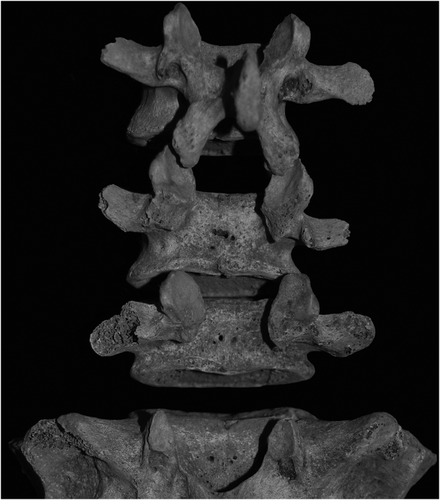
Overall, spondylolysis affected 37.5% of the analysed sample and 38 out of 355 studied vertebrae (Table 1; see Figure 1 for illustration). The incidence is higher in males compared with females, although the difference is statistically insignificant (chi-square value for the individual frequency comparison was 0.845, df = 1, p = 0.358; for the total proportion of the vertebrae affected, it was 0.614, df = 1, p = 0.433). The most frequent site of the pathology was the 24th presacral vertebra (L5), followed by the 23rd (L4); several cases were seen at other lumbar levels. The lumbalized first sacral (25 presacral vertebrae) was present in nine individuals, and in four of these, the vertebra was affected by spondylolysis. 1
| Individual count | True prevalence (by level) | |||||||||||||||
|---|---|---|---|---|---|---|---|---|---|---|---|---|---|---|---|---|
| V20a | V21 | V22 | V23 | V24 | V25b | Total vertebrae affected, % | ||||||||||
| N | n | % | N | n | N | n | N | n | N | n | N | n | N | n | ||
| Males (>12 years) | 28 | 12 | 42.9 | 33 | 1 | 31 | 0 | 35 | 3 | 32 | 2 | 35 | 10 | 7 | 4 | 11.6 |
| Females (>12 years) | 26 | 8 | 30.8 | 26 | 0 | 27 | 0 | 27 | 0 | 28 | 4 | 26 | 8 | 2 | 0 | 8.8 |
| Unsexed (>12 years) | 5 | 3 | 60.0 | 4 | 0 | 4 | 1 | 4 | 1 | 5 | 1 | 5 | 2 | 0 | 0 | 22.7 |
| Unsexed (<12 years) | 5 | 1 | 20.0 | 5 | 0 | 4 | 0 | 5 | 0 | 5 | 0 | 5 | 1 | 0 | 0 | 4.2 |
| All | 64 | 24 | 37.5 | 68 | 1 | 66 | 1 | 71 | 4 | 70 | 7 | 71 | 21 | 9 | 4 | 10.7 |
- a V20 refers to the 20th presacral vertebra etc.
- b Scored in cases of 25 presacral vertebrae.

The analysis of age dynamics gives the following results. Among five children, only a single case of bilateral L5 spondylolysis is observed (Table 1). This was an older child (7–12 years old). In subsequent age groups, the highest percentage of spondylolysis was observed in adolescents (in seven out of 11 individuals) and young adults (in nine out of 22) (Table 2). The frequency drops in middle adulthood (five out of 16) and, especially, in old adulthood (one out of 9). The Spearmen's rank correlation test reveals that decrease in frequency from adolescence to old adulthood is significant at p < 0.05 (rs = −0.317) (age categories scored in the analysis as an ordinal variable: 2, adolescent; 3, young adult; 4, middle adult and 5, old adult). The change in the severity of the stress fracture shows opposite tendencies, with a trend towards increasing severity from the adolescence to the middle adulthood, indicating that the number of affected vertebrae in a single individual and the proportion of bilateral versus unilateral fractures increase with age. However, it was impossible to track this trend into the late adulthood because of the small sample size.
| Adolescents (N = 11) | Young adult (N = 22) | Middle adult (N = 16) | Old adult (N = 9) | |
|---|---|---|---|---|
| % Individuals affected | 63.6 | 40.9 | 31.3 | 11.1 |
| Mean severitya | 1.21 | 1.27 | 1.70 | 1 |
- a Mean number of vertebrae affected per individual, calculated as the sum of the number of affected vertebrae in affected individuals divided by the number of affected individuals. In case of unilateral spondylolysis, it was counted as 0.5 vertebra affected.
4 DISCUSSION AND CONCLUSION
The 38% incidence of spondylolysis in the Ekven sample is in the range of the values reported for the North American and Greenland Inuit groups and is expectedly high against those reported for groups of European and African ancestry. It is close to the frequencies, reported by Stewart for the Alaskan sample North of Yukon (40%), whereas frequencies of spondylolysis in more southern regions of Alaska were substantially lower (15%) (Stewart, 1953). In line with the data on other groups, Ekven males have higher incidence of spondylolysis compared with females. This was also the case for the Alaskan, North Canadian and Greenland groups (Merbs, 2002; Simper, 1986; Stewart, 1953).
Regarding age dynamics, results on Ekven Eskimo are consistent with Merbs' results (1995) on the Canadian Inuit in that he observed a decrease in frequency from young to middle adulthood but different from the data of Stewart (1953), who observed an increase in the incidence of spondylolysis with age. Merbs proposed that the stress fracture begins to develop in the young age, and in young adulthood, it progresses to complete lysis (Merbs, 1995, 2002). Later in life, some of the defects may heal or progress further. The tendencies, observed in the Ekven sample towards increased “severity” of the defects (higher proportion of multiple and bilateral defects) that otherwise decrease in frequency with age, seem to be in line with Merbs' hypothesis of progressing or healing fractures. This age dynamics may be conditioned by the level of individual's activity according to age. For example, the peak fracturing at adolescents may correspond to the beginning active involvement of the individuals into the economic and social life. This is the time when the vertebral column is still developing and may be particularly prone to displacements and traumatization. This susceptibility to lower spine stress fractures may be exaggerated by the insufficient experience in performing some tasks (e.g., hunting). By the time the individuals reach the middle adulthood, they have enough experience and physical training to reduce the risks of new fracturing, and by the old adulthood, direct involvement in daily tasks may be reduced and become more that of supervision. Thus, the progressive healing of the lower spine fractures and decreasing involvement in injurious behaviour would contribute to the decreasing incidence of the spondylolysis with the increasing age.
This age dynamics may certainly be related to other factors, such as a random effect of sample size, or, otherwise, there may be differential survivability of affected and nonaffected individuals. Although modern clinical data suggest that the condition is often asymptomatic, some individuals experience low back pain and neurological symptoms, which may necessitate surgical or conservative treatment (Mansfield & Wroten, 2020). Thus, the possibility of the existence of differential mortality via emerging adaptive constraints is not excluded. This interpretation was proposed by Lessa (2011) with regard to a precolonial coastal group from Southern Brazil, where a similar decrease in spondylolysis incidence was observed from early to late adulthood. Lessa hypothesized that “pain would have been one of the causes of adaptive limitations, such as difficulties to swim, dive and run, which could contribute to the deaths by drowning, or during hunting or warfare activities” (Lessa, 2011, p. 666).
A perspective direction would be to test if the Eskimo/Inuit are characterized by some morphological specificity regarding the lumbar spine, pelvic incidence or sacral angle, similar to those observed for Aleutians (Whitesides et al., 2005). Though modern Aleuts differ significantly from the Alaskan Inuit and the colonization of the Aleutian Islands and their genetic relationship with other North American Native groups is still discussed, they share distant Aleut–Eskimo ancestry and cluster closely with the Siberian Eskimo and Chukchi based on mDNA sequencing (Crawford, Rubicz, & Zlojutro, 2010). Thus, they may share some postcranial skeleton characteristics with other Arctic groups. Previous researches have shown that Eskimo/Inuit axial skeletons have several characteristics that may be linked to morphological traits, adaptive to the Arctic environmental conditions. These are persistent tendencies towards caudal shift in spine patterning in Asian and North American samples (Karapetian & Makarov, 2019; Merbs, 1974; Stewart, 1932); ribs showing less curvature and torsions in Greenland Inuit (García-Martínez et al., 2018) and large, barrel-shaped rib cage in Asian Eskimo (Klevtsova & Smirnova, 1974). It is plausible that there are other characteristics not explored previously.
Though the Eskimo/Inuit groups throughout the Arctic generally used similar strategies of survival, the hunting specialization might differ depending on the area of residence and cultural tradition. For example, archaeological evidence from the Ekven burial site suggests that the transition from the OBS I through the OBS II to the OBS III traditions was characterized by the gradual increase in whale hunting as compared with pinnipeds and possibly the decreasing role of tundra animals. Later, Birnirk tradition was more specialized in hunting seals on ice through the breathing holes, whereas the early Punuk tradition represented an archaic variant of the Thule–Punuk whaling culture (Bronshtein et al., 2016). The Alaskan Inuit might have used similar set of strategies, whereas some of the Inuit groups in Canadian Arctic were more reliant on hunting terrestrial animals, fish and fowl in addition to marine animals (Burch & Csonka, 1999). Interestingly, some precontact coastal groups of Native Indians that were involved in hunting and fishing also show relatively high frequencies of spondylolysis (e.g., Lessa, 2011; Weiss, 2009). Thus, it would be meaningful to perform an analysis of spondylolysis occurrence depending on differences in lifestyle and hunting specialization.
For now, the proposed genetic associations between mutations in some genes and spondylolysis (e.g., in CDMP-1 gene and SLC26A2; Savarirayan et al., 2003; Zheng et al., 2020) cannot explain the high prevalence of this trait in the Arctic groups, as these associations may represent “pathological” spondylolysis because of altered osteogenesis or be a one expression of a hereditary disease. These gene mutations may not target solely the pars interarticularis but may make the individuals prone to other skeletal anomalies. Though the isolation might play a role in the accumulation of certain gene variants, the indigenous circumpolar populations show the high degree of fitness and specialization that would not allow the chance deleterious mutations to spread so widely. It is more plausible that some adaptive morphological characteristics make the Eskimo/Inuit prone to fatigue fractures of the lower spine in the conditions of the specific physical activity.
ACKNOWLEDGEMENTS
This work was financially supported by the Lomonosov Moscow State University Grant for Leading Scientific Schools “Depository of the Living Systems” in frame of the MSU Development Program.
CONFLICT OF INTERESTS
The author has no conflict of interests.
REFERENCES
- 1 In the previous study of this sample (Karapetian & Makarov, 2019), six individuals were reported to have 25 presacral vertebrae. The number of cases is higher in this study because here the inclusion criterion was less strict.


