Ultrasonic modulation of tissue optical properties in ex vivo porcine skin to improve transmitted transdermal laser intensity
Abstract
Background and Objective
Applications of light-based energy devices involving optical targets within the dermis frequently experience negative side-effects resultant from surface scattering and excess optical absorption by epidermal melanin. As a broadband optical absorber, melanin decreases the efficacy of light-based treatments throughout the ultraviolet, visible, and near-infrared spectra while also generating additional heat within the surface tissue that can lead to inflammation or tissue damage. Consequently, procedures may be performed using greater energy densities to ensure that the target receives a clinically relevant dose of light; however, such practices are limited, as doing so tends to exacerbate the detrimental complications resulting from melanin absorption of treatment light. The technique presented herein represents an alternative method of operation aimed at increasing epidermal energy fluence while mitigating excess absorption by unintended chromophores. The approach involves the application of continuously pulsed ultrasound to modulate the tissue's optical properties and thereby improve light transmission through the epidermis.
Materials and Methods
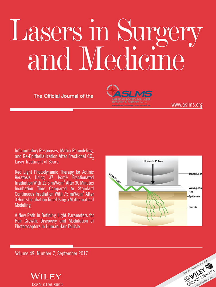
To demonstrate the change in optical properties, pulsed light at a wavelength of 532 nm from a Q-switched Nd:YAG laser was transmitted into 4 mm thick samples of porcine skin, comprised of both epidermal and dermal tissue. The light was transmitted using an optical waveguide, which allowed for an ultrasonic transducer to be incorporated for simultaneous paraxial pulsation in parallel with laser operation. Light transmitted through the tissue was measured by a photodiode attached to an integrating sphere.
Results
Increasing the driving voltage of ultrasonic pulsation resulted in an increase in mean transmitted optical power of up to a factor of 1.742 ± 0.0526 times the control, wherein no ultrasound was applied, after which the optical power increase plateaued to an average amplification factor of 1.733 ± 0.549 times the control.
Conclusions
The increase implies a reduction in light either back-scattered or absorbed within the tissue, which would allow for a greater proportion of incident energy to be delivered to the clinical target, thereby improving procedural efficacy and potentially reducing the severity of detrimental side-effects. Apparatus Lasers Surg. Med. 49:666–674, 2017. © 2017 Wiley Periodicals, Inc.