Greater Saphenous Vein Graft Harvest from Anterolateral Thigh Free Flap Incision: A Novel Technique
Editor's Note: This Manuscript was accepted for publication on May 16, 2023.
Presented at the 2023 Triological Society Combined Sections Meeting, Coronado, California, U.S.A., January 28, 2023.
The authors have no funding, financial relationships, or conflicts of interest to disclose.
Graphical Abstract
The greater saphenous vein can be harvested from the standard incision for an anterolateral free flap and used as a vein graft in complex head and neck reconstruction.

INTRODUCTION
The anterolateral thigh (ALT) free flap is a workhorse in complex head and neck reconstruction. The greater saphenous vein (GSV) is a large-caliber superficial vein running from the ankle to the medial groin and is commonly used for grafting in coronary bypass surgery. The GSV segment within the thigh is traditionally harvested either open (parallel or stair-step incisions) or endoscopically.1 Here we describe a case of harvesting the GSV from an existing ALT free flap incision without the need for additional incisions for two microvascular vein grafts in a previously operated and irradiated neck.
CASE REPORT
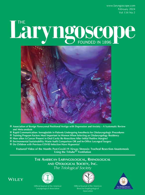
A non-smoking 44-year-old female with a longstanding history of oral lichen planus developed two synchronous primary squamous cell carcinomas (SCC) involving the left maxillary sinus and right oral tongue. Resection of the maxilla mass required maxillectomy, subtotal palatectomy (crossing midline), open ethmoidectomy and sphenoidotomy, and intra-dural dissection to obtain negative margins on the vidian and the maxillary division of the trigeminal nerve. The right posterolateral tongue SCC was also resected, leaving a defect of 6 × 4 cm. She underwent concurrent bilateral neck dissections as well. The maxilla defect was reconstructed with a left fibula free flap with microvascular anastomoses to the left facial artery and left posterior facial vein. A chimeric anterolateral thigh free flap with two perforators was used to reconstruct both the tongue and right soft palate with microvascular anastomoses to the right facial artery, right external jugular vein, and right posterior facial vein. She then underwent adjuvant chemoradiation with 60 Gy and concurrent cisplatin. Eighteen months later she developed three new primary SCCs involving the right maxilla, mandibular alveolar ridge, and left mucosal lower lip. The mucosal lip was wedge excised and closed primarily. The mucosa of the anterior mandibular alveolar ridge was resected and a marginal mandibulectomy performed. The mandible was then covered with a right anterolateral thigh fascia lata free flap and a full thickness skin graft. The recipient vessels for this free flap were the left superficial temporal artery and vein. The right maxilla was resected from the junction of the previous left fibula free flap back to the pterygoids and was reconstructed with a right fibula free flap. The fibula pedicle was short due to a low take off of the peroneal vessels from the tibioperoneal trunk. Microvascular anastomoses were completed using the right superior thyroid artery and external jugular vein as recipient vessels. There was not enough pedicle length for a second venous anastomosis, so the smaller vena comitans was ligated. On postoperative day one, the fibula flap developed venous congestion, and the patient was urgently taken back to the operating room. The venous anastomosis was taken down and thrombus was evacuated. However, this resulted in insufficient venous length for a revision venous anastomosis. Additionally, there were no other available veins in the neck with adequate length and caliber given her significant prior surgery and radiation treatments. At this point, the right ALT incision was opened and dissection performed medially, staying just superficial to the muscular fascia, to locate the GSV (Fig. 1, 2). A 6 cm segment was harvested and divided into two vein grafts for both venae comitantes (Fig. 3). The venous anastomoses were performed with GEM microvascular couplers. The thigh incision was closed primarily. There were no further issues with the venous anastomoses, and the patient has healed well.


DISCUSSION
The autologous reversed GSV graft was first described in 1967 for use in coronary artery bypass.1 Although other options for coronary bypass have since emerged, namely the left internal mammary artery, the GSV graft is still a mainstay for coronary revascularization.1 In the head and neck, the GSV has been used for reconstruction following resection of the carotid artery.2 Chang et al. described one case of using a GSV interposition graft as an arterial conduit to the external carotid artery for simultaneous ALT and fibula free flaps.3 However, the case report did not describe the method of GSV harvest.
To our knowledge, the technique of harvesting the GSV from an existing ALT free flap donor site has not been described. For the patient in this report, dissection was performed medially and superficially to fascia of the thigh musculature through the existing ALT harvest site incision. The GSV was easily encountered in the posteromedial aspect of the thigh. We harvested 6 cm of vein but there was 12+ cm available if additional length was needed. In the coronary literature, 35 cm is obtainable superior to the knee or up to 70 cm along the entire length of the vein down to the ankle.1
The reason for this patient's flap compromise is unclear. While it is preferred to use both vena comitantes for our venous anastomosis to two recipient veins, it is not unusual in our practice to only use one of the peroneal venae comitantes if a second vein is not readily available. The geometry of the pedicle was also relatively favorable in this case. Nonetheless, the flap was able to be salvaged with two vein grafts, suggesting that the greater saphenous vein is a good option for head and neck reconstruction.
CONCLUSION
It is possible and straightforward to harvest the GSV from an existing harvest site of an ALT free flap by extending the dissection medially and superficially to the muscular fascia without the need for additional incisions. The GSV is a valuable option for vein grafting in the case of a short free flap pedicle and/or a vessel-depleted neck.