Erratum
In the Original Research article “Sonographic Visualization of the Posterior Cutaneous Nerve of the Forearm: Technique and Validation Using Perineural Injections in a Cadaveric Model” by Maida et al (J Ultrasound Med 2017;36:1627–1637), the labels in some parts of Figures 3–8 did not appear properly. The affected images have been corrected and are published herewith.
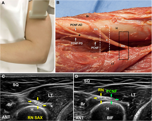
Sonographic identification of the RN and PCNF in the spiral groove region. A, The transducer is placed in an anatomic axial plane on the posterolateral humerus at the level of the spiral groove. B, Relationship of the transducer to the underlying anatomy. H indicates humerus; PCNF-AD, anterior division of the PCNF; and PCNF-PD, posterior division of the PCNF. Bottom is posterior; left, distal; right, proximal; and top, anterior. C, Correlative short-axis (SAX) sonographic view corresponding to the transducer position depicted by the solid black rectangle in B. The RN (yellow arrows) is shown between the LT and adjacent to the humerus. Br indicates brachialis; and SQ, subcutaneous tissue. D, Correlative short-axis sonographic view corresponding to the transducer position depicted by the dashed white rectangle in B. The PCNF (green arrows) arises from the posterior RN. BIF indicates bifurcation. Bottom is deep; left, anterior (ANT); right, posterior; and top, superficial.
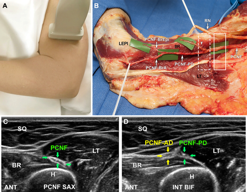
Sonographic visualization of the PCNF at its bifurcation into anterior and posterior divisions. A, The transducer is moved distally in the anatomic axial plane along the posterolateral humerus to follow the PCNF in its short axis. Approximately 7 cm proximal to the LEPI, the PCNF will typically bifurcate into anterior and posterior divisions (PCNF-AD and PCNF-PD). B, Cadaveric dissection showing the relationship of the transducer (rectangles) to the underlying anatomy. Bottom is posterior; left, distal; right, proximal; and top, anterior. C, Correlative short-axis (SAX) sonographic view corresponding to the transducer position depicted by the solid white rectangle in B. The PCNF (green arrows) is shown traveling between the LT and BR muscles. H indicates humerus; and SQ, subcutaneous tissue. D, Correlative short-axis sonographic view corresponding to the transducer position depicted by the dashed white rectangle in B. The PCNF bifurcates into an anterior division (yellow arrows) and posterior division (green arrows). BIF indicates bifurcation. Bottom is deep/medial; left, anterior (ANT); right, posterior; and top, superficial/lateral.
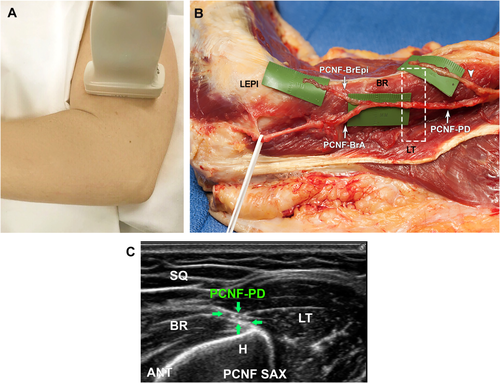
Sonographic appearance of the posterior division of the PCNF (PCNF-PD) within the BR-LT interval. A, The transducer is moved distally in the anatomic axial plane along the posterolateral humerus, distal to the position in Figure 4. B, Cadaveric dissection showing the relationship of the transducer to the underlying anatomy. Bottom is posterior; left, distal relative to the humerus; right, proximal relative to the humerus; and top anterior. C, Correlative short-axis (SAX) sonographic view corresponding to the transducer position depicted by the dashed white rectangle in B. The posterior division of the PCNF (green arrows) is between the BR muscle and anterior to the LT. H indicates humerus; and SQ, subcutaneous tissue. Bottom is medial/deep; left, anterior (ANT); right, posterior; and top, lateral/superficial.
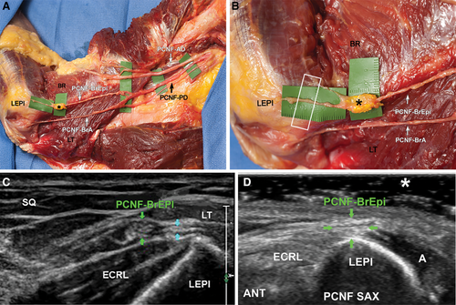
Cadaveric and sonographic views of the posterior division of the PCNF in the region of the LEPI. A, Cadaveric dissection showing the orientation of the posterior division of the as it travels distally toward the LEPI. Latex injectate (asterisk) surrounding the PCNF branch to the epicondyle (PCNF-BrEpi) is noted, with no infiltration to surrounding tissues. PCNF-BrA indicates PCNF anconeus branch; and PCNF-PD, PCNF posterior division. B, Zoomed-in image of the PCNF branch to the epicondyle at the LEPI, documenting successful sonographic identification and injection. Bottom is posterior; left, distal; right, proximal; and top, anterior. C, In this specimen, the PCNF branch to the epicondyle is well seen in the short axis, with a honeycomb appearance representing the hypoechoic nerve fascicles and the interdigitating echogenic fibroadipose connective tissue. A small branch from this branch courses directly over the LEPI (blue arrows). ECRL indicates extensor carpi radialis longus. D, A different example shows the PCNF branch to the epicondyle with a more homogeneous echogenic appearance, blending almost imperceptibly with the subcutaneous adipose tissue. Using a thick layer of gel (asterisk) as a standoff between the transducer and skin and applying a light touch will help in visualizing this branch at this level. SAX indicates short axis; Bottom is deep; left, anterior (ANT); right, posterior; and top, superficial.
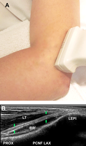
Sonographic long-axis (LAX) image of the posterior division of the PCNF in the supracondylar region, proximal to the LEPI. A, The transducer is placed in the coronal plane along the long axis of the nerve. B, Correlative sonographic image shows the PCNF posterior division (green arrows) traveling between the LT and BR muscles along the supracondylar ridge of the humerus, before becoming subcutaneous in the region of the LEPI. Bottom is anteromedial/deep; left, proximal (PROX); right, distal; and top, posterolateral/superficial.
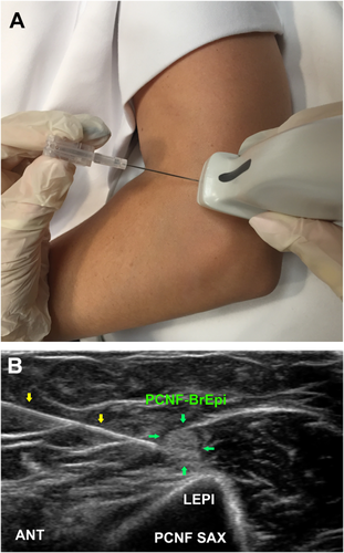
Sonographic short axis (SAX) image of the PCNF branch to the epicondyle (PCNF-BrEpi) proximal to the LEPI. A, Setup for the perineural injection to this branch. The transducer is placed in the anatomic axial plane relative to the arm, along the short-axis of the nerve, with a 22-gauge needle directed in an in-plane anterior-to-posterior approach. B, Sonographic image showing perineural injection to the branch, targeting a small distal epicondylar branch (green arrows) using an in-plane, anterior-to-posterior approach and a 22-gauge needle (yellow arrows). Bottom is medial/deep; left, posterior; right, (ANT); and top, lateral/superficial.


