Circulating miRNAs associated with bone mineral density in healthy adult baboons
Abstract
MicroRNAs (miRNAs) regulate gene expression post-transcriptionally and circulate in the blood, making them attractive biomarkers of disease state for tissues like bone that are challenging to interrogate directly. Here, we report on five miRNAs—miR-197-3p, miR-320a, miR-320b, miR-331-5p, and miR-423-5p—associated with bone mineral density (BMD) in 147 healthy adult baboons. These baboons ranged in age from 15 to 25 years (45–75 human equivalent years) and 65% were female with a broad range of BMD values including a minority of osteopenic animals. miRNAs were generated via RNA sequencing from buffy coats collected at necropsy and areal BMD (aBMD) measured postmortem via dual-energy X-ray absorptiometry (DXA) of the lumbar vertebrae. Differential expression analysis controlled for the underlying pedigree structure of these animals to account for genetic variation which may drive miRNA abundance and aBMD values. While many of these miRNAs have been associated with the risk of osteoporosis in humans, this finding is of interest because the cohort represents a model of normal aging and bone metabolism rather than a disease cohort. The replication of miRNA associations with osteoporosis or other bone metabolic disorders in animals with healthy aBMD suggests an overlap in normal variation and disease states. We suggest that these miRNAs are involved in the regulation of cellular proliferation, apoptosis, and protein composition in the extracellular matrix throughout life; and age-related dysregulation of these systems may lead to disease. These miRNAs may be early indicators of progression to disease in advance of clinically detectible osteoporosis.
1 INTRODUCTION
Research interest in microRNAs (miRNAs) has exploded in the past decade alongside recognition of the potential for these small circulating RNAs to act as biomarkers of disease state or cellular messengers. As posttranscriptional regulators, these small noncoding RNAs are uniquely able to regulate gene expression in the cytoplasm and may travel among cells to do so. This unique ability to travel between cells—sometimes in membrane-bound extracellular vesicles1—allows for the detection of potentially functional miRNAs in the peripheral blood in contrast to other omic technologies like transcriptomics and proteomics which require access to the relevant tissues. The relatively noninvasive nature of peripheral blood miRNA assessments makes them attractive biomarkers for clinical applications. Indeed, a panel of miRNA biomarkers aimed at osteoporosis diagnosis in postmenopausal women is now commercially available.2 In human studies, circulating miRNAs have been implicated in bone formation, turnover and homeostatic maintenance, osteoporosis, and fracture risk.3-7
While a number of studies have focused on the potential of miRNAs as biomarkers for osteoporosis and fracture risk, little is understood about miRNAs in the context of normal variation in spinal bone mineral density (BMD) among healthy adults. Here we report on five circulating miRNAs that are associated with variation in BMD within the healthy range for middle-aged and older baboons.
The baboon is a unique model for age-related bone loss, as it is closely related phylogenetically to humans, is relatively large bodied, and exhibits intracortical remodeling throughout life, unlike rodent models of skeletal aging. The adult bone remodeling and skeletal fracture properties of baboons are also more similar to humans than are other mid- to large-sized mammals.8, 9 Like humans, female baboons undergo bone loss associated with late-in-life hormonal shifts and sex and age influence BMD10 leading to osteopenia in approximately 25% of older females.11 Furthermore, biomechanical properties directly relevant to fracture, including vertebral trabecular bone mechanical properties,12 femoral cortical bone microstructure,13 and femoral bone shape,14 are strongly heritable in the baboon, making pedigreed animals ideal for assessing the effects of genetic and nongenetic factors related to bone biomarkers and bone fragility.15 We leveraged the unique anatomical and genetic advantages of the baboon as a model of aging bone to identify circulating miRNAs associated with BMD across a broad range of mildly osteopenic and healthy adult baboons while controlling for underlying genetic factors.
2 METHODS
2.1 Study population
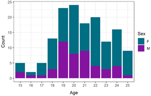
We generated miRNA and BMD data for 147 middle-aged and older baboons (hybrid Papio hamadryas species) drawn from a larger pedigree of baboons housed at the Southwest National Primate Center (SNPRC) at Texas Biomedical Research Institute.16 The sample is 65% female and ranged in age from 15 to 25 years of age at necropsy which is the equivalent of approximately 45–75 human years (Figure 1).
During life, all animals were housed outdoors in large social group cages and maintained on commercial monkey chow to which they had ad libitum access. Animals with medical conditions known to influence bone metabolism (e.g., diabetes, chronic renal disease) or a history of traumatic fracture were excluded. No animals were sacrificed for this study; all were euthanized for other reasons. All animal procedures were approved by the Institutional Animal Care and Use Committee at Texas Biomedical Research Institute and conducted in Association for Assessment and Accreditation of Laboratory Animal Care approved facilities at the SNPRC. Blood samples were drawn in EDTA tubes at necropsy and processed buffy coat layers stored at −80°C until RNA was extracted. Lumbar spines were frozen immediately after necropsy and stored at ‒20°C until thawed for analysis.
2.2 Dual-energy X-ray (DXA)
DXA absorptiometry scans were performed postmortem on thawed lumbar vertebrae17, 18 using a Lunar DPX 6529 (General Electric) scanner and areal BMD (aBMD) was determined in the anterior-posterior (AP) direction for L4-L2 using the manufacturer's software for adults. To account for sexual dimorphism in aBMD, we performed z-scoring on male and female aBMD values separately and combined the scaled values for analysis.
2.3 miRNA sequencing and analysis
Total RNA was isolated from buffy coat using TRIzol (Invitrogen) and the Qiagen miRNeasy Mini Kit as described previously.19 RNA quality was assessed using an Agilent Bioanalyzer 2100 with RNA Integrity Number (RIN) values greater than or equal to 8.0 for all samples. RNA was enriched for small noncoding RNAs (sncRNAs) by using the Ambion mirVana miRNA Isolation Kit. Complementary DNA (cDNA) libraries were generated with the Illumina Small RNA Prep Kit v1.5 following the manufacturer's protocol and sequenced in Illumina's Genome Analyzer (GAIIx).20 mirDeep221 was used to align reads to the known human miRbase version 2122, 23 mature and hairpin miRNAs and to identify novel miRNAs. We aligned to the human miRNAs because miRNAs remain relatively poorly annotated in all nonhuman primate species. We required a log-odds score of 4 or greater for sequence matching to ensure that miRNAs with low homology between humans and baboons were not included in the analysis.
Raw miRNA counts were analyzed using the R statistical package DESeq2 to identify individual miRNAs associated with aBMD.24, 25 DESeq2 has the advantage of incorporating shrinkage estimators for dispersion and fold change which better handles low-abundance miRNAs compared to other methods. After z-scoring, weight and age were no longer associated with aBMD so these values were not included as covariates. miRNAs significantly associated with aBMD at a false discovery rate (FDR) < 0.05 were further tested to determine if this association was driven by underlying genetic variation in the pedigree. A likelihood ratio test was calculated to compare linear mixed-effects models for z-scored aBMD as the outcome variable fit with lmekin in the coxme R package26 with and without miRNA abundance as a fixed effect while including pedigree-based kinship as a random effect.
2.4 miRNA target prediction
A major challenge in understanding the potential functional role of a circulating miRNA is that most are computationally predicted to bind dozens of different mRNAs. To better evaluate roles these circulating miRNAs could play in bone biology, we used the R package multiMiR27 to query three experimentally validated miRNA target databases—miRecords,28 miRTarBase,29 and TarBase.30 To be considered a potential target, we required experimental validation of miRNA-mRNA interaction via luciferase, quantitative reverse transcription-polymerase chain reaction, or western blot experiments as well as the presence of the target protein in baboon bone (Table S1).
3 RESULTS
3.1 Bone mineral density
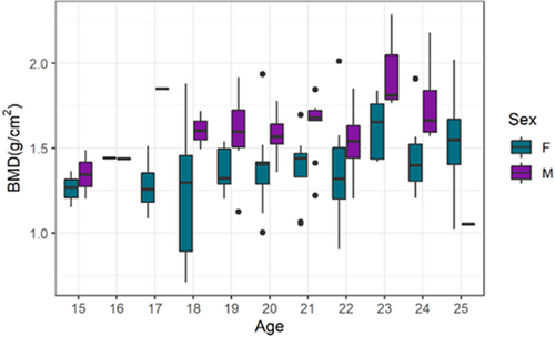
Baboons exhibit high levels of sexual dimorphism in weight and body size leading to significant differences in mean aBMD (0.22 g/cm2, p = 1 × 10‒6, Figure 2). The distribution of aBMD in these animals is consistent with previously published reference standards from more than 650 baboons.10 Within the 10-year agespan of our study cohort, there is no significant decline in aBMD. Nevertheless, the distribution of aBMD across the sample is broad, ranging from 4.3 standard deviations below to 8.4 standard deviations above the healthy, young adult mean for females and ‒3.5 to +9.3 standard deviation for males.
3.2 miRNAs associations
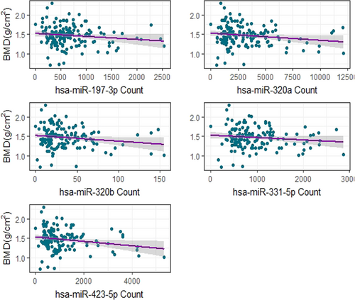
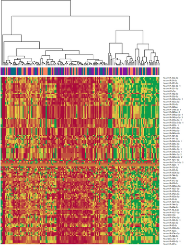
After FDR correction, 15 miRNAs were significantly associated with aBMD in the DESeq2 analysis of which five were associated (likelihood ratio test p < 0.1) after correcting for the genetic relatedness of the animals (Table 1). All five of these highly associated miRNAs—miR-197-3p, miR-320a, miR-320b, miR-331-5p, and miR-423-5p—are negatively correlated with aBMD (Figure 3), suggesting that increased levels of these miRNAs, which would be expected to decrease mRNA expression, are indicative of a decline in aBMD. Individually, these miRNAs do not account for a large proportion of the overall variance in aBMD as highlighted by the broad distribution of aBMD among individuals with similar miRNA counts. However, these miRNAs are part of broader patterns of differential miRNA expression (Figure 4).
| β | Mean Expression | p | LRT p | |
|---|---|---|---|---|
| hsa-miR-423-5p | ‒0.28 | 886 | 2.4×10‒4 | 0.03 |
| hsa-miR-320a | ‒0.22 | 2561 | 3.9×10‒3 | 0.04 |
| hsa-miR-339-5p | ‒0.19 | 1697 | 6.2×10‒3 | 0.22 |
| hsa-miR-3173-5p | ‒0.38 | 16 | 3.0×10‒2 | 0.14 |
| hsa-miR-371a-5p | 0.6 | 7 | 3.0×10‒2 | 0.40 |
| hsa-miR-16-2-3p | ‒0.44 | 137 | 3.3×10‒2 | 0.27 |
| hsa-miR-197-3p | ‒0.18 | 577 | 3.5×10‒2 | 0.09 |
| hsa-miR-21-5p | 0.16 | 30,037 | 3.5×10‒2 | 0.68 |
| hsa-miR-331-5p | ‒0.09 | 808 | 3.5×10‒2 | 0.07 |
| hsa-miR-374a-3p | 0.26 | 231 | 3.5×10‒2 | 0.88 |
| hsa-miR-374a-5p | 0.19 | 4255 | 3.5×10‒2 | 0.92 |
| hsa-miR-424-3p | ‒0.16 | 86 | 3.5×10‒2 | 0.10 |
| hsa-miR-484 | ‒0.13 | 2334 | 3.5×10‒2 | 0.19 |
| hsa-miR-320b | ‒0.21 | 28 | 3.6×10‒2 | 0.05 |
| hsa-miR-574-3p | ‒0.16 | 94 | 4.1×10‒2 | 0.25 |
- Note: Bolded miRNAs remained significantly associated after correction for pedigree structure.
3.3 Predicted miRNA targets
Based on our screening criteria, we identified 60 genes present in bone and experimentally validated as potential targets of our five significant miRNAs (Table S2). Analysis in DAVID31 identified several significantly enriched functional annotations within the 60 genes. The top five enriched gene ontology terms representing more than 10% of the genes on the list are myelin sheath (26.4-fold enrichment, FDR = 6.1 × 10‒10), focal adhesion (11.2-fold enrichment, FDR = 2.9 × 10‒7), extracellular matrix (11.1-fold enrichment, FDR = 3.6 × 10‒5), cadherin binding involved in cell-cell adhesion (9.5-fold enrichment, FDR = 9.0 × 10‒4), and cell–cell adherens junction (9.0-fold enrichment, FDR = 5.4 × 10‒4). Additionally, these putative targets include seven genes previously linked to osteoporosis in genome-wide association studies based on data drawn from the Public Health Genomics and Precision Health Knowledge Base (v7.1)32 (Table 2).
| Gene name | miRNA |
|---|---|
| FGB | hsa-miR-197-3p |
| IGFBP5 | hsa-miR-197-3p |
| SOD1 | hsa-miR-197-3p |
| SOD2 | hsa-miR-197-3p |
| hsa-miR-331-5p | |
| GAPDH | hsa-miR-320a |
| hsa-miR-423-5p | |
| MIF | hsa-miR-320a |
| MMP9 | hsa-miR-320a |
4 DISCUSSION
Most previous research on bone density and miRNAs has focused on patients with osteoporosis or other disorders of bone metabolism rather than aBMD variation among healthy individuals. This bias in the literature is apparent in the fact that many of our healthy aBMD-associated miRNAs have been suggested as potential clinical biomarkers in humans.
Of the miRNAs we identified, literature linking the miRNA-320 family to bone fragility is the most robust. miR-320b is more common in women with a recent fracture compared to age-matched controls.33 This may be due to its role in osteoblast differentiation. miR-320b overexpression prevents osteoblast differentiation, while inhibition promotes bone matrix mineralization and differentiation via BMP-2.34 Additionally, researchers studying low-energy fractures in premenopausal, postmenopausal, and idiopathic osteoporosis patients found miR-320a was upregulated in these patients compared to controls35. Prior analysis of trabecular bone tissue showed significant dysregulation of miR-320a in patients with osteoporosis, possibly via regulation of osteogenesis by targeting bone-forming genes such as CTNNB1 (B-catenin) and RUNX2.36 These findings led to the inclusion of miR-320a on the commercially available OsteomiR panel (Tamirna).
Similarly, in a study of plasma miRNAs associated with fracture risk, miR-423-5p expression was significantly negatively associated with FRAX score, but not BMD, in osteoporosis patients.37 This finding was reinforced by research on facial bone atrophy where miR-423-5p was shown to promote bone proliferation via prevention of apoptosis in mesenchymal stem cells.38 While there is little additional evidence of a role for this miRNA in bone, it has been linked to the regulation of apoptosis in cardiomyocytes,39 kidney cells,40 retinal pigment epithelial cells,41 and colon cancer cells.42
Circulating miR-331-5p levels have been identified as a potential biomarker for osteoporosis and subsequent bone fracture,43 although little is known about the role of this miRNA in bone. Hints to its function come from studies of vascular smooth muscle cells, where miR-331-5p is induced by BMP2-PPARγ signaling to regulate cellular proliferation.44 We predict a role for miR-331-5p in regulating SOD2 expression which directly induces a BMP2 response under hypoxic conditions.45
While miR-197-3p has not previously been linked to adult bone metabolism, it was identified in the downregulation of osteogenesis in human amniotic membrane-derived mesenchymal stem cells due to its suppression of SMAD2 in the TGF-β pathway during osteoblast differentiation.46 We predict that miR-197-3p may target expression of both SOD1 and SOD2, antioxidative enzymes that respond to vitamin D levels 47 and are thought to play a critical role in the regulation of cellular senescence.48, 49
Taken together, our aBMD-associated miRNAs and their putative targets point toward the regulation of extracellular matrix proteins, apoptosis, and cell proliferation—three key components in the maintenance of bone homeostasis. Our findings demonstrate the overlap in miRNAs associated with bone mineral density and related traits in humans and nonhuman primates, highlighting the utility of this model for understanding aging bone. The association of these miRNAs with variation in areal bone mineral density within the healthy, nonosteoporotic, range suggests they may be useful in identifying the earliest stages of preclinical metabolic shifts in bone. While the impact of epigenetic regulation of gene expression is evident in bone tissue, the use of miRNAs as biomarkers for low aBMD and high fracture risk is still relatively new. It is apparent more research is needed to better understand these molecular pathways. Future work should focus on identifying the presence and functional role of these miRNAs in bone tissue to solidify their promise as biomarkers.
ACKNOWLEDGMENTS
The authors would like to thank Lorena Havill for her role in the development of this project. This study was supported by the R01 AR064244 to Todd L. Bredbenner and K01 AG056663 to Ellen E. Quillen. Care of animals in the SNPRC pedigree was supported by grant P51 OD011133.
AUTHOR CONTRIBUTIONS
Ellen E. Quillen, Laura A. Cox, and Todd L. Bredbenner designed the study. Jaydee Foster, Anne Sheldrake, and Jeremy Glenn acquired the data. Ellen E. Quillen and Maggie Stainback drafted the manuscript. All authors approved the final version of the manuscript.


