Donor age and sex have limited effects on the mechanical and microstructural properties of human connective tissues
Abstract
Connective tissues, such as tendons, ligaments, and capsules, play a large role in locomotion and joint stability and are often subjected to traumatic injuries and degeneration. The purpose of this study was to evaluate if the mechanical and microstructural properties of connective tissues correlate with the age and sex of the human donor. Dissected samples were prepared for mechanical testing, consisting of 10 cycles of preconditioning, a stress-relaxation ramp and hold, and a quasi-static ramp to failure. During the testing protocol, the microstructural organization of tissues was analyzed using quantitative polarized light imaging. A linear mixed model was used to assess whether tissue type, donor age, or donor sex were significantly associated with mechanical and microstructural tissue properties. Tissue type had a significant effect on all parameters, while donor age and sex did not. Groupings by tissue type (i.e., tendon vs. ligament vs. capsule) were evident for microstructural data, with tendons having a tighter grouping and ligaments having a larger spread of values. The interaction of tissue type and age yielded a significant effect for linear modulus only (p = 0.007), with the palmaris tendon appearing to have the largest contribution to this effect. There were no significant interaction effects between sex and tissue type or donor age. Donor age appears to affect linear modulus in some, but not all, tissue types. Otherwise, age and sex do not have significant effects on the mechanical and microstructural properties of the range of connective tissues that were analyzed in this study.
1 INTRODUCTION
Tendon and ligaments are vital structures in locomotion and joint stability and are subject to traumatic injury and chronic degeneration. The mechanical and microstructural properties of these tissues are clinically relevant in terms of their vulnerability to injury and disease as well as their role as grafts for reconstructive procedures. Several past animal studies have shown differences in mechanical and material properties of various tissues based on age and sex.1-3 However, while the findings of these animal studies are valuable, the translation to human tissues is not a given, emphasizing the importance of studying human soft tissue. Previous studies investigating human soft tissues have shown some variance in tissue properties with age, although these changes were typically not statistically significant.4-7 As a result, it is difficult to know if results from biomechanical research performed using more readily available older specimens are generalizable to younger patients. In addition, the effects of donor sex on tissue properties are not well understood.
Adding to the complexity of understanding structure-function relationships of connective tissues is the variety of anatomical locations within the body. Collagenous tissues like tendons, ligaments, and capsule tissue are present throughout the human body and in every extremity. Following injury, native tissues in the knee such as the anterior cruciate ligament (ACL) and posterior cruciate ligament (PCL) are typically reconstructed using tissues from several other locations in the lower extremity.8 When torn, the ulnar collateral ligament (UCL) in the elbow is often reconstructed with grafts from the forearm or the lower extremity.9 Graft choice is typically made after considering several factors, including but not limited to, primary versus revision, donor-site morbidity, potential bone integration, and surgeon preference.10, 11 Since grafts are used from varying locations that often differ from the native tissues they are being used to replace, it becomes critical to understand how material properties vary by location and tissue type, particularly related to age and sex, to optimize graft-based surgical reconstruction.
Considering the aforementioned roles of connective tissue as clinical grafts and in biomechanical studies, understanding variations in the mechanical and microstructural properties of tendons, ligaments, and capsular tissues are important to optimize graft selection and properly interpret biomechanical studies of cadaveric tissues. The current study evaluated a collection of previously published data that applied a polarized light imaging technique to measure the dynamic microstructural properties of several human soft tissues of interest, while simultaneously quantifying mechanical properties of tissues under load.12-18 This new analysis sought to assess the association of age, sex, and tissue type with material properties and test the hypothesis that age and sex will significantly influence the microstructural and biomechanical properties of cadaveric connective tissues.
2 METHODS
2.1 Sample acquisition
All sample acquisition, sample preparation, mechanical and microstructural testing, and data collection were completed in previously published studies.12-18 Samples of 11 different tissues were acquired from a total of 91 donors (48 females, 43 males) with an average age of 56 ± 16 years. The examined tissues, harvested from both the arm and leg (Figure 1), included five ligaments (ACL, PCL, UCL, lateral collateral ligament [LCL], anterolateral ligament [ALL]), five tendons (bone-patellar tendon-bone [BPTB], hamstring tendon [HS], quadriceps tendon [QT], palmaris [PL], and gracilis [GR]), and one capsule (anterolateral capsule [ALC]). These sample groups do not comprise a comprehensive set of connective tissues, but instead represent a subset of clinically relevant soft tissues that were previously studied to address tissue-specific structure-function hypotheses. The motivation to combine these data in the present study was to enable a deeper examination of general structure-function relationships using a data set that spanned multiple tissue types, donor ages, and both sexes. A breakdown of total sample numbers for each tissue type is shown in Table 1 (note: multiple tissues were collected from single donors). Samples were acquired with no identifiable patient information; therefore, Institutional Review Board approval was not required.

| Tissue type | Male | Female | Average age | Age range |
|---|---|---|---|---|
| BPTB | 2 | 5 | 44 ± 6 | 35–50 |
| QT | 6 | 4 | 51 ± 13 | 24–68 |
| HS | 2 | 6 | 45 ± 6 | 35–50 |
| GR | 2 | 6 | 45 ± 6 | 35–50 |
| PL | 8 | 5 | 65 ± 10 | 44–81 |
| UCL | 14 | 18 | 67 ± 17 | 37–97 |
| ACL | 7 | 4 | 41 ± 9 | 24–53 |
| PCL | 8 | 5 | 43 ± 4 | 36–48 |
| LCL | 5 | 11 | 45 ± 7 | 36–59 |
| ALL | 5 | 11 | 45 ± 7 | 36–59 |
| ALC | 5 | 11 | 45 ± 7 | 36–59 |
2.2 Dissection and mechanical testing
Limbs were acquired from several different donor programs and stored at −20°C before use. After limb acquisition, specimens were thawed at room temperature for 24 h before tissue harvesting. Great care was taken during the dissection and extraction processes to ensure samples were kept hydrated with phosphate-buffered saline (PBS) and freeze-thaw cycles were kept to a minimum. When possible, samples were dissected into multiple regions (e.g., BPTB samples were split into lateral, central, and medial thirds) for a more in-depth analysis of region-specific mechanical and microstructural properties. Detailed information on the dissection techniques of each individual tissue type are included in previous publications.12-18 Once harvested, all tissues underwent the same sample preparation procedure before testing. Samples were thinned on a freezing stage (Physitemp Instruments) sledge microtome (Leica 1400, Leica) to a thickness of 0.6-1.0 mm to ensure ample light penetration for collection of the QPLI data: degree of linear polarization (DoLP) and angle of polarization (AoP). During the laser measurement portion of the sample preparation, cross-sectional area was also determined for use in later calculations of modulus and stress. After thinning, sandpaper tabs were attached to the ends of each sample for increased friction between the tissue and clamp interface and four small beads (0.8-mm diameter) were glued to the surface for optical strain tracking. Samples were then submerged in a tissue bath containing PBS and loaded into a mechanical testing machine (TestResources). The mechanical testing protocol consisted of a preload to 0.1 N, 10 cycles of preconditioning between 1.5% and 4.5% clamp-to-clamp strain, a ramp-and-hold stress-relaxation test at 5% strain for 300 s, and a ramp to failure at a 1% strain/s. Mechanical and microstructural data were simultaneously collected during testing (i.e., toe modulus, linear modulus, transition stress, transition strain, AVG DoLP, and STD AoP). DoLP measures the strength of alignment on a pixel-by-pixel basis. We report the average DoLP across the selected region of interest (ROI), where small AVG DoLP values (~0) represent unaligned collagen and larger values represent greater strength of alignment. AoP values represent the angle of collagen (averaged through the thickness) on a pixel-by-pixel basis for the ROI. We report the standard deviation (STD AoP) which indicates the spread of the histogram of angular values of pixels in the ROI. Samples with low STD AoP have more aligned collagen (i.e., smaller variation) while samples with large STD AoP have less aligned collagen (i.e., larger variation). Toe modulus is the slope of the stress-strain curve during the early portion of the ramp to failure (before the transition point) and the linear modulus is the slope of the linear portion at higher strain values (after the transition point). The transition stress and strain values represent the point of the stress-strain data when the curve transitions from early loading to full engagement. Analysis and results of mechanical and microstructural data of the 11 tissues were published previously.12-18
2.3 Statistical analysis
A primary goal of this study was to determine if there were any statistical effects of donor age, sex, or tissue type on the measured mechanical and microstructural properties. A linear mixed model was used to compare the impact of age and sex across tissue types for various output parameters (JMP, SAS Institute Inc.). The linear mixed model accounted for within-tissue correlation (i.e., multiple subsamples from a single tissue) and within-donor correlation (i.e., multiple tissues from the same donor) since some donors provided multiple tissue samples. The statistical analysis evaluated whether direct factors (e.g., tissue type, age, and sex) or interactions (e.g., tissue type*age, tissue type*sex, and age*sex) had statistically significant effects on each of the measured parameters.
2.4 Additional data analysis
Based on outcomes from the statistical analysis (see Section 3), several additional approaches were employed to further examine differences by tissue type. To determine how similar or different each tissue type was to the overall data set, normalized heat maps were created using group-averaged values (GraphPad Prism 9, GraphPad Software, www.graphpad.com). For each parameter, an overall average value was computed across all tissue types, then used to normalize each specific tissue's parameter value. In the resultant heat maps, standard deviation was used to create a color scale which indicates how close the tissue was to the overall average. White boxes indicate values near the overall average, with blue coloring indicating that specific tissue type's parameter value was below the average (lighter blue = closer to average, darker blue = further from average), and red coloring indicating a value above the overall average (lighter red = closer to average, darker red = further from average). The data from several tissues (i.e., UCL, ACL, and PCL) are displayed in the heat maps as separate tissue bands/bundles rather than as grouped whole tissues (UCL: anterior and posterior band; ACL: anteromedial and posterolateral bundles; PCL: anterolateral and posteromedial bundles) because previous results indicated significant differences between parameter values for each subsection.12-18 Results for other tissues that were split into sub-regions for testing are shown as grouped data due to more homogeneous parameter values across tissue regions (i.e., BPTB, QT, HS, GR, PL, and LCL). ALL and ALC samples were originally tested as single regions and thus are also displayed on the heat maps as single regions. Heat maps were generated for mechanical and microstructural data typically recorded for this testing protocol and for measures of dynamic collagen realignment during mechanical loading. Specifically, changes in the microstructure between zero and the transition point (Z-T) and between the transition point and linear region (T-L) during the ramp to failure were computed to quantify the relative amount of microstructural reorganization that occurs in the early and late portions of loading, corresponding roughly to the toe and linear modulus, respectively. In addition, data for AVG DoLP and STD AoP were averaged for the three strain levels (zero, transition, and linear) and averaged for each individual tissue type. The AVG DoLP was then plotted against the STD AoP to show microstructural changes through time during the ramp. Data were color-coded for each individual tissue type and then color-coded to show groupings of tendons, ligaments, and capsules.
3 RESULTS
3.1 Effects of age, sex and tissue type
A summary of the statistical analysis shows results for the evaluation of individual factors (i.e., tissue type, age, and sex) and interaction effects on each mechanical and microstructural parameter (Table 2). Tissue type had a significant effect on all evaluated parameters (p < 0.001), while age and sex exhibited no significant effects on any parameter. The interaction of tissue type*age showed a significant effect for linear modulus (p = 0.007) but no other mechanical or microstructural parameters. Since this was the only significant effect (besides tissue type alone), linear modulus values were plotted versus donor age (Figure 2) to explore this interaction further. To this end, linear moduli for each individual sample were plotted, with data points color-coded based on tissue type along with group-specific linear regressions showing 95% confidence intervals. This plot is shown as two panels, with panel A containing tendon samples and panel B containing ligament and capsule samples. For most tissue types, linear modulus values exhibited flat curves (no effect) when plotted versus age. However, there were a few tissues (e.g., PL) that demonstrated increasing or decreasing curves with age (i.e., showed slight age effects). Other interaction effects of tissue type*sex and age*sex showed no statistically significant effects for any parameter that was analyzed.
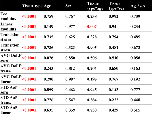 |
- Note: Individual fixed effects as well as paired fixed effects are shown and any significant differences (p < 0.05) are red. Overall, there were few significant correlations, which may have been potentially skewed by one or two tissue types only.

3.2 Normalized data in heat maps
Toe and linear moduli (Figure 3) show that BPTB, QT, GR, PL, PCL (both bundles), and LCL had values that were above average, while HS, UCL (both bundles), ACL (both bundles), ALL, and ALC exhibited below-average values. This pattern was consistent for AVG DoLP and STD AoP values as BTPB, QT, GR, PL, and LCL were more aligned than average (i.e., larger AVG DoLP values and smaller STD AoP values), while UCL, ACL, ALL, and ALC were less aligned. STD AoP values typically have an inverse relationship with AVG DoLP values (i.e., less spread in the distribution of angular orientation typically corresponds with greater strength of alignment in those directions), which was confirmed by examination of the parameter heat maps (Figure 3). For example, where AVG DoLP values were below average (i.e., blue squares), STD AoP values were conversely above average (i.e., red squares). For transition strain and stress, the UCL AB and PL values appeared to be closest to average, while PCL and BPTB exhibited values that were the most above and below average, respectively. The measures of transition stress and strain indicate the tissues ability to deform or undergo microstructural realignment under early loading. Higher values indicate tissues that are less susceptible to quick failures. Considering all parameters, QT and LCL samples exhibited the largest moduli and most strongly aligned microstructural properties. In contrast, the weakest and least aligned tissues within this data set were the ALL and ALC, as their moduli were well below average, with alignment data that also appeared to be the furthest from the average.

The HS, UCL AB, and LCL all exhibited less change in AVG DoLP and STD AoP compared to an average sample during early (Z-T) and late loading (T-L; Figure 4). Samples such as BPTB, PL, UCL PB, and PCL AL showed larger values for Z-T boxes for AVG DoLP compared to corresponding T-L, indicating larger changes in early loading than later loading within these tissue types. Similarly, QT, PCL AL, ALL, and ALC showed larger changes in STD AoP in the Z-T compared to T-L, demonstrating more collagen realignment during the toe-region compared to the linear region of the stress–strain curve. For the latter portion of the test (T-L), many tissue types exhibited light colored boxes indicative of average amounts of change, while some samples (e.g., QT, HS, ACL AM, and LCL) showed at least one dark T-L box indicating larger changes compared to the average.

3.3 Tissue type comparisons
A simultaneous consideration of AVG DoLP and STD AoP values (Figure 5A) yielded additional insights into differences in microstructural alignment by tissue type as well as unique patterns of reorganization under load (i.e., as QPLI values changed from zero to transition to linear). The typical inverse relationship between DoLP and AoP values was evident, where increasing strength of alignment (i.e., larger AVG DoLP values) corresponded to decreasing spread of angular orientation (i.e., smaller AoP values). Qualitatively, each tissue type demonstrated fairly unique microstructural alignment and loading-dependent behavior, with data points appearing in different locations of the QPLI dataspace (Figure 5B). Data for tendons were bunched more closely together compared to the vast spread evident in the various ligament samples. Also, relatively close proximity of datapoints was observed for some tissue groups (e.g., ACL-AM vs. ACL-PL, PCL-AL vs. PCL-PM, ALL vs. ALC), demonstrating similar organization between these samples. In terms of microstructural realignment under load, datapoints for some tissues did not change much from one point to the next (e.g., ALL, ALC UCL). In contrast, some tissues changed fairly consistently between the Z-T and T-L portions of the stress-strain curve (e.g., ACL and PL), while a majority of the tissues exhibited substantial changes from zero to transition but only minor reorganization (i.e., change in DoLP and AoP values) from transition to linear (e.g., PCL, HS, QT, BPTB, GR, and LCL). In the latter case, these samples appeared to undergo most of their microstructural alignment during the early/low-strain region of the loading curve before limiting reorganization during the late/high-strain loading, which may be indicative of their in vivo functional requirements.

4 DISCUSSION
Contrary to our hypothesis, donor age and sex do not significantly impact the majority of measures in this study. The analysis across tissue types demonstrated relatively little effect of age or sex on the microstructural and biomechanical properties of human soft tissues. These findings are consistent with prior investigations which have also found little or no sex-based differences in mechanical properties.19, 20 The lack of any age associated differences also align with previous studies which show limited to no differences caused by age.21, 22 Tissue type showed statistically significant effects on all evaluated parameters. Thus, tissue type was the primary factor that was responsible for variation in material properties considered in this study. It is possible that the properties of other tissues not tested in this study could vary with age and/or sex. One study found differences in properties across age in the Achilles tendon, which was not included in our current study23 Several animal studies of the Achilles and supraspinatus tendons have also found age and sex related differences.1-3
Qualitatively comparing the full data set, there were no obvious patterns that distinctly separated out samples by tissue type (i.e., tendon, ligament, and capsule), particularly for AVG DoLP and STD AoP values. However, the tendon samples demonstrated more consistent values as a group than the wide range of responses observed for different ligaments. For example, within ligament samples, the UCL PB showed very little microstructural reorganization between the zero, transition and linear portions of the stress-strain curve. In contrast, the PCL AL exhibited a large change in QPLI parameters from the zero to transition point, then no change in organization from the transition to linear points (Figures 4 and 5). Such data demonstrate that two different ligaments can have vastly different microstructural properties and dynamic realignment with loading, perhaps due to adaptations to withstand physiological loading depending on their unique anatomical locations. The UCL may be more of a checkrein ligament which stabilizes the joint and undergoes little changes during normal motion, while the ACL and PCL see much larger loads and changes with typical activity.14-16 This illustrative example of very different properties in two ligaments from disparate anatomic locations could help explain at least in part why there is a wider range of values across ligaments.
The evaluated tendons, on the other hand, demonstrated relatively similar microstructural properties and dynamic realignment (Figures 3-5). Tendons showed much more uniform values for AVG DoLP and STD AoP as a group than the ligament samples (Figure 3), while also exhibiting larger initial changes in microstructure and small changes once the transition point had been reached (Figure 5B; tendons grouped closer together in the upper left of the plot). Unlike ligaments, which appear to have more variable properties depending on anatomical locations, the tendons we tested did not appear to change as dramatically with varying anatomical location (e.g., GR and BPTB have different anatomical locations/orientations but very similar microstructural properties).
Differences in mechanical properties across various anatomic locations may result from a number of factors such as differences in mechanical loading, cellular response to stress and local environment (i.e., synovial vs. extra-synovial). Unfortunately, the present study cannot identify all of the possible factors or assess their contribution to this variance. Further research on these tissue types is necessary to better understand the factors contributing to differences across anatomic locations. Another unanswered question is why ligaments have a wider range of values than tendons. While both types of tissue vary greatly in shape, length, and composition in vivo,24, 25 shape and length were all made consistent for testing and QPLI imaging in this study, suggesting that compositional differences should be the reason for the disparity between ligaments and tendons.
While biomechanical studies may desire to investigate samples across a wide range of demographical backgrounds (e.g., varying age and sex), this is not always feasible given limitations on human donor availability, cost, and realistic sample numbers. Therefore, the lack of effect of age or sex on mechanical and microstructural properties shown in this study is an important finding. As a result, biomechanical studies of cadaveric tissues are unlikely to suffer from large bias due to donor age or sex, and such studies are likely to still be relevant even if the donor demographics (e.g., age and sex) vary from the patient population of interest. These findings may also have value for clinicians performing surgical repair or reconstruction of connective tissues. For example, samples from donors with different demographics compared to recipient patients (e.g., varying age or sex) may not negatively impact the overall success of the procedure.
The results of this study also highlight the variability of mechanical and microstructural properties between tissue types, which is important to consider when evaluating graft selection for reconstruction techniques. These data provide insight into how closely graft properties match the native tissues they are commonly used to replace. For example, our previous study showed that linear modulus was larger for QT and BPTB (both tendons) than the two ligaments they are commonly used to replace, namely the ACL and PCL.18 Other such examples can be observed from the composite data set presented herein (e.g., Figures 3-5). While microstructural properties can and should be taken into consideration when choosing a graft, they are obviously not the only factor for making that decision. Examination of differences between tissue types is important for considering time-zero mechanical and microstructural properties. These results also suggest that donor age and sex may be less important in terms of graft selection.
This study was not without limitations. First, samples were removed from their anatomical location and thinned to allow for light transmission. While this was done so we could collect quantitative microstructural data, tissues were no longer in their anatomical location/orientation. However, since this study compared the material properties and the same procedure was used for all samples, this aspect of the protocol does not negatively impact the results. Second, data were not collected specifically for the current study and thus, the analyzed samples do not form a complete set of data across all ages within each tissue. Additionally, some samples came from a narrower middle-age range, while for other tissues (i.e., PL), samples were from the mid to upper age range. The inconsistent range in ages for each tissue could potentially skew the data; however, if additional work permitted increasing the age range of all tissues, the single observed effect from age might decrease or even disappear. Only a single type of capsule tissue was evaluated across all our previous studies; thus, no definitive conclusions could be drawn about capsule tissues in general compared to tendons and ligaments. While there was no correlation of age with the material properties we studied, it is possible a nonlinear relationship exists. Finally, all cadaveric donor samples were from skeletally mature donors; properties of tissue from skeletally immature donors may differ significantly. Future studies could be used to confirm these results and explore additional questions.
5 CONCLUSION
The results of this study demonstrate that the mechanical and microstructural properties of connective tissues from skeletally mature cadaveric donors were not significantly affected by age or sex. In contrast, evaluated samples showed a diverse range of properties based on tissue type. The unique properties of each tissue type could be a result of distinct anatomical locations and different physiological loading requirements. Further research into these tissues with a more complete range of ages for each sex would help confirm the results seen in this study. A better understanding of mechanical and microstructural properties enables clinicians and investigators to better assess and treat complex clinical questions and problems.
AUTHOR CONTRIBUTIONS
All authors have read and approved the final submitted manuscript. Ryan M. Castile contributed to research design, acquiring and analyzing data, and drafting bulk of manuscript. Paul C. Cannon contributed to data analysis and creation of statistical models. Matthew V. Smith contributed to research design, result interpretation, and manuscript draft. Robert H. Brophy contributed to research design, result interpretation, and manuscript draft. Spencer P. Lake contributed to research design, interpretation of data, and manuscript draft.