Relation between diameter of a lateral screw and pull-out strength in distal clavicle fracture in plates with different geometry: A cadaveric biomechanical study
Abstract
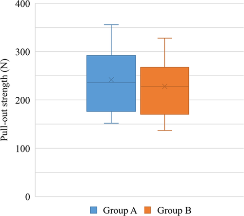
Plate fixation has recently gained popularity among the various surgical methods used to treat Neer type II distal clavicle fractures. The use of a low-profile distal clavicle locking plate is logically considered a better option when there is no significant difference in the fixation strength between insertions of 3.5- and 2.7-mm diameter screws. Therefore, the purpose of this biomechanical study was to investigate any differences in fixation strength among varying sizes of screws that are used to treat distal clavicle fractures. The study was performed with 20 paired shoulder girdles from 10 fresh frozen cadavers. To create a type IIA fracture of Neer classification, osteotomy was performed perpendicularly to the longitudinal axis of the clavicle at the medial end point of the conoid ligament. Two custom-made fixtures designed to be attached to both upper and lower sides of the Instron were fabricated for the evaluation. The mean maximum pull-out strength for fixation using 3.5-mm diameter screws was 241.9 ± 67.8 N, whereas the mean pull-out strength in fixation with 2.7-mm diameter screws was 228.1 ± 63.0 N. There was no statistically significant difference between the two groups. Distal fragment fixation with distal clavicle locking plates using two 2.7-mm diameter screws showed comparable biomechanical pull-out strength at the time-zero setting to fixations with a hook plate using two 3.5-mm diameter screws. Therefore, the fixation of the distal fragment with a low-profile plate and 2.7-mm screws may be preferred as an alternative option if the distal fragment of the fractured clavicle is not extremely small.
1 INTRODUCTION
Clavicle fractures account for 2.6% of all fractures and 44% of fractures involving the shoulder girdle.1-4 Of these clavicle injuries, the fractures involving the distal portion comprise about 15% and are the second most frequently injured site after injuries involving mid-shaft portion of the bone.5, 6 Among distal clavicle fractures, those that are type II of Neer classification have considerable displacement and are unstable. Such fractures are indicated for surgical treatments since nonoperative treatments of type II fractures have been shown to result in up to 47% malunion and 22%–45% nonunion in previous studies.7-9
There have been many descriptions of various surgical methods to treat Neer type II distal clavicle fractures. Among them, plate fixation has become more popular in recent times. There are two types of plates that can be used for a distal clavicle fracture. One type contains a hook attached to the distal portion of a plate, and the other type utilizes locking head screw fixation without an attached hook.
The most common screw sizes used in clavicle fractures are 2.7 and 3.5 mm in diameter, and distal clavicle locking plates have been designed to use 2.7-mm diameter screws so that multiple screw insertions can be used in the distal portion. Generally, it is known that screws with a larger diameter provide more fixation strength.10-13 The distal clavicle fracture is unique in that it is close to a joint where multiple structures, such as the acromioclavicular (AC) and coracoacromial (CC) ligaments, are inserted; it differs from mid-shaft clavicle fractures and even from other long bone fractures.14, 15 Therefore, due to the unique characteristic of the distal clavicle fracture, most fixation failures seem to arise from pull-out of screw rather than plate breakage,16-18 and it is difficult to predict the fixation strength of screws with varying diameters. To date, there has been no evaluation to measure the strength of screws with different diameters.
In Neer type II distal clavicle fractures, if there is no significant difference in the fixation strength between insertions of 3.5- and 2.7-mm diameter screws, then the use of the low-profile distal clavicle locking plate is considered a better option, since this low-profiled plate may diminish the incidence of implant-related skin irritation and lessen the subsequent need for removal. However, 3.5-mm diameter screws can also be considered in lateral fixation to provide stronger fixation strength, despite the presence of several disadvantages found in this particular screw size.
The purpose of this biomechanical study was to investigate any differences in fixation strength among different sizes of screws and plates that are used to treat distal clavicle fractures. The hypothesis of this study was that 2.7-mm diameter screws used for fixation of the distal clavicle locking plate would provide comparable pull-out strength to that of 3.5-mm diameter screws of the hook plate. This planned investigation compared the aforementioned two fixation methods by reproducing distal clavicle fracture in the shoulder girdle of cadaveric models.
2 MATERIALS AND METHODS
2.1 Sample preparation and measuring bone density
The study was performed with 20 paired shoulder girdles from 10 fresh frozen cadavers. There were seven male and three female cadavers, and the mean age of the specimens was 58.7 years (range: 37–78 years). Samples were excluded from further evaluation if there was a surgical history, deformity, or fracture of any bone in the shoulder girdle, including the clavicle. The scapula and clavicle from the shoulder girdles were separated in one piece. All of the rotator cuff muscles, including supraspinatus, infraspinatus, subscapularis, and teres minor muscles that were attached to the scapula, were removed while preserving the AC and CC ligaments. All specimens were thawed at room temperature 24 h before the experiment. Due to the possibility that bone density might greatly influence the results of the planned investigation, quantitative computed tomography (qCT) (LightSpeed VCT 64; GE Healthcare) was performed to analyze the bone qualities of each sample. To align the distal end of the clavicle in the 12 O'clock position, individual samples were fixed horizontally, and axial scans of 30 slices of 3-mm thickness each were performed over the range of 8–12 cm at 120 kV and 150 mAs. All of the data from qCT were analyzed with PRO Software version 4.2.3 (Mindways Software, Inc.). For each region of interest, bone density was evaluated up to 2 cm medially with reference to the AC joint. Then, using a bone cutting saw to create a type IIA fracture of the Neer classification, osteotomy was performed perpendicularly to the longitudinal axis of the clavicle at the medial end point of the conoid ligament, where the ligament inserts into the clavicle.
2.2 Fixation method and evaluation of pull-out strength
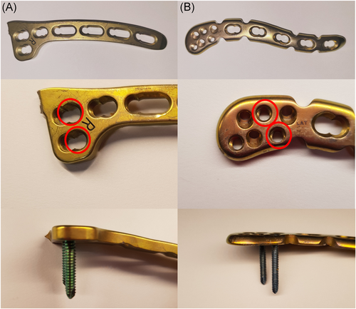
The hook plate for the 3.5-mm diameter screw fixation was chosen as it is easily obtainable, due to the much wider clinical usage, and its shape resembles the distal portion of the clavicle more closely. However, since the hook itself has a stabilization effect and substantially affects the pull-out strength, it was detached before fixation of the plate to the bone. The samples that were fixed with the hook plate (3.5-mm diameter screw, Clavicle hook plate; Synthes) were classified as Group A, and the rest that were fixed with the distal clavicle locking plate (2.7-mm diameter screw, Superior anterior clavicle plate with lateral extension; Synthes) were categorized as Group B. To facilitate the comparison, one type of plate was fixed to the shoulder from a cadaver, and the other plate type was fixed to the contralateral side of the same cadaver. In both plates, two 3.5-mm locking screws were used to fix the medial fragment of the fracture. The two screws were inserted into holes that were the most lateral to the medial fragment of the fracture. To fix the lateral fragment of the fracture, two 3.5-mm locking screws were used at the most lateral portion of the hook plate. Likewise, two 2.7-mm locking screws were inserted into holes that are perpendicular to the longitudinal axis of the clavicle among the six screw holes available. These screws were locked to both plates with identical strength (1.5 Nm) using the torque limiter attached to the screw driver (Figure 1).
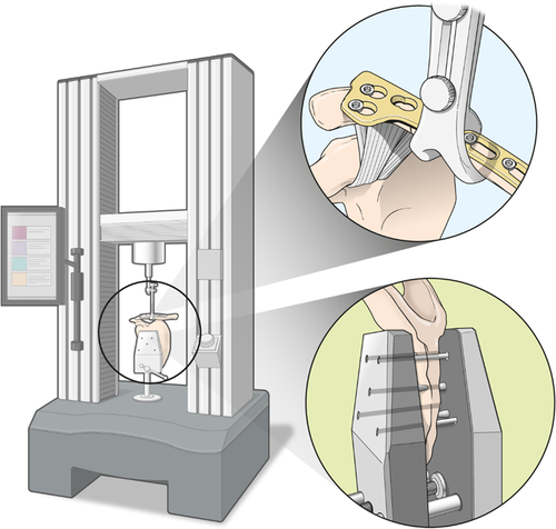
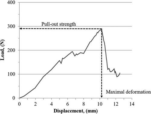
Instron measuring equipment, with two custom-made fixtures to be attached to the upper and lower sides of the equipment, was used to measure the pull-out strength (Instron 3366, Instron Co., Ltd.). The scapula of a cadaver was firmly fixed to the lower fixture, and the upper apparatus was fixed to a site where the locking head screws were inserted on the medial fragment of the fracture (Figure 2). Then, zeroing of the Instron equipment was performed to provide tensile pulling at a 90° angle. To find an ultimate tensile strength, tension was delivered at a velocity of 30 mm/min, and pull-out strength was defined as the measured maximum tensile strength at the moment of separation of the laterally inserted locking head screw from the bone (Figure 3).
2.3 Sample size calculation and statistical analysis
A pilot study was conducted before this study since there was no previous literature that examined the difference in fixation strength among various diameters of screws. The pilot study used three cadavers (six shoulder girdles) and showed that the mean pull-out strength was 212.8 ± 38.2 N. Power analysis using a type 1 error of less than 5% (α = .05) and a statistical power of 80% indicated that further evaluation would require eight samples from each group.
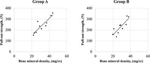
The Wilcoxon Signed-Rank test was used to compare bone mineral density and pull-out strength between the two groups. Spearman's correlation coefficient was used to assess the correlation between bone marrow density and pull-out strength. A significant difference was determined to be present for p < .05. The SPSS statistics version-25.0 program (IBM) was used for the statistical analyses.
3 RESULTS
Bone density at the region of interest where screws would be inserted was 31.6 ± 7.8 mg/cc in Group A and 28.8 ± 6.2 mg/cc in Group B. No statistically significant difference was found between the two groups. During this biomechanical study, there were no failed experiments due to screw breakage and avulsion of the CC ligament. Furthermore, tensile strength measured just before the occurrence of screw pull-out was determined to be the maximum pull-out strength in all specimens. The mean maximum pull-out strength for Group A was 241.9 ± 67.8 N, and that for Group B was 228.1 ± 63.0 N. No statistical significance was identified between the two groups (Figure 4). In addition, there was a significantly positive correlation between bone mineral density and pull-out strength in both Group A (r = .867, p < .001) and Group B (r = .891, p < .001) (Table 1 and Figure 5).
| Correlation coefficient | p value | |
|---|---|---|
| Group A (n = 10) | 0.867 | .001 |
| Group B (n = 10) | 0.891 | .001 |
- Note: Group A: fixed with the clavicle hook plate and two 3.5-mm locking screws. Group B: fixed with the distal clavicle locking plate and two 2.7-mm locking screws.
4 DISCUSSION
The purpose of this study was to determine any difference in the pull-out strength related to different diameters of screws used for the fixation of distal clavicle fracture. The results of this study confirm our hypothesis that 2.7-mm locking screw fixation yielded comparable pull-out strength to 3.5-mm locking screw fixation.
The hook plate provided stable fixation for Neer type II distal clavicle fractures, even when the distal fragment of the fractured clavicle was severely comminuted or considerably small in size. However, erosion of the acromion undersurface has often been noted, and this problem certainly necessitates early removal of the plate within a few months after the fixation.19-21 Also, the occurrence of clavicle fracture just medial to the plate has been observed in older patients with osteoporosis.22 Although there are several sizes of plates and depths of the hook and the portion for screw fixation of the hook plate that is placed onto the clavicle can be bent manually according to the contour of distal clavicle, it is difficult to bend the hook itself, which is expected to be placed underneath of the acromion. Therefore, it is difficult to avoid this complication. The anatomical morphology of the clavicle is characterized by varying contours along the bone. The distal portion of the clavicle is more flattened and has a widened anteroposterior length compared to the mid-portion of the bone. Therefore, if a distal fragment of the fractured clavicle is not extremely small, it may be possible to fix the fracture with a plate that does not contain the hook, and the distal clavicle locking plate with holes available for the insertion of 2.7-mm diameter screws into the distal portion may be another option in these fractures.
However, the counter-pull of the trapezius and sternocleidomastoid muscles on the proximal fragment occasionally causes fixation failure that the distal portion of the plate is pulled out from the distal fracture fragment.23-25 Although the distal clavicle locking plate without a hook allows several screw fixations for the distal portion, an adequate number of screw fixations can be hindered in type II distal clavicle fractures with comminution or small distal fragments. Furthermore, the screw diameter is 2.7 mm. Therefore, the question was whether fixation with these small diameter screws would produce biomechanical properties comparable to those of 3.5-mm diameter screws, which are the typical size for fixation of a distal clavicle fracture.
Previously, several biomechanical studies for distal clavicle fracture evaluated and compared the fixation strength between non-anatomical and anatomical plates and between locking head and non-locking head screws.26-30 However, no investigation has examined the relation between varying screw diameters and fixation strength of the distal clavicle fracture. Although Hungerer et al.11 performed a biomechanical study to compared the fixation of 3.5- and 2.7-mm diameter screws and noted that 3.5-mm sized screws show superior fixation strength, their study examined the distal humerus fracture. The outcomes for distal humerus fractures can be substantially different from the outcomes of treating distal clavicle fractures, since the anatomy of the distal clavicle is much more intricate due to the presence of the AC and CC ligaments. In addition, as reported in the previous literatures, there was a positive correlation between bone mineral density and pull-out strength in both groups.
In the current study, a simple pull-out testing model to enable a direct comparison of plate fixation using two different sized screws was possible since most cases of distal clavicle fractures present with upward displacement of a proximal fragment due to sternocleidomastoid and trapezius muscles. In contrast, mid-shaft clavicle fractures are affected by multiaxial forces, such as torsion or compression. In addition, previous studies reported that about 400–600 N strength is required for failure of fixation due to pull-out.26, 28, 31 Considering the ultimate pull-out strength of 235 N in this study, the distal clavicle locking plate can only be used in cases that allows three or more screws in lateral fixation, due to the significant possibility of failure with lateral fixation using only two screws.
In this study, the pull-out strength was higher in the 3.5-mm diameter screw fixation; however, there was no significant difference compared to the strength measured for the 2.7-mm diameter screw. One advantage of the 2.7-mm screw over the 3.5-mm screw is that, due to its smaller size, more screws can be used in a plate with an identical area. Also, the anatomical plate that uses 2.7-mm diameter screws has screw holes that are designed with various insertion angles for the screw, which may provide improved fixation strength. However, further studies are necessary to discover how much the size of lateral clavicle fragment is necessary to securely apply this type of plate and how many screws are required to clinically prevent fixation failure.
The present study had several limitations. First, many Neer type IIA fractures are categorized as a vertically fractured pattern, yet the identical pattern used in this study actually presented with a multiplanar or oblique pattern. Such clinically different patterns may affect the outcomes of this study. Second, the current study is a biomechanical study and had a limited plane of applying force, but the fracture can actually be affected by dynamic forces from various planes in vivo. However, despite the potential presence of multiplanar dynamic forces in vivo, the authors contend that the pull-out of laterally fixed screws is the most important source of fixation failure, as the CC ligament is attached to the distal fragment in Neer type II distal clavicle fractures. Third, this biomechanical study did not consider the influence of each plate's geometry and thickness. Despite the benefits of using the hook plate with 3.5-mm locking screws, its complications cannot be overlooked. Therefore, we designed this study to prove the biomechanical noninferiority of using the distal clavicle plate with 2.7-mm locking screws to avoid the use of the hook plate. Finally, the current study is a time-zero cadaveric study, including any biologic process that occurs during fracture healing.
5 CONCLUSION
For Neer type IIA distal clavicle fractures, distal fragment fixation with 2.7-mm diameter screws in a distal clavicle locking plate showed comparable biomechanical pull-out strength at time-zero setting as fixation with 3.5-mm diameter screws in the hook plate. Therefore, if the distal fragment of the fractured clavicle is not extremely small, fixation of a distal fragment with a low-profile plate and 2.7-mm screws may be an alternative option.
ACKNOWLEDGEMENT
The authors thank Medical Illustration & Design, part of the Medical Research Support Services of Yonsei University College of Medicine, for all artistic support related to this work. This study was supported by a faculty research grant from Yonsei University College of Medicine (6-2019-009912).
AUTHOR CONTRIBUTIONS
Tae-Hwan Yoon: Research design and acquisition, analysis and interpretation of data, writing—original draft, approval of the submitted and final versions. Chong‑Hyuk Choi: research design and acquisition, analysis and interpretation of data, approval of the submitted and final versions. Yun-Rak Choi: research design, supervision, approval of the submitted and final versions. Hun-Jin Ju: research design, supervision, approval of the submitted and final versions. Yong-Min Chun: research design and acquisition, analysis and interpretation of data, writing—review and revised, approval of the submitted and final versions. All authors have read and approved the final version of the manuscript for submission.


