Association of COVID-19 vaccination before conception with maternal thyroid function during early pregnancy: A single-center study in China
Yan Zhao and Yongbo Zhao contributed equally to this study.
Abstract
Despite the high vaccination coverage, potential COVID-19 vaccine-induced adverse effects, especially in pregnant women, have not been fully characterized. We examined the association between COVID-19 vaccination before conception and maternal thyroid function during early pregnancy. We conducted a retrospective cohort study in Shanghai, China. A total of 6979 pregnant women were included. Vaccine administration was obtained from electronic vaccination records. Serum levels of thyroid hormone were measured by fluorescence and chemiluminescence immunoassays. Among the 6979 included pregnant women, 3470 (49.7%) received at least two doses of an inactivated vaccine. COVID-19 vaccination had a statistically significant association with both maternal serum levels of free thyroxine (FT4) and thyroid stimulating hormone (TSH). Compared with unvaccinated pregnant women, the mean FT4 levels were lower in pregnant women who had been vaccinated within 3 months before the date of conception by 0.27 pmol/L (β = −0.27, 95% confidence interval [CI], −0.42, −0.12), and the mean TSH levels were higher by 0.08 mIU/L (β = 0.08, 95% CI, 0.00, 0.15). However, when the interval from vaccination to conception was prolonged to more than 3 months, COVID-19 vaccination was not associated with serum FT4 or TSH levels. Moreover, we found that COVID-19 vaccination did not significantly associate with maternal hypothyroidism. Our study suggested that vaccination with inactivated COVID-19 vaccines before conception might result in a small change in maternal thyroid function, but this did not reach clinically significant levels.
1 INTRODUCTION
Several coronavirus disease 2019 (COVID-19) vaccines have been made available to mitigate the ongoing epidemic.1 According to the World Health Organization (WHO) COVID-19 dashboard, as of July 1, 2022, more than five billion persons have been vaccinated with at least one dose of the COVID-19 vaccine worldwide.2 Despite the high vaccination coverage, potential vaccine-induced adverse effects, especially in vulnerable populations, have not yet been fully characterized.3 Pregnant women are at increased risk of severe COVID-19 and have been recommended to receive COVID-19 vaccines in more than 100 countries.4 However, safety data on COVID-19 vaccination among pregnant women remain limited.5, 6
It is well known that maternal thyroid function during pregnancy is critical for a successful pregnancy and fetal brain development.7 Disrupted maternal thyroid function, especially during early pregnancy when the fetal thyroid gland does not produce any thyroid hormone (TH), has been associated with an increased risk of adverse neurodevelopmental outcomes in children.8-14 Recently, concerns have been raised regarding the potential of COVID-19 vaccination in inducing thyroid dysfunction, as cases of Graves' disease15-20 and subacute thyroiditis15, 18, 21-23 have been frequently reported among vaccinated people. However, no study to date has assessed the impact of COVID-19 vaccination on the thyroid function of pregnant women.
In China, pregnant women are not recommended to receive COVID-19 vaccines.24 Therefore, the aim of this study was to examine the association between COVID-19 vaccination before conception and maternal thyroid function during early pregnancy. Since anti-thyroglobulin antibody (TgAb) and thyroid peroxidase antibody (TPOAb) have been suggested as good markers of early thyroid autoimmune diseases,25-27 and pregnancies complicated by thyroid autoantibody positivity require more care,28 we also examined whether pregnant women with early autoimmune damage were more susceptible to the potential alterations of thyroid function caused by COVID-19 vaccination.
2 METHODS
2.1 Study population
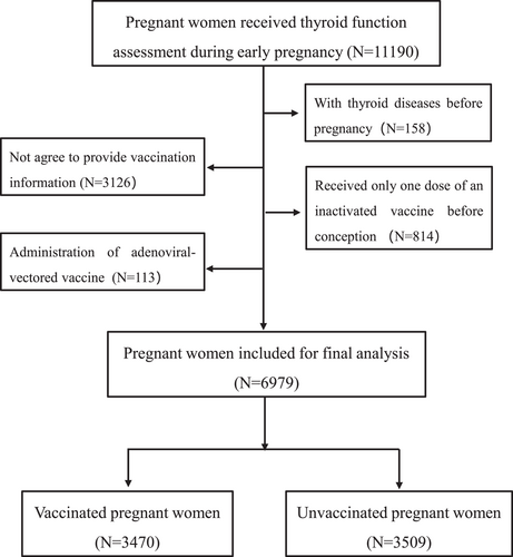
This retrospective cohort study was conducted in a tertiary-care hospital, which is one of the largest prenatal healthcare providers in China, serving more than 45 000 inpatients per year. Participants were pregnant women who were issued with an antenatal card during their first prenatal visit and received routine antenatal care from September 2021 to January 2022. The flowchart of participant enrollment is presented in Figure 1. Briefly, a total of 11 190 singleton pregnant women who had no history of SARS-CoV-2 infection and received a thyroid function assessment during early pregnancy were screened for eligibility. Participants with a known history of thyroid disease before pregnancy (N = 158), those who did not agree to provide their vaccination information (N = 3126), or those who received an adenoviral-vectored vaccine (N = 113) and only one dose of an inactivated vaccine before pregnancy (N = 814) were excluded. Ultimately, 6979 pregnant women were included in this analysis. This study was approved by the hospital's Human Ethics Committee (KS22259, March 26, 2022), and was performed in accordance with Helsinki Declaration.

2.2 Exposure
Vaccination information, including the vaccine type and manufacturer, number of dosages, and date of vaccination, was obtained from each participant's electronic vaccination record. Participating pregnant women were categorized as having exposure if they received at least two doses of an inactivated vaccine before conception.
2.3 Clinical variables
For each participating pregnant woman, we extracted demographic characteristics regarding maternal age (MA), weight and height before the current pregnancy, and age at menarche from the initial prenatal visit record. Reproductive history, including maternal parity, abortion history, and mode of conception, was extracted from the electronic medical record system of the hospital. Prepregnancy body mass index (BMI) was estimated as measured body weight in kilograms divided by height in meters squared.
2.4 Outcomes
The outcome of interest was maternal thyroid function, which was assessed by measuring serum levels of free thyroxine (FT4), thyroid stimulating hormone (TSH), TPOAb, and TgAb during early pregnancy. Serum concentrations of TH were measured by fluorescence and chemiluminescence immunoassays using ADVIA Centaur instruments and kits (Siemens). The laboratory reference ranges of FT4, TSH, TPOAb, and TgAb were 12.25–19.74 pmol/L, 0.07–4.08, 0–60, and 0-60 IU/ml, respectively. Hypothyroidism, both clinical and subclinical, was defined as the presence of elevated TSH with decreased FT4 or normal FT4 and was diagnosed based on the American Thyroid Association recommendation.29 Participating pregnant women were categorized as TPOAb negative (≤60 IU/ml) or positive (>60 IU/ml) and TgAb negative (≤60 IU/ml) or positive (>60 IU/ml).
2.5 Statistical analysis
All participating pregnant women were categorized into vaccinated and unvaccinated groups, and the general characteristics were compared between the two groups. Continuous serum FT4 and TSH levels are presented as mean ± SD and were compared using the two-sample t-test. Because serum TPOAb and TgAb levels are below the limit of detection in nearly half of the participating pregnant women, they are treated as categorical variables (%) and were compared using the chi-square test.
Multiple linear regression models were conducted to assess the associations of COVID-19 vaccination with serum FT4 or TSH levels. We adjusted for the following potential confounders: gestational age at the time of the thyroid function assessment, MA, parity, age at menarche, mode of conception, abortion history, and pregnancy BMI. Given that one study observed that the binding and neutralizing antibodies declined over time 3 months after receiving two doses of an inactivated virus vaccine,30 based on the time intervals from complete vaccination to the date of conception, vaccinated pregnant women were further divided into two subgroups: ≤3 months and >3 months since vaccination. The association between the time intervals and serum TH levels was estimated by a separate linear regression analysis, in which vaccination status (unvaccinated, ≤3 months, or >3 months since vaccination) was fitted into the model as an independent variable, whereas maternal thyroid function was fitted as the dependent variable. In addition to the analysis among all participants, we stratified the study population by thyroid autoantibody status and assessed the association of vaccination with maternal TH levels in thyroid autoantibody positive and negative women separately.
Multivariable log-binomial regression analysis was performed to estimate the relative risk (RR) and 95% confidence interval (CI) for the associations of COVID-19 vaccination with incident hypothyroidism with adjustment of the aforementioned confounders. Similar to linear regression analysis, the associations between the time intervals from complete vaccination to the date of conception and maternal hypothyroidism were also estimated. A separate log-binomial regression analysis was conducted and vaccination status (unvaccinated, ≤3 months or >3 months since vaccination) was fitted into the model as an independent variable, whereas maternal hypothyroidism (yes/no) was fitted as the dependent variable. In log-binomial regression analysis, the unvaccinated participating pregnant women served as a reference. Additionally, we stratified all participants by thyroid autoantibody status and assessed the association of vaccination with maternal hypothyroidism in autoantibody-positive and negative women separately.
As a sensitivity analysis to address the confounding effect of thyroid dysfunction in previous pregnancies, we replicated the multiple linear regression analysis by restricting the participants to nulliparous women. Another sensitivity analysis was conducted to ensure that the observed associations were not confounded by the mode of conception. In a third sensitivity analysis, we examined the potential confounding effects of abortion history by restricting the multiple linear regression analysis to pregnant women with no abortion history. All statistical analyses were performed using R statistical software version 3.2.3 (R Project for Statistical Computing), and a two-sided p < 0.05 was considered significant.
3 RESULTS
3.1 Demographic characteristics of the participants
Of the 11 190 eligible pregnant women, 6979 were included in the final analysis. As shown in Supporting Information: Table S1, the included and excluded subjects did not differ in MA, parity, age at menarche, prepregnancy BMI, or mode of conception. Of the 6979 included pregnant women, 49.7% (3470/6979) received at least two doses of an inactivated vaccine. The characteristics of the participants according to vaccination status are summarized and compared in Table 1. Compared with unvaccinated pregnant women, vaccinated pregnant women were more likely to be multiparous and less likely to have an abortion history or to conceive by assisted reproduction technology (ART) during the current pregnancy.
| Characteristics | Total (N = 6979) | Vaccination | p Valuea | |
|---|---|---|---|---|
| Yes (N = 3470) | No (N = 3509) | |||
| Maternal age (year) | ||||
| <35 | 5868 (84.1%) | 2944 (84.8%) | 2924 (83.3%) | 0.084 |
| ≥35 | 1111 (15.9%) | 526 (15.2%) | 585 (16.7%) | |
| Parity | ||||
| Nulliparous | 5581 (80.0%) | 2541 (73.2%) | 3040 (86.6%) | <0.001 |
| Multiparous | 1398 (20.0%) | 929 (26.8%) | 469 (13.4%) | |
| Age at menarche (year) | ||||
| ≤12 | 1381 (19.8%) | 700 (20.2%) | 681 (19.4%) | 0.434 |
| 13–14 | 3845 (55.1%) | 1885 (54.3%) | 1960 (55.9%) | |
| ≥15 | 1753 (25.1%) | 885 (25.5%) | 868 (24.7%) | |
| Pregnancy BMI (kg/m2) | ||||
| <18.5 | 873 (12.5%) | 447 (12.9%) | 426 (12.1%) | 0.063 |
| 18.5–24.9 | 5326 (76.3%) | 2665 (76.8%) | 2661 (75.8%) | |
| ≥25 | 780 (11.2%) | 358 (10.3%) | 422 (12.0%) | |
| Mode of conception | ||||
| The use of ART | 230 (3.3%) | 39 (1.1%) | 191 (5.4%) | <0.001 |
| Natural conception | 6749 (96.7%) | 3431 (98.9%) | 3318 (94.6%) | |
| Abortion history | ||||
| Yes | 1705 (24.4%) | 940 (27.1%) | 765 (21.8%) | <0.001 |
| No | 5274 (75.6%) | 2530 (72.9%) | 2744 (78.2%) | |
- Note: Data are presented as numbers (%).
- Abbreviations: ART, assisted reproduction technology; BMI, body mass index.
- a p-values were calculated by chi-square test.
3.2 Distribution of serum thyroid hormone levels
Serum FT4, TSH, TPOAb, and TgAb levels were measured and are presented in Table 2. The mean (±SD) levels of FT4 and TSH of the 6979 participating pregnant women were 16.6 ± 2.8 pmol/L and 1.4 ± 1.4 mIU/L, respectively. Among the 6979 participating pregnant women, 760 (10.9%) were identified as TPOAb positive, and 539 (7.7%) were identified as TgAb positive.
| Characteristicsa | Total (N = 6979) | Vaccination | p Valueb | |
|---|---|---|---|---|
| Yes (N = 3470) | No (N = 3509) | |||
| FT4 (pmol/L) | 16.6 ± 2.8 | 16.6 ± 2.8 | 16.7 ± 2.9 | 0.417 |
| TSH (mIU/L) | 1.4 ± 1.4 | 1.4 ± 1.6 | 1.4 ± 1.1 | 0.395 |
| No. of TPOAb positive | 760 (10.9%) | 415 (12.0%) | 345 (9.8%) | <0.001 |
| No. of TgAb positive | 539 (7.7%) | 297 (8.6%) | 242 (6.9%) | <0.001 |
- Abbreviations: TgAb, thyroglobulin antibody; TPOAb, thyroid peroxidase antibody.
- a Values are shown as mean ± standard deviation or numbers (percentages).
- b p-values were calculated by Student's t test or chi-square test.
3.3 Association of COVID-19 vaccination with serum thyroid hormone levels
Analyses of potential differences in serum TH levels between unvaccinated and vaccinated pregnant women showed that the two groups did not differ in serum FT4 or TSH levels (Table 2). However, the proportions of TPOAb-positive women (12.0% vs. 9.8%, p < 0.001) and TgAb-positive women (8.6% vs. 6.9%, p < 0.001) were significantly higher in the vaccinated groups than in the unvaccinated groups.
The adjusted associations of COVID-19 vaccination with serum FT4 and TSH levels are presented in Table 3. In all participants, COVID-19 vaccination was associated with lower serum FT4 levels. Compared with unvaccinated pregnant women, the mean serum FT4 levels were lower in pregnant women who had been vaccinated within 3 months before the date of conception by 0.27 pmol/L (β = −0.27, 95% CI, −0.42, −0.12). Regarding TSH, COVID-19 vaccination was associated with higher serum TSH levels. Compared with unvaccinated pregnant women, the mean serum TSH levels were higher in pregnant women who had been vaccinated within 3 months before the date of conception by 0.08 mIU/L (β = 0.08, 95% CI, 0.00, 0.15). However, when the interval from complete vaccination to the date of conception was prolonged to more than 3 months, COVID-19 vaccination was not significantly associated with serum FT4 or TSH levels.
| Unvaccinated | Vaccinated | Time intervalc | ||
|---|---|---|---|---|
| ≤3 months | >3 months | |||
| All participants | ||||
| FT4 | 0 (Reference) | −0.20 (−0.33, −0.07)** | −0.27 (−0.42, −0.12)** | 0.11 (−0.28, 0.06) |
| TSH | 0 (Reference) | 0.05 (−0.01, 0.12) | 0.08 (0.00, 0.15)* | 0.02 (−0.07, 0.11) |
| Autoantibody positivea | ||||
| FT4 | 0 (Reference) | 0.05 (−0.33, 0.43) | 0.27 (−0.18, 0.71) | 0.22 (−0.27, 0.72) |
| TSH | 0 (Reference) | −0.06 (−0.42, 0.30) | −0.15 (−0.55, 0.26) | −0.01 (−0.46, 0.44) |
| Autoantibody negativeb | ||||
| FT4 | 0 (Reference) | −0.24 (−0.38, −0.10)** | −0.31 (−0.48, −0.15)** | −0.14 (−0.32, 0.04) |
| TSH | 0 (Reference) | 0.06 (0.00, 0.11)* | 0.09 (0.03, 0.15)** | 0.01 (−0.05, 0.07) |
- Note: Models adjusted for gestational age at the time of thyroid function assessment, maternal age, maternal parity, age at menarche, mode of conception, abortion history, and prepregnancy body mass index.
- Abbreviations: CI, confidence interval; FT4, free thyroxine; TgAb, thyroglobulin antibody; TPOAb, thyroid peroxidase antibody; TSH, thyroid stimulating hormone.
- a TPOAb or TgAg positive.
- b TPOAb and TgAg negative.
- c Time interval from complete vaccination to the date of conception.
- * p < 0.05
- ** p < 0.01.
We further performed stratified analysis among thyroid autoantibody positive and negative women separately. Results showed that the associations of vaccination with FT4 or TSH in autoantibody-negative women were generally similar to those observed in all participants. However, COVID-19 vaccination before conception was not significantly associated with serum FT4 or TSH levels in autoantibody-positive women.
3.4 Association of COVID-19 vaccination with maternal hypothyroidism
As we found the features of COVID-19 vaccination and maternal thyroid function to be consistent with those of hypothyroidism, a condition whereby increased TSH levels occur with (clinical hypothyroidism) or without (subclinical hypothyroidism) a concomitant decrease in FT4 levels, we investigated the association of COVID-19 vaccination with maternal hypothyroidism. As shown in Table 4, COVID-19 vaccination was not significantly associated with maternal hypothyroidism. Additionally, results of the stratified analysis also showed that vaccination was not significantly associated with maternal hypothyroidism in both autoantibody-positive and negative women.
| Unvaccinated | Vaccinated | Time intervalc | ||
|---|---|---|---|---|
| ≤3 months | >3 months | |||
| All participants | ||||
| RR (95% CI) | 1.00 (Reference) | 1.08 (0.81, 1.45) | 1.28 (0.92, 1.78) | 0.87 (0.59, 1.28) |
| p-value | / | 0.536 | 0.174 | 0.629 |
| Autoantibody positivea | ||||
| RR (95% CI) | 1.00 (Reference) | 0.88 (0.52, 1.48) | 1.01 (0.57, 1.80) | 0.68 (0.32, 1.44) |
| p-value | / | 0.631 | 0.963 | 0.319 |
| Autoantibody negativeb | ||||
| RR (95% CI) | 1.00 (Reference) | 1.16 (0.82, 1.64) | 1.31 (0.87, 1.98) | 0.99 (0.63. 1.56) |
| p-value | / | 0.410 | 0.189 | 0.980 |
- Note: Models adjusted for gestational age at the time of thyroid function assessment, maternal age, maternal parity, age at menarche, mode of conception, abortion history, and prepregnancy body mass index.
- Abbreviations: CI, confidence interval; TgAb, thyroglobulin antibody; TPOAb, thyroid peroxidase antibody.
- a TPOAb or TgAg positive.
- b TPOAb and TgAg negative.
- c Time interval from complete vaccination to the date of conception.
3.5 Sensitivity analysis
In sensitivity analysis, when restricting the multiple linear regression analysis to nulliparous pregnant women, the associations of COVID-19 vaccination with FT4 or TSH were similar to those observed in all participants (Supporting Information: Table S2). In addition, after restricting the analysis to pregnant women who conceived spontaneously, the observed associations also did not appreciably change (Supporting Information: Table S3). Furthermore, by restricting the multiple linear regression analysis to pregnant women with no history of abortion, the association of COVID-19 vaccination with maternal TH levels remained unchanged (Supporting Information: Table S4).
4 DISCUSSION
Because of the expression of angiotensin-converting enzyme 2 (ACE-2), the thyroid gland is a potential target of attack by severe acute respiratory syndrome coronavirus 2 (SARS-CoV-2).31 As several studies have reported COVID-related subacute thyroiditis32-34 and autoimmune thyroid disorders,35-38 it is reasonable to postulate that COVID-19 vaccination may also be associated with a spectrum of thyroid dysfunction.
In this study, we examined the impact of vaccination with inactivated vaccines before conception on maternal thyroid function during early pregnancy. We found that COVID-19 vaccination within 3 months before the date of conception was associated with a small decrease in FT4 levels and a small increase in TSH levels. However, when the interval from complete vaccination to the date of conception was prolonged to more than 3 months, COVID-19 vaccination was not associated with serum thyroid hormone levels. Moreover, we found that COVID-19 vaccination before conception did not significantly associate with maternal hypothyroidism. Our findings suggested that vaccination with inactivated COVID-19 vaccines before conception might result in a small change in maternal thyroid function, but this did not reach clinically significant levels.
Adverse impacts of COVID-19 vaccination on thyroid function have been reported among the general population. Several studies reported a series of patients who presented with new onset or relapses of Graves' disease15-20 and subacute thyroiditis15, 18, 21-23 shortly after receiving the COVID-19 vaccine. Both Graves' disease and subacute thyroiditis patients presented hyperthyroidism. Adjuvants-induced autoimmune/inflammatory syndrome and molecular mimicry were proposed to be the mechanisms for vaccination-induced subacute thyroiditis and Graves' disease.39 However, in our study, COVID-19 vaccination, especially vaccination more than 3 months before the date of conception, had no impact on maternal thyroid function. The inconsistent findings are mainly ascribed to the differences in vaccine types and population groups. In previous case reports, we noticed that subacute thyroiditis or Graves' disease mainly occurred after vaccination with messenger RNA (mRNA) COVID-19 vaccines in the general population. In our study, all vaccinated pregnant women received inactivated vaccines. Compared with mRNA vaccines, inactivated vaccines may have lower immunogenicity.40
To our knowledge, this is the first study to assess the impact of inactivated COVID-19 vaccines on the thyroid function of pregnant women. Our findings provide additional safety evidence that vaccination with inactivated COVID-19 vaccines does not alter maternal thyroid function. Moreover, our findings may help inform decision-making about COVID-19 vaccination by women who are trying to conceive.
Bearing certain limitations, our results should be interpreted with caution. First, the retrospective nature of our study prevented us from assessing the antibody levels of each participating pregnant woman. However, we reviewed their medical records and ensured that no participants had any prior history of SARS-CoV-2 infection. Second, data regarding the prior history of autoimmune diseases and prevaccination thyroid autoantibody levels were not available, and residual confounding from unavailable confounders remained. Third, in our study, all vaccinated pregnant women received inactivated COVID-19 vaccines, and we could not compare the effect of different types of vaccines on maternal thyroid function. Finally, all participants were from one of the largest cities in China, which may limit the generalizability of our findings.
In conclusion, our study provided the first evidence that vaccination with inactivated COVID-19 vaccines before conception might result in a small change in maternal thyroid function, but this did not reach clinically significant levels. Healthcare providers are encouraged to remain vigilant about other non-thyroid disorders or non-thyroid autoimmune diseases in pregnant women after exposure to inactivated COVID-19 vaccines. Further larger prospective cohort studies or randomized controlled trials are needed to validate our findings.
AUTHOR CONTRIBUTIONS
The study was designed by Liping Jin and Yan Zhao. Acquisition, analysis, or interpretation of data were done by Liping Jin and Yan Zhao. Drafting of the manuscript was done by Yan Zhao and Yongbo Zhao. Statistical analysis was done by Yan Zhao and Yongbo Zhao. Yicheng Zhou, Ziyi Zhang, Yijun Zhang, Mengyuan Li, and Xin Su provided administrative, technical, and material support. Liping Jin obtained the funding and had full access to all the data in the study and took responsibility for the integrity of the data and the accuracy of the data analysis. All authors contributed to the reviewing and approved the final version.
ACKNOWLEDGMENTS
This study was supported by the Strategic Collaborative Research Program of the Ferring Institute of Reproductive Medicine, Ferring Pharmaceuticals and Chinese Academy of Sciences (FIRMSCOV02), the National Natural Science Foundation of China (81730039, 82071653, 82271701, 82173533), and the Shanghai Rising-Star Program (21QA1407300).
CONFLICT OF INTEREST
The authors declare no conflict of interest.
Open Research
DATA AVAILABILITY STATEMENT
The data that support the findings of this study are available upon reasonable request from the corresponding author.


