Brain metastases from breast cancer using magnetic resonance imaging: A systematic review
Abstract
Despite improvements in imaging and treatment approaches, brain metastases (BMs) continue to be the primary cause of mortality and morbidity in about 20% of adult cancer patients. This research aimed to review the magnetic resonance imaging (MRI) and clinical characteristics of BMs resulting from breast cancer (BC). A systematic review of original research articles published from January 2000 to June 2023. We selected studies that reported MRI findings of BMs in BC patients. We excluded reviews, case reports, books/book chapters, animal studies and irrelevant records. We identified 24 studies that included 1580 BC patients with BMs. T1-weighted (T1-w) (pre- and postcontrast), T2-weighted (T2-w), fluid-attenuated inversion recovery (FLAIR) and T2*-weighted (T2*-w) was used to measure the lesion size, shape and area. In other studies, advanced structural techniques including diffusion-weighted imaging (DWI), diffusion tensor imaging (DTI) and susceptibility-weighted imaging (SWI) were used to more precisely and sensitively evaluate the pathological area. Furthermore, functional and metabolic techniques like functional MRI (fMRI), magnetic resonance spectroscopy (MRS) and perfusion-weighted imaging (PWI) have also been utilised. The MRI findings of BMs varied depending on the MRI technique, the BC subtype, the lesion size and shape, the presence of haemorrhage or necrosis and the comparison with other brain tumours. Some MRI findings were associated with prognosis, recurrence or cognitive impairment in BC patients with BMs. MRI detects, characterises and monitors BMs from BC. Findings vary by MRI technique, BC subtype, lesion characteristics and comparison with other brain tumours. More research should validate emerging MRI techniques, determine the clinical implications of findings and explore the underlying mechanisms and biology of BMs from BC. MRI is a valuable tool for diagnosis, targeted therapy and studying BC metastasis.
Introduction
As a multi-step process, metastasis involves the dissemination of cancer cells from the initial tumour to the target organ.1, 2 Brain metastases (BMs) remain the leading cause of death and morbidity in about 20% of adult cancer patients, despite advances in imaging and treatment modalities.3 Having BMs is a disastrous consequence for patients with solid malignancies such as breast cancer (BC), lung cancer and melanoma, and is often linked with a poor prognosis and decreased quality of life.1, 4
Because of its specialised cells, anatomical characteristics, metabolic restrictions and immunological surroundings, the brain microenvironment exerts a vastly different selection pressure on tumour cells than extracranial lesions do, which in turn influences the metastatic process and treatment responses.5
Approximately 15%–25% of patients with BC may develop central nervous system (CNS) metastases.1, 6 The ability of BC cells to metastasize to the brain is influenced by their receptor status. In fact, as compared to hormone receptor-positive individuals, human epidermal growth factor receptor 2 (HER2)-positive and triple-negative BC (TNBC) subtypes had the highest association with CNS metastases, with the frequencies of BMs reaching values as high as 30–40%.1, 7, 8
Accurate identification of the cause of BMs is critical for developing an appropriate treatment plan to enhance the patients prognosis.9 Computed tomography (CT) scans and magnetic resonance imaging (MRI) scans are the most common imaging techniques used to diagnose brain malignancies.10-15 The high signal-to-noise ratio (SNR), contrast-to-noise ratio (CNR), spatial resolution, contrast resolution and the abundance of MRI sequences available to define intracranial lesions allow MRI to give more detailed, localisation and characterisation of BMs.16, 17 To be more specific, T1-weighted (T1-w) and T2-weighted (T2-w) sequences outline the morphologic and anatomical de-alignment of tissue brought about by the tumour.18
The purpose of this systematic review was to document the utility of reported MRI techniques in imaging BC, as well as the MRI and clinical features of BMs from BC to aid in the diagnosis and treatment of similar individuals in the future.
Material and Methods
Search strategy
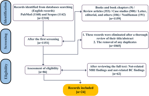
This systematic review followed the PRISMA statement for reporting systematic reviews and meta-analyses.19 The aim of this review was to synthesise the available evidence on BMs from BC using MRI. We searched PubMed and Scopus databases from January 2000 to June 2023 using the following combination of keywords: "MRI," "breast cancer," "brain metastases," and "brain metastasis." We restricted our search to articles published in English that reported MRI and clinical data on BMs in BC patients. The detailed search strategy for PubMed is provided in Table S1.
Study selection
We selected the titles and abstracts of the retrieved records for eligibility and excluded reviews (including literature reviews, systematic reviews and meta-analyses), case reports, books and book chapters, animal studies and other irrelevant records. We obtained the full texts of the potentially eligible records and assessed their inclusion based on the following criteria: (1) original research article (2); MRI findings of BMs in BC patients; and (3) related BC findings associated with MRI. We excluded articles that did not meet these criteria or did not provide sufficient MRI data. We also checked the reference lists of included articles for additional relevant studies. After reviewing the full texts and removing any records not related to the MRI / BC findings, we identified 86 records that may meet our inclusion criteria.
Data extraction and quality assessment
Three independent reviewers (S.Gh., M.M. and S.M.) performed the study selection process and any disagreements were resolved by consensus or consultation. We extracted the following data from each included article: authors, year of publication, number of BC patients with BMs, MRI techniques used, anatomical locations of BMs and imaging findings. Table 1 summarises the characteristics and findings of the included studies. Other reviewers used Microsoft Excel (version 2016) to record the data. Two reviewers (S.Gh. and M.M.) independently extracted the data and cross-checked for accuracy. Any discrepancies were resolved by discussion or consultation with a third reviewer (S.M.). The quality of the included studies was assessed using the Cochrane Risk of Bias tool. Figure 1 shows the PRISMA flow diagram of the study selection process.
| First author (year) | Number of Patients | MRI Technique(s) | Anatomical Location(s) | MRI Findings |
|---|---|---|---|---|
| Xue (2023)20 | 4 | T1-w and T2-w | Superficial parenchyma lesions and deep lesions |
|
| Young (2023)21 | 34 | CE MRI | Frontal, temporal, perietal, occipital and cerebellum |
|
| Reibelt (2022)22 | 15 | 3D T1-w MPRAGE | L. cerebellum WM, R. pallidum, L. thalamus, L. choroid plexus, L. Lat. ventricle and total GM |
|
| Young (2021)23 | 38 | T1-w and T1-w post | Frontal, temporal, perietal, occipital and cerebellum |
|
| Santos (2020)24 | 147 | T1-w, T1-w post, T2-w, DWI and T2*-w | Hippocampal and hydrocephalus |
|
| Zhang (2019)25 | 3 | T1-w, T1-w post, T2-w, DWI, ADC and FLAIR | NA |
|
| Mayinger (2019)26 | 851 | 3D T1-w | Bilateral and central/median subcortical structures |
|
| Kniep (2019)27 | 37 | T1-w, T1-w post and FLAIR | NA |
|
| Ortiz-Ramón (2018)28 | 17 | 3D T1-w | NA |
|
|
Skogen (2018)29 |
5 | DTI | Intracranial lesions without visible haemorrhage, multiple lesions and infratentorial lesions |
|
| Muto (2018)30 | 13 | DSC | Temporal lobe |
|
| Kyeong (2017)31 | 100 | 3D T1-w | HER2-positive type: occipital, temporal lobes and cerebellum / Luminal type: frontal, occipital lobes and cerebellum |
|
| Kesler (2017)32 |
74 |
rsfMRI, T2*-w GRE, HR3D IR, FSPGR GRE T1-w and DTI | R. inferior parietal lobe, R. middle inferior orbital frontal gyrus, R. medial superior frontal gyrus, R. inferior and middle frontal gyri, bilateral postcentral gyri, R. precuneus, L. inferior temporal gyrus, L. middle occipital gyrus, R. parietal lobule, R. cuneus, R. superior temporal gyrus and R. inferior temporal gyrus. |
|
| Bette (2017)33 | 13 | T2-FLAIR | NA |
|
| Fan (2017)34 | 13 | T1-w FLAIR and DWI (before enhancement) and T1-w FLAIR | R. occipital lobe |
|
| Franceschi (2016)35 | 38 | T1-w post and SWI | NA |
|
| Kesler (2015)36 | 36 | DTI and T1-w | L. corpus callosum, bilateral inferior longitudinal fasciculus, L inferior fronto-occipital fasciculus, and bilateral temporal and frontal lobe white matter |
|
| Yeh (2015)37 | 62 | T1-w, T1-w post, T2-w fast spin echo (FSE), FLAIR and DWI | Parietal lobe, R. frontal lobe and L. cerebellum |
|
| Quattrocchi (2014)38 | 42 | T1w, T1-w post and T2-w FLAIR | Parieto-occipital lobes and cerebellum |
|
| Huang (2010)39 | 17 | Multivoxel 2D-CSI MRS and DCE (n = 21 BC) | NA |
|
| Hakyemez (2010)40 | 5 | T2-w and PWI for rCBV | NA |
|
| Takeda (2008)41 | 13 | 2D T1-w SE, 3D MPRAGE T1-w post, 2D T1-w SE post and 2D T2-w SE | Posterior fossa, middle fossa and supratentorial |
|
| Kremer (2003)42 | 2 | T1-w, T2-w, T2*-w, R2* and DCE | NA |
|
| Geijer (2002)43 | 1 | T1-w, T2-w, DWI and ADC | Lateral wall of the posterior horn of the L. lateral ventricle and falx cerebri |
|
- 2D T1-w SE, two-dimensional T1-weighted spin echo; 2D T2-w SE, two-dimensional T2-weighted spin echo; 3D T1-w MPRAGE, 3D T1-weighted magnetisation prepared rapid acquisition gradient echo; ADC, apparent diffusion coefficient; BC, breast cancer; BMs, brain metastases; CBV, cerebral blood volume; CE MRI, contrast-enhanced MRI; CE, contrast-enhanced; DCE, dynamic contrast-enhanced; DSC, dynamic susceptibility contrast; DTI, diffusion tensor imaging; DWI, diffusion-weighted imaging; EVR, enhanced volume ratio; FLAIR, fluid-attenuated inversion recovery; FSPGR, fast spoiled gradient recalled; GBM, glioblastoma multiforme; HER2, human epidermal growth factor receptor 2; HIF-1, hypoxia-inducible factor-1; HR3D IR high-resolution 3D inversion recovery; L., Left; LAVA, liquid attenuation inversion recovery; LITT, laser interstitial thermal therapy; MPRAGE, magnetisation prepared rapid acquisition gradient echo; MRI, magnetic resonance imaging; MRS, magnetic resonance spectroscopy; Multivoxel 2D-CSI MRS, multivoxel two-dimensional chemical shift Imaging magnetic resonance spectroscopy; PWI, perfusion-weighted imaging; R., right; R2*, transverse relaxation rate; rCBV, relative cerebral blood volume; rsfMRI, resting-state functional MRI; SWI, susceptibility-weighted imaging; T1-w post, T1-weighted postcontrast; T1-w, T1-weighted; T2*-w, T2-star weighted; T2-w, T2-weighted; TNBC, triple-negative breast cancer; WMH, white matter hyperintensities.
- a Hurst exponent measures a time series' memory, with values >0.5 showing trends and <0.5 indicating mean reversion.

Results
Twenty-four studies that investigated BMs from BC were included in the analysis (Table 1). The studies included a total of 1580 patients with BMs from BC and used various MRI techniques such as T1-w, T2-w, contrast-enhanced (CE), diffusion-weighted imaging (DWI), fluid-attenuated inversion recovery (FLAIR), dynamic susceptibility contrast (DSC), resting-state functional MRI (rsfMRI), susceptibility-weighted imaging (SWI), magnetic resonance spectroscopy (MRS) and perfusion-weighted imaging (PWI). The anatomical locations of BMs varied across studies, but some common regions were the frontal, temporal, parietal and occipital lobes, the cerebellum and the ventricles. The MRI findings of BMs also varied depending on the MRI technique, the BC subtype, the lesion size and shape, the presence of haemorrhage or necrosis and the comparison with other brain tumours such as glioblastoma multiforme (GBM). Some MRI findings were associated with prognosis, recurrence or cognitive impairment in BC patients with BMs. The table provides a comprehensive overview of the current state of knowledge on BMs from BC using MRI.
The MRI findings for BMs from BC based on the studies in Table 1 indicate that the most common location for metastases is in the cerebral hemispheres. The majority of metastases are solid-enhancing masses with irregular or poorly defined margins. The number of patients in each study ranged from 1 to 851. The enhancement pattern of BMs was reported to be variable, with some studies reporting homogeneous enhancement, while others reported heterogeneous or rim enhancement.
Discussion
The aim of this systematic review was to summarise the MRI techniques, anatomical locations and MRI findings of BMs from BC using different MRI modalities. The MRI findings from these studies can be broadly categorised into lesion identification, characterisation and prediction of prognosis or response to treatment.
MRI is a useful and versatile tool for detecting, characterising and monitoring BMs from BC, as it can provide information on the lesion size, shape, location, composition, perfusion, metabolism and connectivity.26, 32, 41 MRI techniques can vary in their sensitivity and specificity for BMs from BC, depending on the contrast agent, the sequence parameters, the image processing and the interpretation criteria.21, 34, 35 Some MRI techniques such as CE MRI, DWI, ADC, DSC and SWI can help to differentiate BMs from BC from other brain tumours such as GBM or ischemic lesions.29, 30, 34, 35, 40, 42 Other MRI techniques such as postcontrast T1-w and FLAIR can help to assess the response to treatment or the recurrence of BMs from BC.33-35 Some MRI techniques such as DTI and rsfMRI can help to evaluate the cognitive impairment or the functional connectivity of BMs from BC.29, 32, 36
MRI findings of BMs from BC can vary depending on the BC subtype, such as HER2-positive, luminal or triple-negative. Different BC subtypes can have different patterns of BM distribution in the brain, different lesion contours and compositions on MRI, and different associations with prognosis or imaging features. For example, HER2-positive BMs tend to occur in the occipital lobe and cerebellum and have a smooth contour and a solid composition on MRI. Triple-negative BMs tend to be evenly distributed in the brain and have a lobulated contour and a cystic necrotic composition on MRI.21, 23, 31, 37
MRI findings of BMs from BC can also vary depending on the lesion size and shape, the presence of haemorrhage or necrosis and the comparison with other brain tumours such as GBM or ischemic lesions. Larger BMs tend to have more haemorrhage and necrosis than smaller BMs. Haemorrhage is more prevalent in melanoma than in BC BMs. Necrosis is more common in GBM than in BMs.30, 35 ADC-based texture analysis can help to distinguish between solitary BM and GBM by measuring the heterogeneity of the lesion.25 rCBV can help to distinguish between metastatic lesions and gliomas by measuring the perfusion of the lesion and the surrounding oedema.40, 42
MRI techniques have also been used to predict prognosis and treatment response in BC BMs.24 Xue et al. observed an increase in tumour volume on MRI scans following laser interstitial thermal therapy (LITT) and suggested that an enhanced volume ratio (EVR) greater than 40% on the 30-day MRI could indicate a potential for tumour recurrence.20 Reibelt et al. found that subcortical volume changes after radiotherapy were sensitive indicators of neuroanatomical modifications and brain atrophy,22 while Muto et al. demonstrated the clinical utility of DSC in distinguishing between tumour recurrence, tumour necrosis and pseudoprogression in cerebral metastases.30 These findings underscore the potential of MRI in monitoring treatment response and predicting outcomes in BC BM patients.
The strengths of this review are that it provides a comprehensive overview of the current state of knowledge on BMs from BC using MRI. It covers a wide range of MRI techniques, anatomical locations and MRI findings that are relevant for clinical practice and research. It also identifies some knowledge gaps and limitations that need further investigation.
This review has some limitations that should be acknowledged and addressed in future research. The studies included in the analysis may have been subject to selection bias, as they were not randomly selected and may not represent the broader population of patients with BMs. The patients included in the studies varied in terms of tumour type, size and location, as well as demographic and clinical characteristics. This heterogeneity could introduce variability into the findings and limit the generalisability of the results. The studies used different MRI techniques and protocols, which could affect the accuracy and reliability of the findings. The studies used different criteria for defining and classifying MRI findings, which could lead to inconsistencies and difficulties in comparing results across studies. Some studies had limited follow-up periods, which could limit the ability to assess longer-term outcomes and the recurrence of BMs.
Based on the findings and limitations of this review, some recommendations for future research can be made. First, more studies are needed to validate some MRI techniques for BMs from BC, such as ADC-based texture analysis, rCBV measurement, machine-learning classifier, and structural and functional connectome analysis. These techniques have shown promising results in distinguishing BMs from BC from other brain tumours or predicting their prognosis or cognitive impairment, but they need further confirmation and refinement in larger and more diverse samples. Second, more studies are needed to explore the clinical implications of MRI findings for BMs from BC, such as their impact on treatment decision-making, survival outcomes, quality of life or functional recovery. These studies can help to translate the MRI findings into meaningful and actionable information for patients and clinicians. Third, more studies are needed to investigate the underlying mechanisms and pathways of BMs from BC using MRI, such as their molecular and genetic characteristics, their interaction with the brain microenvironment, or their response to therapy. These studies can help to elucidate the pathophysiology and biology of BMs from BC and identify potential targets or biomarkers for diagnosis or treatment.
Conclusion
Diseases may be diagnosed earlier and more precisely with the use of medical imaging, especially MRI. This review shows that MRI is a valuable tool for detecting, characterising and monitoring BMs from BC using different MRI techniques. MRI can be used to examine the structure, function, metabolism and important MRI biomarkers. MRI can provide information on various aspects of BMs from BC such as their size, shape, location, composition, perfusion, metabolism and connectivity. MRI findings of BMs from BC can vary depending on the MRI technique, the BC subtype, the lesion size and shape, the presence of haemorrhage or necrosis, and the comparison with other brain tumours.
MRI can aid in the early diagnosis of tumours and their spread in the initial phases of their growth and metastasis. In addition to its better sensitivity, MR imaging's unique characteristics (such as its high SNR, CNR, spatial resolution and contrast resolution) make it a valuable tool for prognosis and early identification of disease, all of which are critical for implementing targeted therapy. Further studies are needed to validate some MRI techniques for BMs from BC and to explore their clinical implications.
Conflict of Interest
The authors declare no financial or other conflicts of interest.
Open Research
Data Availability Statement
The data that support the findings of this study are available on request from the corresponding author.


