Activated Microglia Mediate the Motor Neuron-, Synaptic Denervation- and Muscle Wasting-Changes in Burn Injured Mice
Shingo Yasuhara and JA Jeevendra Martyn equal contribution by co-senior authors.
ABSTRACT
Background
Muscle wasting (MW) of burn injury (BI) remains unresolved. Microglia-mediated inflammatory cytokine/chemokine release, motor neuron loss (MNL) and MW is observed after BI but connection of the central changes to synaptic-denervation and MW is unelucidated. Stimulation of microglia α7acetylcholine receptors (α7AChRs), a Chrna7gene-product, exhibits anti-inflammatory properties (decreased cytokine/chemokines). Hypothesis tested was that exploitation of the microglia α7AChR anti-inflammatory properties mitigates cytokine inflammatory responses, MNL, synaptic-denervation and MW of BI.
Methods
Wild-type or α7AChR knockout (A7KO) mice received 30% body BI or Sham BI (SB) under anaesthesia with and without selective α7AChR agonist, GTS-21. Lumbar spinal cord tissue and hindlimb muscles were harvested. Immunohistochemistry, TUNEL assay for apoptosis and/or Nissl staining were used to examine microglia (Iba1 staining), MNL (NeuN staining) and synapse morphology (synaptophysin for nerve and α-bungarotoxin for muscle α7AChR). Spinal cytokine/chemokine transcripts, inflammatory transducer-protein expression and tibialis, soleus and gastrocnemius muscle weights were measured.
Results
BI to Wild-type mice caused significant microgliosis (5.8-fold increase, p < 0.001) and upregulated TNF-α, IL-1β, CXCL2, MCP-1 transcripts, and inflammatory transducer-protein (STAT3 and NF-κB, p < 0.01) expression together with increased transcripts of iNOS (p < 0.01) and CD86 (p < 0.01) at day 14 reflective of inflammatory M1 microglia phenotype. Significant apoptosis, MNL (32.2% reduction, p < 0.05), increased spinal caspase-3 expression (> 1100-fold, p < 0.05) and synaptic denervation were observed with BI. The tibialis muscle endplates (synapse) of SB had a smooth pretzel shaped appearance with good apposition of presynaptic nerve to postsynaptic muscle. In BI mice, the normal pretzel-like appearance was lost, and the endplates were fragmented with less nerve to muscle apposition. Tibialis, soleus, and gastrocnemius mass were decreased 31.7% (p < 0.01), 23.4% (p < 0.01) and 27.5% (p < 0.01) relative to SB. The A7KO mice with SB showed significant MNL loss (61.5% reduction, p < 0.05), which was aggravated with BI, accompanied by significantly higher expression of STAT3 and Nf-kB (p < 0.05). GTS-21 ameliorated the spinal expression of above enumerated cytokines/chemokines, inflammatory transducer-proteins (p < 0.05) together with mitigated MNL (p < 0.05), synaptic denervation (p < 0.05) and decreased MW of tibialis (25%), gastrocnemius (15%) and soleus (20%) relative to untreated wild type BI mice (p < 0.01). GTS-21 beneficial effects were absent in the A7KO mice.
Conclusions
Microglia-mediated inflammatory responses play pivotal role in MNL as decrease of inflammatory responses improved MNL; α7AChR stimulation also mitigated synaptic denervation and MW changes of BI. α7AChRs have a role in spinal homeostasis even in uninjured state.
Abbreviations
-
- α7AChRS
-
- acetylcholine receptors: α7acetylcholine receptors
-
- A7KO
-
- α7acetylcholine receptor knockout
-
- BCA
-
- bicinchoninic acid
-
- BI
-
- burn injury
-
- CXCL2
-
- C-X-C motif chemokine ligand 2
-
- DAPI
-
- Diamidino-2-Phenylindole
-
- GAPDH
-
- glyceraldehyde-3-phosphate dehydrogenase
-
- IHC
-
- immunohistochemistry
-
- IL-1β
-
- inteukin-1β,
-
- Iba1
-
- ionised calcium-binding adapter molecule 1
-
- L3-L4 segments
-
- lumbar spinal cord
-
- MNL
-
- motor neuron loss
-
- MCP-1
-
- monocyte chemoattractant protein
-
- NF-κB
-
- nuclear factor-kappa B
-
- OCT
-
- optimal cutting temperature
-
- SB-BI
-
- Sham burn Injury
-
- RT-PCR
-
- real-time quantitative polymerase chain reaction
-
- STAT
-
- signal transducer and activator of transcription3
-
- TUNEL
-
- terminal deoxynucleotidyl transferase dUTP nick-end labelling
-
- TNF-α
-
- tumour necrosis factor-α
1 Introduction
Burn injury (BI) occurs globally in ~11 million people per year and is the leading cause of injury in children [1]. Despite advances in critical care and survival, the muscle wasting (MW) and neuromuscular dysfunction of BI remains unresolved [2-4], and have serious public health consequences affecting quality of life, morbidity and mortality even 5 years after BI [5, 6]. Most previous studies, focusing on correcting the aberrant anabolic/catabolic signalling changes and/or use of enhanced nutritional/anabolic supplements, have not successfully rectified the MW of BI [2, 7, 8]. Thus, further understanding of molecular mechanisms of loss of muscle mass or MW of BI is a key area of study that needs novel approaches in order to provide effective corrective therapeutics.
Previous studies from this laboratory have indicated that BI induces a denervation-like state evidenced as increase in muscle acetylcholine receptors (AChRs), a pathognomonic biochemical feature of denervation [9, 10]. The aetiology of the denervation-state, whether it is due to factors central or peripheral in origin, has never been elucidated. Both impaired motor neuron to muscle anterograde signalling and peripheral muscle to nerve retrograde signalling can cause synaptic denervation [11, 12]. Even following a small localised (1%) BI, there is a time-dependent microgliosis (proliferation of microglia) on the dorsal horn of the spinal cord supplying the area of injury [13, 14]. Major body BI also leads to a time-dependent microgliosis, microglia activation (increased inflammatory cytokine release) in both dorsal and ventral horns together with motor neuron loss (MNL) [15] suggestive of a central nervous system (CNS)-component to MW but a cause-and-effect relationship between the microglia activation and motor neuron changes to MW has not been established.
Microglia are the resident macrophages of the CNS, representing 5–10% of total CNS cells [16]. Microglia, originally thought to be supportive structures, are now confirmed to play a major role in neuro-pathophysiology [17, 18]. As immune-competent macrophage-like cells, the microglia maintain CNS homeostasis by constant surveillance and are activated by pathologic events (e.g., injury or pathogens).
Furthermore, many systemic inflammatory diseases can lead to neuro-inflammation together with microglia activation [19-22]. although the typical signs of inflammation, namely edema (tumour) and heat (calor), are usually absent. Microgliosis and/or activated microglia occurs in conjunction with release of inflammatory cytokines and chemokines, which can modulate function and even survival of neurons as shown in neuropathologic MW states of encephalitis, amyotrophic lateral sclerosis (ALS), and Alzheimer's disease [19, 21, 22]. Thus, normalisation of innate immune microglia dysfunction and associated neuro-inflammatory responses could benefit MNL and MW of BI.
The α7 nicotinic acetylcholine receptors (α7AChRs), encoded by Chrna7 gene, plays an important role in the brain and innate immune system signalling including macrophages and microglia [23]. The innate immune macrophage and microglia cells constitutively express α7AChRs, which when stimulated exhibit anti-inflammatory properties and improved pain responses of BI [13, 14]. Hypothesis tested was that microglia-mediated cytokine and chemokine release plays a pivotal role in the motor neuron changes and distant synaptic disintegration and MW of BI and mitigation of microglia activation by agonist stimulation of α7AChRs attenuates microglia-mediated inflammatory cytokine/chemokine responses, motor neuron, synaptic (junctional) disintegration/denervation and MW of BI.
2 Materials and Methods
2.1 Materials and Reagents
RNeasy plus Universal Kit was obtained from Qiagen (Germantown, MD). High-Capacity RNA-to-cDNA Kit, Platinum SYBR Green qPCR Super Mix, ViiA7 Real-Time PCR System, Pierce 660 nm Protein Assay kit, Secondary antibodies conjugated with Alexa Fluor 488, Alexa Fluor 594, DyLight 680, and DyLight 800 were from Thermo Fisher Life Technologies (Grand Island, NY). ApopTag fluorescein kit and primary antibody against NeuN were from Millipore Sigma (Burlington, MA). Nova Ultra Nissl stain kit was from IHC World (Woodstock, MD). Iba1 antibody was from FUJIFILM Wako Pure Chemical Corporation (Japan), primary antibody against unphosphorylated and phosphorylated signal transducer and activator of transcription3 (STAT, pSTAT3), unphosphorylated and phosphorylated nuclear factor-kappa B (NF-κB. pNF-κB) were obtained from Cell Signalling Technology Inc (Danvers, MA). Alexa Flour 594-labelled α-bungarotoxin for labelling of synaptic acetylcholine receptor was from Thermo Fisher Life Technologies (Grand Island, NY). Protease inhibitor Cocktail were obtained from Sigma (St. Louis, MO). The selective α7AChR agonist was synthesised at the University of Florida (Gainesville, FL, William E. Kem, Ph.D.). The pharmacological properties of GTS-21 including its anti-inflammatory properties and its brain penetration have been previously described [24, 25]. GTS-21 was dissolved in physiological saline (pH 6.5), kept refrigerated, administered (4 mg/kg) twice daily I.P.
2.2 Animals, Burn Injury Model and Experimental Design
The study protocol was approved by the Institutional Animal Care Committee at Massachusetts General Hospital (Protocol # 2013 N000193). Wild-type (WT, C57BL6/J) and mice deficient for a7AChR (A7KO) on C57BL6/J background (B6.129S7-Chrna7tm1Bay, Stock No. 003232) were purchased from The Jackson Laboratory (Bar Harbour, Maine). Mice homozygous for the a7AChR null allele are viable and fertile. Neuropathological and histochemical assessment of brains from α7nAChR knockout mice reveal no abnormalities and have high-affinity [I-125] alpha-bungarotoxin binding sites in the brain. Breeding of homozygous knockout mice or wild-type mice was established to obtain progenies. All animals (20–25 g body weight) were habituated to environment for 1 week before the experimental procedures. Mice (n = 80) were randomly divided into sham-burn (SB) control or BI group, with or without treatment with selective partial α7AChR agonist, GTS-21. The lumbar 3–4 spinal cord ventral horn and skeletal muscle tissues were harvested on day 7 and 14 after perturbations.
BI was produced under anaesthesia using a mixture of ketamine (100 mg/kg, body weight) and xylazine (5 mg/kg) mixture given intra-peritoneally. Third degree BI was produced to achieve a 30% total body surface area injury, by immersing the back side and both flanks of body for 5 s and abdominal side for 4 s in 80°C water, as previously described [15]. Fluid resuscitation was performed by injecting 1 mL of normal saline to both groups. Buprenorphine (0.1 mg/kg, subcutaneously) was provided as analgesia every 8 h for the first 3 days and was administered to both BI and Sham BI mice. The age- and weight-matched SB group were treated in the same manner as the BI group, except that they were immersed in lukewarm water. BI mice were permitted to consume food ad libitum and pair feeding of sham-burned (SB) mice were attempted; no obvious differences in food intake were observed, however, between groups. At day 7 or 14 after BI and treatments, the animals were euthanized and the muscle (gastrocnemius, soleus and tibialis anterior) and spinal cord (Lumbar 3–4 ventral horn) segments were harvested.
2.3 Immunofluorescent Staining
Mice were euthanized and tissues were perfuse-fixed with 4% paraformaldehyde in PBS followed by further immersion fixation in 4% paraformaldehyde in PBS at 4°C for overnight, and subsequently transferred to 15% and 30% sucrose solution. Lumbar spinal cord (L3-L4 segments) and skeletal muscle tissues were collected and embedded in an optimal cutting temperature (OCT) compound for frozen sections and cut into 10 or 30 μm thick slices. The sections were quenched in 0.1% sodium borohydride and incubated in 1% BSA solution for 1 h at room temperature. They were then incubated overnight with ionised calcium-binding adapter molecule 1 (Iba1, a microglia marker, 1:300; Wako, Japan) and NeuN primary antibody (neuron marker, 1:300; Merck Millipore, Bedford, MA) for staining spinal cord ventral horn sections. Synaptophysin antibody (Abcam, 1:300) was used for nerve terminals and α-bungarotoxin (α-BTX, 1:500, Thermofisher) for AChRs in muscles. The sections were washed with PBS before being incubated with Alexa Fluor 568- and Alexa Fluor 488-conjugated secondary antibodies and mounted in Vectashield mounting medium with 4′,6-Diamidino-2-Phenylindole (DAPI) for nuclei counter-staining. Images were recorded by Nikon Eclipse microscope E800 and ZEISS LSM800 Confocal microscope.
2.4 Nissl Staining
To observe the morphology and number of motor neurons, lumbar L3–4 spinal cord segments were cryo-sectioned at 10 μm, dehydrated through 100% and 95% alcohol, rehydrated in distilled water, and stained according to Nova Ultra Nissl stain kit protocol (IHC World, Woodstock, MD). Subsequently, the slices were immersed in 95% ethyl alcohol for 5 min, dehydrated in 100% alcohol and cleared in xylene for 5 min. Spinal cord slices were mounted and observed under Nikon Eclipse E800 light microscope. The numbers of motor neurons in the spinal ventral horn (neurons with a diameter ≥ 25 μm) were counted in each animal.
2.5 Apoptosis Testing
L3-L4 segments of lumbar spinal cords were collected and embedded in OCT compound cryosectioned into 10 μm thick slices. Apoptotic cell death was detected using the in-situ terminal deoxynucleotidyl transferase dUTP nick-end labelling (TUNEL) assay according to the manufacturer's instructions (Millipore, ApopTag fluorescein in situ apoptosis detection kit). Sections were then incubated with NeuN (neuronal marker) primary antibody (1:300; Merck Millipore, Bedford, MA) and Alexa Fluor 568-conjugated secondary antibody for counter-staining against neurons. Nuclear-staining agent, 4′,6-diamidino-2-phenylindole (DAPI) was added and mounted on glass slides. Images were recorded by Nikon Eclipse E800 microscope for the whole tissue reconstitution and the TUNEL/NeuN positive cells were counted in each section at 200x magnification.
2.6 Real-Time Quantitative PCR (RT-PCR)
To determine if BI-induced microglia activation results in increased inflammatory responses in the spinal cord, real-time quantitative PCR was performed to analyse mRNA expression of cytokines and chemokines in lumbar spinal cord segments. Total RNA was extracted using RNeasy plus kit (Qiagen, Germantown, MD) from the ventral horn of the spinal cord. The first strand cDNA was synthesised using RNA-to-cDNA Kit (Thermo Fisher Life Technologies) and served as the template for real-time PCR. Real-time RT-PCR reactions were carried out using Platinum SYBR Green RT-PCR Super Mix (Thermo Fisher Life Technologies) on a 7 ViiA7 Real-Time PCR System (Life Technologies). Each reaction was performed in duplicate independently with endogenous controls (mouse GAPDH).
2.7 Western Blot
Spinal cord (L3-L4 segment) and gastrocnemius muscles were homogenised in tissue lysis buffer (Sigma, St. Louis, MO) containing protease inhibitors. After centrifugation of homogenates at 12 000 g for 15 min at 4°C, the supernatants were extracted, and the protein concentrations determined using the bicinchoninic acid (BCA) protein assay (Pierce, Grand Island, NY). Equal protein amounts were separated by 4–12% SDS-PAGE and transferred onto polyvinylidene difluoride membranes. The membranes were blocked in blocking buffer for 1 h, incubated with primary antibodies for glyceraldehyde-3-phosphate dehydrogenase (GAPDH) as loading control and pStat3 and pNF-κB overnight at 4°C. After three washes, the membranes were incubated with fluorescent-conjugated anti-rabbit or anti-mouse antibody for visualization by LiCor Odyssey Imaging System and the detected bands were quantified and normalised to the loading control, GAPDH.
2.8 Muscle Weight Measurement
After removing connective tissues and tendons, the harvested skeletal muscle samples were dehydrated for 18 h by incubating at 60°C. Muscle protein weight was then normalised to day 0 (pre-burn) body weight. This was because after BI, both body and muscle continue to lose mass with time and therefore normalising muscle mass relative to the day of harvesting would not reflect true protein loss.
2.9 Statistical Analysis
All data are expressed as the mean ± SD and were analysed using Prism (Graph Pad Software, Inc., La Jolla, CA, USA). Comparisons differences between groups were performed using one-way ANOVA and subjected to Tukey's multiple comparison tests. A p-value < 0.05 was considered statistically significant.
3 Results
3.1 Burn Injury Causes Microgliosis, Decreased Neuronal Expression and Apoptosis in Ventral Horn
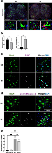
Confocal images (20X magnification) were used after triple immunofluorescence staining of lumbar 3–4 (L3–4) segments of spinal cord ventral horn using microglia-marker, 1ba1 (red), neuronal maker, NeuN (green), nuclear maker, DAPI (blue) at Day 14 after sham burn (SB) or burn injury (BI). Motor neurons numbers appeared decreased and were smaller in size together with prominent microgliosis at day 14 after BI (inset of dotted white box and magnified at 63X bottom of Figure 1a). (At that magnification, a threshold higher than 25 μm was used to identify motor neuron numbers) [15]. When motor neuron numbers per field were counted, there was a significant decrease in the motor neuron numbers after BI (Figure 1b left). Quantitation of the occupied microglia area revealed a significant dorsal and ventral horn microgliosis in the BI mice (Figure 1b right). Some of the microglia processes (red) in BI injured mice were juxtaposed to the green stained neurons (magnified images in bottom right of Figure 1a). To examine if the MNL was due to apoptosis, in situ TUNEL assay was performed in the ventral horn together with counter staining of neurons (NeuN) and nuclei (DAPI) to quantify the extent of neuronal apoptotic cell death (Figure 1c). Compared to SB, BI mice showed increased TUNEL positive nuclei within the neuron's indicative of neuronal cell death (Figure 1c, white arrows point to positive TUNEL staining within neurons in the merged figure indicative of neuronal apoptosis). The spinal cord sections were also immunofluorescent-stained against activated cleaved caspase-3, a pivotal enzyme causing apoptotic cell death (Figure 1d). BI mice showed significantly increased of caspse-3 at day 14; (Figure 1d). The merged figure shows that the increased caspace-3 activity was within the neuron. RT-PCR of caspase-3 normalised to GAPDH (internal control) showed significantly increased transcripts in the ventral horn of BI mice at day 14 (Figure 1e). Overall, these immunofluorescent staining data indicate significant microgliosis, increased apoptosis, and decreased motor neuron numbers together with caspase-3 mediated apoptosis of neuronal death in the ventral horn of BI mice.
3.2 BI Leads to Microglia Inflammatory Cytokine Release in the Spinal Cord Ventral Horn
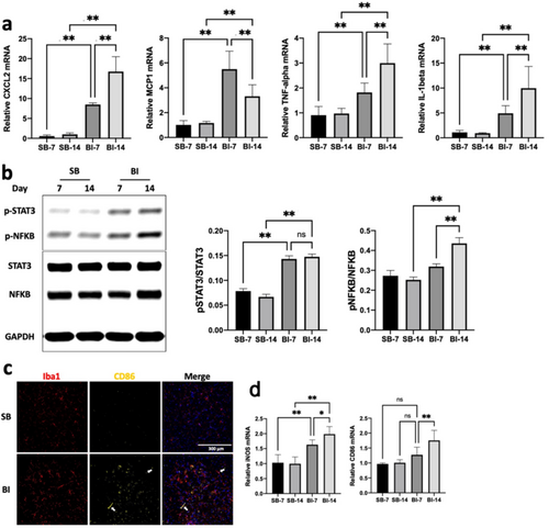
In the following studies, we examined if the microgliosis induced by BI leads to microglia activation evidenced as pro-inflammatory cytokine and chemokine release; the release of inflammatory cytokines can lead to neuronal damage including motor neuron loss as shown in pathologic state of encephalitis, ALS and other motor neuron diseases [18-22]. The microgliosis-mediated cytokine and chemokine release was, therefore, measured by RT-PCR and included pro-inflammatory IL-1β, TNF-α, CXCL2 and MCP-1(a.k.a., CCR2) at day 7 and 14 after BI. All cytokines and chemokines enumerated above were significantly upregulated at day 7 and day 14 (CXCL2: p < 0.0001, p < 0.0001, MCP1: p < 0.0001, p = 0.0023, TNF-α: p = 0.0057, p < 0.0001, IL-1β: p = 0.0003, p < 0.0001, for day7 and day14, respectively, Figure 2a). Additionally, innate immune inflammatory-transducer-proteins, pSTAT3 and pNF-κB, were significantly upregulated at both 7 and 14 days after BI (Figure 2b). Furthermore, the microglia expression of CD86, a pro-inflammatory M1 phenotype marker (Figure 2c), was increased in the ventral horn of BI mice in a time-dependent manner along with increased transcripts both CD86 and iNOS, both pro-inflammatory markers of M1 phenotype (Figure 2d).
3.3 Burn Injury Induces Neuromuscular Junction (NMJ) or Synaptic Denervation and Muscle Mass Loss
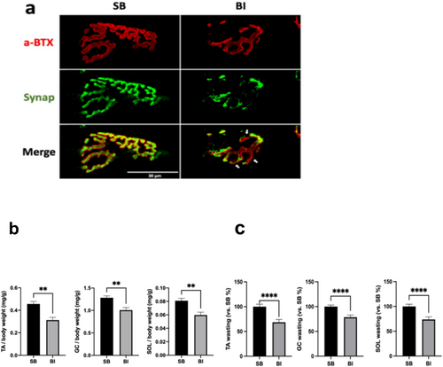
To explore if MNL observed in the ventral horn of BI mice has distant downstream effects, the morphology of NMJ (synapse) was examined. In SB mice, the NMJ in the tibialis anterior muscle showed the typical pretzel-like shape with smooth boundary, suggesting the normal aggregates of acetylcholine receptors (Figure 3a, labelled by α-bungarotoxin). The presynaptic nerve terminal stained with synaptophysin showed good apposition of nerve to muscle AChRs confirming normal innervation (merged Figure 3a bottom). After BI, however, the muscle and nerve (synapse) margins disintegrated and fragmented with irregular edges. In some parts of the NMJ, staining of presynaptic nerve terminal was absent suggesting partial denervation in BI. In the SB mice, the area of post-synaptic AChR occupancy (stained with α-BTX) closely matches the area of presynaptic nerve terminals (stained with synaptophysin), with an occupancy rate of 71.8%. In contrast, BI mice exhibit a significantly reduced occupancy rate of 39.6% of the nerve terminal with the AChRs. These changes in NMJ morphology indicated that BI caused disruption of the NMJ or synapse, mimicking a denervation state which could have implications for anterograde and retrograde signals to and from muscle to nerve and vice versa. [11, 12] Further the downstream effects of MNL and synaptic disruption on muscle were next examined by assessing muscle mass in the tibialis anterior (TA), soleus (SOL), and gastrocnemius (GC) at day 14 after BI. To exclude the effects of edema on muscle mass, the dry muscle weight was measured relative to pre-burn body weight. Compared with SB group, the muscle mass of the TA, SOL, and GC normalised to pre-burn body weight were significantly (Figure 3b). Conversion of the muscle masses relative to SB controls, the TA, SOL, and GC were decreased 31.7%, 23.4%, and 27.5%, respectively at day 14 after BI (Figure 3c).
3.4 GTS-21 Mitigates Microgliosis and Inflammatory Phenotype in BI WT but Not in the A7KO Mice
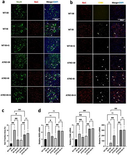
Previous studies have documented the expression of α7AChR on the microglia and stimulation of α7AChR leads to mitigation of inflammatory cytokine and chemokine release [13, 14, 26]. GTS-21, a selective α7AChR agonist, was administered in vivo to exploit the anti-inflammatory properties exhibited by α7AChRs expressed in microglia. GTS-21 effects on microglia cytokines and chemokines, MNL and nuclear changes were examined in the ventral horn. In order to minimise rodent numbers, the effects of GTS-21 were tested in wild type and A7KO mice with and without BI only. (The effects of GTS-21 on WT-SB and A7KO were not performed). As previously described in Figure 1, BI caused spinal microgliosis compared to SB; the administration of GTS-21, however, reduced the BI-induced microgliosis in the wild type mice (Figure 4a). The microgliosis was more significant (p < 0.05) in the α7KO mice even with SB (126% more than WT-SB). BI-associated increased protein expression of inflammatory markers and CD86 was suppressed by GTS-21 (Figure 4b). The microgliosis with BI was further exaggerated with BI to A7KO mice (84.4% increase from A7KO-SB) and was unchanged by GTS-21 treatment (Figure 4c). The succeeding studies examined if the mitigation of microgliosis with GTS-21 was associated with suppression of inflammatory phenotype. Consistently, the increased transcriptional expression of iNOS, and CD86 were significantly mitigated by GTS-21 (Figure 4d). The marker for anti-inflammatory subtype of microglia, Ym-1 [27] was increased by GTS-21 treatment in the WT-BI animals; GTS-21 did not change the expression of Ym-1 in A7KO mice with BI (Figure 4d).
3.5 Acute Anti-Inflammatory Effects of GTS-21 on Spinal Inflammatory Responses
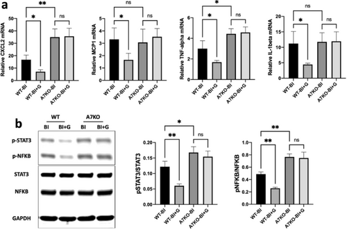
These experiments explored further if the beneficial effects of GTS-21 affected the inflammatory cytokines and the other inflammatory transducer proteins. Quantitative RT-PCRs for mRNA expression in the ventral horn were performed and normalised GAPDH in WT-BI and A7KO with and without GTS-21 at day 14. The administration of GTS-21 significantly ameliorated inflammatory cytokine and chemokine expression of IL-1β, TNF-α, and CXCL2, MCP-1, respectively, in the wild type mice (Figure 5a). The beneficial effect of GTS-21 was, however, nullified in the A7KO mice. Immunoblots of lumbar spinal cord (L3-L4 segments) isolated from BI mice were probed for STAT3 and NF-κB and normalised to GAPDH as loading control. The probed immunoblots were quantified to measure band intensities of target proteins (phosphorylated vs. total molecule). The increased levels of the pSTAT3 and of pNF-κB of BI were significantly reduced by GTS-21 treatment in the wild-type (WT) mice. GTS-21 treatment of BI A7KO mice showed no mitigation of the STAT3 and NF-kB phosphorylation indicating the specificity of GTS-21 (Figure 5b,c).
3.6 GTS-21 Alleviates BI-Induced Motor Neuron Loss and Neuronal Apoptosis
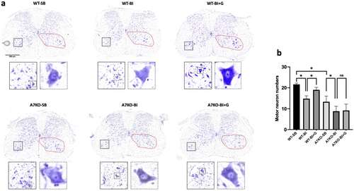
Microglia activation with its concomitant cytokine release results in deleterious effects on the motor neuron in many neurological diseases including amyotrophic lateral sclerosis and Parkinson's disease [17, 18]. In the following studies, the anti-inflammatory properties exhibited by α7AChRs in microglia were capitalised using GTS-21 as agonist to mitigate microglia and neuronal changes. Nissl-stained spinal cord dorsal and ventral horn of the SB and BI mice with or without GTS-21 were examined in wild type or A7KO mice. In the ventral horn of the wild-type SB mice, the typical dark blue staining of the peripheral cytoplasmic portion due to the presence of Nissl bodies was observed (Figure 6a, inset below- 400X magnification); in the wild type with BI without GTS-21 treatment, the spinal cord central motor neuron nucleus was lightly stained and less obvious, the overall cell shape was swollen and the clear cytoplasm around the nucleus was absent (Figure 6b- boxed inset 400X magnification). The numbers of morphologically normal motor neurons were fewer (31.6% reduction compared to SB, satisfying threshold criteria of 25 μm or higher). GTS-21 treatment partially reverted these pathologic changes (improved to 12.3% reduction from SB, central nucleus was more visible together with the central cytoplasm Figure 6b). Of note, the Nissl-stained spinal cord of A7KO mice with SB showed reduced motor neuron numbers (61.5% of WT even with SB). BI exacerbated the pathologic morphology in the A7KO mice (33.8% reduction from A7KO-SB, Figure 6a). The favourable effects of GTS-21 were not observed in the A7KO mice suggesting an important role of α7AChRs in the preservation and/or reversal of the damaging effects of microglia on neurotransmission from motor neuron to muscle.
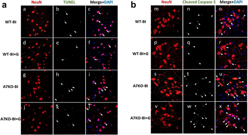
Next, to understand the mechanisms of MNL and its reversal by α7AChR stimulation by GTS-21, the extent of neuronal apoptosis in the ventral horn was analysed using in situ TUNEL assay. GTS-21 treatment reduced the extent of neuronal cell death in WT mice, but not in A7KO mice (Figure 7a). Similarly, cleaved caspase-3, as shown by the immunofluorescent staining showed suppression of caspase-3 activation by GTS-21 treatment in the WT, but not in A7KO mice (Figure 7b).
3.7 Prevention of Motor Neuron Loss by GTS-21 Mitigates NMJ Disintegration and Muscle Wasting
These experiments explored the downstream effects of prevention of MNL by GTS-21 treatment on both the synaptic denervation and MW induced by BI. As compared to BI mice without treatment, GTS-21 rescued the disintegration of the endplate, and mitigated the partial denervation in the WT mice, but not in the A7KO mice. In wild-type BI mice not treated with GTS-21, the area of presynaptic nerve terminals (stained with synaptophysin) was reduced relative to the area of postsynaptic AChR occupancy (stained with α-BTX), indicating partial denervation (occupancy rate: 27.4%). Treatment with GTS-21 significantly ameliorated this denervation, restoring occupancy to 71.3%. However, in A7KO knockout mice, the reduced occupancy observed after burn injury (31.4%) showed no significant improvement following GTS-21 treatment (35.8%) (Figure 8a). It should be noted that a 70% occupancy in terms of area size comparison corresponds to near-perfect apposition of the pre- and postsynaptic apparatus, as postsynaptic receptors typically occupy a larger anatomical area because AChRs extend to the primary and secondary clefts on the muscle.
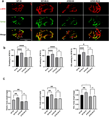
Furthermore, muscle weights of tibialis, soleus and gastrocnemius were significantly increased with GTS-21 treatment of the wild-type BI mice, but not in A7KO mice (Figure 8b). In other words, the treatment of BI wild type mice with GTS-21 significantly increased the muscle of tibialis, gastrocnemius and soleus muscle weights by 25%, 15%, and 20%, respectively (Figure 8c). These results indicate that GTS-21 mitigates microglia-mediated neuroinflammation and reduced MNL by apoptosis, which in turn had beneficial effects on the synaptic denervation and MW changes of BI (Figure 8a-c).
4 Discussion
The salient findings of this study are that: 1) BI leads to microgliosis together with ventral horn MNL due to apoptosis mediated at least in part by caspase-3 activation (Figure 1); 2) The microgliosis and microglia activation of BI increased the expression of transcripts of pro-inflammatory markers IL-1β, TNF-α, CXCL2 and MCP-1 (a.k.a., CCR2), CD86, and iNOS and protein expression of pSTAT3 and pNF-κB, reflecting the inflammatory status of the spinal microglia (Figure 2); 3) synaptic disintegration mimicking denervation and muscle protein loss occurred in hind limb muscles distant from body burn (Figure 3); 4) GTS-21 decreased microgliosis and microglia activation exemplified by the decreased expression of inflammatory markers enumerated above; 5) GTS-21 decreased pro-inflammatory responses and increased the anti-inflammatory marker Ym-1 (Figure 4); 6) Neuronal apoptosis was also mitigated by GTS-21 (Figure 7); 7) The typical dark blue staining of the cytoplasm due to Nissl bodies in motor neurons was less obvious due to lighter staining and cell was swollen in WT-BI mice (Figure 6). Neuron numbers were also decreased (31.6% reduction compared WT-SB mice, Figure 6). GTS-21 ameliorated the ventral horn neuron loss in the WT mice (improved to 12.3% reduction from untreated WT-BI); 8) GTS-21 ameliorated synaptic disintegration and muscle mass loss (Figure 8). The beneficial anti-inflammatory effects of GTS-21 were absent in the A7KO mice.
Most previous studies on MW of BI, focusing on reversal of the muscle hyper-catabolic and hypo-anabolic state and/or providing enhanced nutritional/anabolic supplements, have not rectified the MW of BI [5-8]. Ma et al., previously provided evidence that microglia activation and MNL but their relationship to synaptic disintegration and MW was not provided [15]. The current studies using GTS-21 to take advantage of the anti-inflammatory properties exhibited by α7AChRs expressed by immune microglia cells and demonstrated that GTS-21 decreased microglia activation led to mitigated MNL, which in turn alleviated the synaptic disintegration and muscle mass changes. Since α7AChRs are expressed in macrophages too, it is possible that some of the improvements in muscle mass could be attributed to anti-inflammatory properties exhibited by macrophages directing on muscle. However, the decreased MNL with GTS-21 cannot be attributed to decreased macrophage inflammation but attributable to the decreased microglia activation by GTS-21. This reveals a cause-and-effect relationship between decreased the spinal inflammatory proteins and improved motor neuron changes. The fact that even in the non-injured (SB) state, the MNL and muscle mass was lower in the A7KO mice relative to age-matched non-BI wild type mice suggest that even in the basal non-injured (burned) state, the α7AChRs have a protective role in the maintenance of spinal homeostasis. One may pose the question as to why there was no overt evidence of muscle weakness in A7KO mice in the basal or BI injured despite the MNL and muscle wasting. This is most likely related tremendous margin of safety of neuromuscular transmission. This scenario is akin to that seen early in the aged where motor neuron changes and denervation occurs but muscle wasting is not evident till late in the disease [28].
A fraction of CD86 positive cells in the spinal cord were Iba1-negative (Figure 2c); infiltrating macrophages/monocytes could account for this and contribute augmented spinal neuroinflammation of BI. Furthermore, the circulating innate immune macrophages also constitutively express α7AChRs. Our studies presented here do not distinguish GTS-21 effects on the circulating inflammatory macrophages. The cross talk between systemic macrophages and muscle has also received minimal attention both in BI and other MW conditions and needs exploration. Future studies [29, 30] could verify the pivotal roles of macrophages vs. microglia in the spinal cytokine, neuronal and muscle changes.
The altered inflammatory phenotype of microglia was manifested by the significantly upregulated transcript-markers of inflammation (Figure 2). This inflammatory soup probably affects neuronal homeostasis and ultimately even survival as demonstrated in many neuropathologic states such as ALS, encephalitis, and Alzheimer's disease where neuro-inflammation also leads to muscle changes [31-33]. The microglia-mediated MNL after BI may explain the well documented phenomenon that even 5 years after BI, despite continued anabolic therapy and exercise, the MW of BI has not been reversed to normal [2-8].
In the normal mature muscle, in addition to anterograde signals from motor neuron to muscle, muscle factors acting retrograde induce differentiation signals to the presynaptic motor axon to maintain not only synaptic and nerve integrity but also motor neuron stability [34-36]. In some disease states (e.g., ALS) a significant increase in the expression of inflammatory markers (CD11b and CD68) and inflammatory cytokines (IL-1β and TNF-α) was observed in muscle together with degenerative processes in the neuromuscular junctions very early in disease development [37]. Concordantly, interleukin-6 (IL-6) release effecting bone morphogenic protein (BMP) function leads to loss of protective muscle to nerve signals with denervation and MW in cancer [12, 38]. These lines of evidence suggest that BI, cancer and neurodegenerative diseases share some common traits regarding synaptic denervation and MW and support the mutual dependency of nerve terminal and skeletal muscles for maintenance of their integrity involving anterograde and retrograde trophic signalling. Thus, it is possible that some of the synaptic changes are related to direct effects of systemic inflammation on muscle; a contributory direct effect of systemic inflammation on muscle to cause the synaptic disintegration cannot, therefore, be discounted.
Our current studies demonstrate that BI-induced systemic inflammation can lead to neuro-inflammation and caspase-3 mediated apoptotic MNL, which affects distant muscles. The stimulation of α7AChRs by selective agonist, GTS-21, ameliorates not only the spinal inflammatory changes and MNL by decreasing cleaved caspase-mediated apoptosis, but also improves synaptic denervation and MW. These beneficial effects by GTS-21 were completely nullified in α7 AChR knockout mice, confirming the specific protective role of α7 AChR stimulation and the anti-inflammatory therapeutic effects of GTS-21. The α7AChR has modulatory effects even in uninjured state as demonstrated by greater spinal and muscle changes in the uninjured A7KO mice. GTS-21 administration in BI patients may prove useful for mitigation of MW.
Author Contributions
JY. C contributed to the design and conduction of this study, data analysis, and writing the final manuscript. Y. K. and F. X. participated in establishing the animal model and recording results. WRK provided the GTS-21 and advised on GTS-21 therapeutics. ZY and HL helped with spinal cord biochemical measurements and analysed the data. S. Y was involved with morphology experiments and JAJM initiated the project and continued give suggestions on next experiments. SY and JAJM read the final version of the manuscript. All authors read and approved the final manuscript.
Acknowledgements
JAJM was supported in part by Grants from NIH GM RO1-142042 and Shriners Hospitals Research Philanthropy (Fund # 85300 and #85500). Dr. Yasuhara is supported in part by grants from the NIH RO1-142042 and Shriners Hospital Research Philanthropy (Fund # 85124).
Conflicts of Interest
The authors declare that they have no known competing financial interests or personal relationships that could have appeared to influence the work reported in this paper.


