Lung CT image enhancement based on total variational frame and wavelet transform
Funding information: This work was supported in part by the National Natural Science Foundation of China (62005285) and Youth Innovation Promotion Association CAS (2017429). The authors would like to thank the Cancer center of Sichuan province for the permission to use data.
Abstract
Benefitting from the development of computer vision, computed tomography (CT) images have been used for assisting doctor's clinical diagnosis and improving the diagnostic efficiency. However, there exist some issues in medical images, such as low contrast, obscure detail, and complex noise due to the restriction of the system and the equipment in the process of imaging, medical images. To resolve these issues, a novel lung CT image enhancement method based on total variational framework combined with wavelet transform is proposed. Firstly, low-frequency structure layer with low contrast and high-frequency details layer with complex noise signals are acquired by decomposing the original image using total variational framework. Then, through the analysis of the histogram distribution characteristics of CT image, structure layer requires contrast enhancement; at the same time, the detail layer performs wavelet transform adaptive threshold denoising to remove noise. Finally, weight fusion of processed structure layer and details layer is performed to obtain the final fusion enhancement CT images. Experimental results show that the proposed method can enhance the contrast of clinical lung CT images, improve the clarity of details, and effectively suppress artifacts and complex noises. Contrast and sharpness—objective indicators—prove the proposed method's advantages. Subjectively, the proposed method performs superior over other existing CT enhancement methods, which achieves a better visual recognition.
1 INTRODUCTION
Due to the inherent properties of medical imaging and the restrictions of system equipment, there are several prominent features of lung computed tomography (CT) images, such as low contrast, noisy mask, and blurred details, which are not conducive to the recognition and analysis of clinicians. As a consequence, clinicians suffer from image noise for a long time. Hence, the improvement of the human visual perception of the lung CT image is conducive for doctor to carry out more accurate clinical diagnosis of lesions and reduce the misdiagnosis rate. Consequently, lung CT image enhancement acting as a preprocessing procedure can increase the accuracy and efficiency in actual medical applications, and has important clinical application value. Many scholars have conducted extensive research in this field.1-10
This paper reviews mainstream image enhancement methods, including traditional histogram stretch methods,11-14 image domain transformed methods,15, 16 image enhancement methods based on joint framework, and so on. Traditional image enhancement methods are mainly divided into spatial enhancement and frequency enhancement, which are used to enhance simple binary image. Existing image domain transformed enhancement methods, for instance, wavelet transform, have no conspicuous enhancement effect on lung CT images. Hence, this paper has a conclusion that single framework was unable to resolve the problem well of blurred details caused by complex noise. Meanwhile, this work finds joint frameworks received more and more attention in recent years, for its capability of integrating the advantages of several algorithms and better solution for complex noise model.
Complex noise is defined by integrating the multiplicative noise and the additive noise, including environment noise, equipment noise, transmitted noise, artificial artifacts, and so on, which contribute to CT image noisy mask, blur tissue boundary, low contrast, and unclear details.17-19
Existing CT image enhancement methods that deal with noise models are insufficient, such as only considering single noise model, or taking no account of artifacts produced by contrast enhancement unintentionally.18 Thus, a novel enhancement algorithm based on total variational framework and wavelet transform is proposed in this paper. The proposed framework has considered several noise models, such as environment noise, equipment noise, and artificial artifacts, and those noise models are the so-called complex noise uniformly. Experiments show that the proposed algorithm suppresses the complex noise effectively, improves the sharpness and clarity of details, and enhances the contrast of lung CT images. The objective indicators prove the proposed method is superior compared with other existing CT enhancement techniques.
Contributions of this work are presented as follows: a more complete noise model than existing medical image enhancement methods; proposes a new adaptive polarization adjustment method; applies total variation with wavelet transform on the CT image enhancement.
The details of the proposed algorithm as well as comparisons with other methods are discussed in the following sections. Then, comparing the experimental results of the proposed method with other recent algorithms. Conclusion and discussion about this work in the end.
2 RELATED WORK
The related work reviews and discusses relevant work in the area of image enhancement methods.
2.1 Spatial enhancement methods
In 2020, Yuebin Zhou et al. proposed a new improved optimized method for medical image enhancement, based on global and local enhancement. The algorithm achieves great contrast enhancement but ignores medical image noise.5 In 2020, Julio et al. proposed a medical image enhancement method using multiscale top-hat transform by reconstructing multilevel features. The method improves contrast and detail, but performs poor generalization ability on some images with abundant details.7 In 2021, Acharya U K et al. proposed a genetic algorithm based adaptive histogram equalization (GAAHE) technique for medical image enhancement. The algorithm gave better results on enhancing contrast than other histogram equalization techniques, but without the consideration of any noise model.3
2.2 Frequency enhancement method
The frequency-domain enhancement method is a kind of method that performs the Fourier transform on image and then uses the filter to handle it. Wavelet transform (WT) is a widely used enhancement method in the frequency domain. It takes advantage of multiscale transformation and good time-frequency analysis characteristics.15 In 2020, Zhi Li et al. proposed an efficient and high-quality medical CT image enhancement algorithm, which utilizes a two-dimensional WT. Though the algorithm performs better visual effect on some fuzzy medical images, it has a simple denoising part.4 In 2020, Ullah Z et al. proposed a hybrid image enhancement method based on two-dimensional WT and feature reduction using color moments. The approach is not friendly to image details due to applying median filter and histogram equalization.9
2.3 Joint framework
In recent years, the combination of multiple image enhancement techniques has become one of the research hotspots in the field of image processing. The joint framework can effectively solve complex problems.
In 1992, Rudin and Osher et al. proposed a nonlinear partial differential algorithm model based on noise removal,20 which was also known as the ROF model. This model introduced total variation into the field of image denoising for the first time and was able to separate most of the noise while preserving the majority of image details.
In 2017, a multi-channel block-matching denoising algorithm for spectral photon-counting CT images was proposed, which had consideration for a photon multiplicative noise model.21 In 2018, Wending Xiang et al. proposed an underwater image enhancement method based on red channel weighted compensation and gamma correction model, which effectively corrected the image distortion caused by serious attenuation of red channel information in underwater images.22 In 2019, Zhang Hong et al. proposed an image denoising method based on wavelet threshold and total variational model, which could overcome the problem of image edge blurriness while removing image noise.23 In 2019, Chunyan Li et al. proposed a non-convex regularization method based on total variation and wavelet framework to remove the multiplicative noise, which could preserve the edges and details of the image more effectively than any single regularization method.24 In 2021, Janan et al. proposed a method for quantitative mammographic image enhancement from the perspective of density. This algorithm might perform better combined with more precise denoising method.6
3 METHODOLOGY
3.1 Characteristics analysis of lung CT image
3.2 Total variational framework
Total variational image denoising model represents the image as an energy functional; denoising is realized by subtraction of the energy of the image noise under the constraint conditions. Therefore, the image denoising problem is transformed into an energy minimization problem under the total variational framework. The image denoising algorithm based on the total variational framework can not only process the additive noise of the image26 but also be suitable for processing the multiplicative noise of the image.24
Where the first item on the right is the data fidelity item to ensure the approximation degree between the denoised image and the original image. The second term is regularization term, which has the function of denoising and smoothing. λ is the regularization parameter for balancing the data item and the regularization item.
It is well known that a common practice in resolving computational photography problems is to decompose images into different layers and recombine them; total variational framework is good at decomposing image.
The detail layer ID is obtained by calculating the difference between the input image I and the structure layer IS, ID = I − IS. λ is the regularization parameter that controls the separation of structure layer and detail layer, and its value is related to the compression artifact.
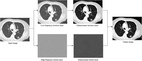
As shown in Figure 1, a lung CT image is resolved into the low-frequency structure layer IS and high-frequency detail layer ID through total variational decomposition from the perspective of image decomposition.18 As we can see, the structure layer retains the main structure of the image, while the detail layer reflects a deeper level of detail. The low-frequency structure layer has removed complex noise, but retained the characteristics of low contrast, thus there needs to perform contrast enhancement on the structure layer, which contributes to the recognition of lung CT image well for clinician. Meanwhile, the original detail layer contains the deep level image detail and the entire complex noise, and there needs more refinement manipulation to deal with the detail layer.

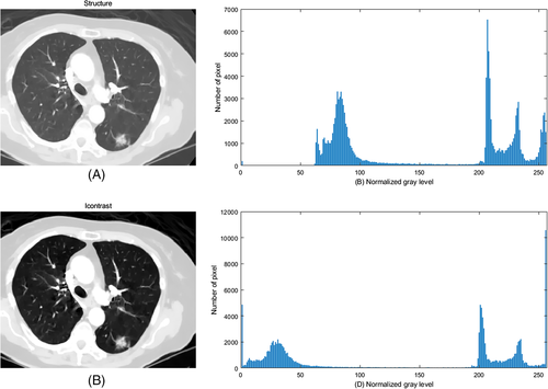
Through the analysis of the histogram of the original CT image, as shown in Figure 2, the reason for low contrast is that pixels are concentrated in the middle of dark and bright regions. An adaptive polarization adjustment method was proposed to improve contrast straightforwardly by referring to studies in the literature,28, 29 and Figure 2 shows its efficiency.
3.3 Wavelet transform
Where ψ and Φ are wavelet function and scale function, respectively.
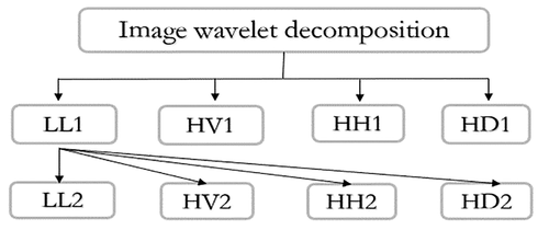
As shown in Figure 3, the four sub-images are low-frequency subband map LL1, horizontal high-frequency subband map HH1, vertical high-frequency subband map HV1, and diagonal high-frequency subband map HD1, respectively. Likewise, wavelet decomposition is performed on LL1 again to obtain LL2, HH2, HV2, and HD2. The low-frequency subband image retains the main structural information and the majority of complex noise, while the high-frequency subband map mainly preserves the details of the original image.31
For lung CT images, the different singularities of signal and noise in the wavelet domain indicate that the wavelet coefficient of effective signal is larger than that of noise. Therefore, the noise can be effectively removed by selecting appropriate thresholds of wavelet coefficient for filtering. Thresholding was performed only on the wavelet coefficients that do not correspond to any image features. Wavelet threshold denoising is to set the coefficient whose absolute value is lower than the threshold to zero to obtain the estimated wavelet coefficient after processing, and then use the estimated wavelet coefficient to carry out wavelet signal reconstruction, and the obtained signal is the denoised signal.23
3.4 The proposed method
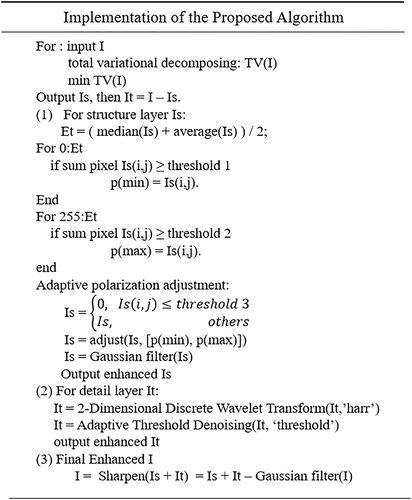
From the perspective of image decomposition, the proposed frame adopts the total variation model to carry out the decomposition on the original image to obtain high-frequency detail layer and low-frequency structure layer, which reduces the artifacts significantly. Then two-dimensional discrete wavelet transforms and an adaptive threshold denoising are applied to the high-frequency detail layer that suppresses complex noise, and contrast enhancement and Gaussian filter are performed on the low-frequency structure layer, which improves the contrast. Finally, the results of two parallel channels are weighted processed sub-images to acquire the final enhanced image.
In detail, note that complex noise is widely distributed on the original CT image or high frequency the details layer through the analysis of image noise model, while structure layer keeps low contrast, further refined processing is necessary.25, 32 The artificial artifacts produced by contrast enhancement unintentionally can be processed by thresholding and Laplace filtering; complex noise can be suppressed through wavelet transform and adaptive threshold denoising in the meantime.
The specific implementation of the proposed framework is shown in Figure 4.
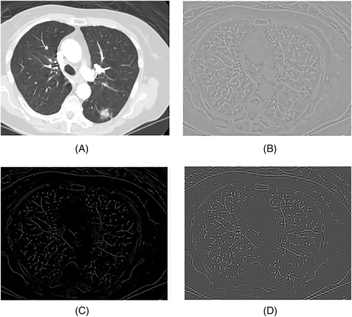
The ROF algorithm can filter out any compression artifacts effectively while generating the structure layer, consequently the detail layer retains complex noise.18 Therefore, the low-frequency structure layer can be directly enhanced, while the high-frequency detail layer needs to conduct more refined denoising. As shown in Figure 5, image (C) is obtained by performing wavelet transform adaptive threshold denoising algorithm on the image (B), then image (D) is acquired through denoising and sharpening the image (C).
Where HS(•) denotes adaptive polarization adjustment, which stretches the dark part and the bright part to polarization. GF(•) denotes Gaussian filter, operator is the Laplacian with parameter zero. DWT(•) denotes two-dimensional discrete wavelet transform where applies the wavelets harr. ATD(•) denotes adaptive threshold denoising, threshold parameter is 0.05. SP(•) denotes sharpen operator, Gaussian filter operator is the Laplacian with parameter zero.
The overview of lung CT image enhancement algorithm is shown in Figure 6; meanwhile, the flowchart of the algorithm is shown in Figure 7.

4 EXPERIMENT RESULTS AND ANALYSIS
The experimental platform is Intel(R)Core (TM) I7-7700HQ processor, the images are from the open data set LIDC-IDRI. To evaluate and verify the performance of the proposed algorithm, this work selects a few lung CT images and chooses several state-of-art enhancement algorithms as comparison.
The proposed algorithm is compared with genetic algorithm based adaptive histogram equalization technique (GAAHE),3 low-dose lung CT image enhancement algorithm based on image decomposition (IDE),25 and a CT image enhancement algorithm based on wavelet transform and histogram equalization (WTHE).4 In the experiment, the experimental parameters are as follows: the iteration of total variational decomposing is 200, lambda is 0.055, the wavelet basis is harr. The experimental results of the above algorithms are compared and analyzed as follows.
4.1 Subjective evaluation
Medical image enhancement aims to enhance the image information, such as tissue, organ, vessel, and lesions, which doctors ar e interested in. Therefore, the algorithm should retain complete detailed information as much as possible while enhancing human visual perception, to help doctors to reduce the clinical misdiagnosis rate. No matter the image is clear or distorted, the image shall eventually be reviewed by doctors, whose subjective image quality evaluation is necessary.33
Figure 8 shows the results of three different lung CT foreground images processed by image enhancement algorithms, the first column (a) are original images. Through subjective observation and analysis, GAAHE method improves contrast but blurs details, resulting in poor visual effect, whereas the proposed method shows superior on these aspects. Then, IDE method enhances contrast while preserves image detail information as much as possible, but it fails to deal with detail sharpening in the lung region and overall contrast, leaving behind blurriness still, whereas the proposed method does well in removing artifacts.
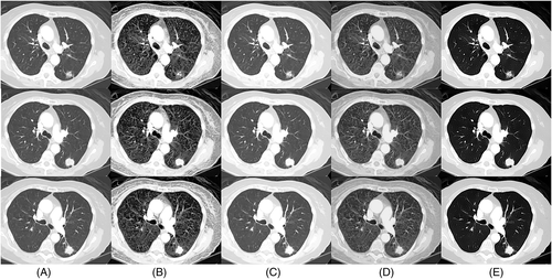
Next, WTHE method enriches the details, whereas this causes misleading possibly for true information, which doctors need recognition, whereas the proposed method makes the interesting details clearer. According to the subjective observation and true assessment of doctors, the proposed method performs well than three compared algorithms in keeping details clear, removing artifacts, denoising, and global sharpness.
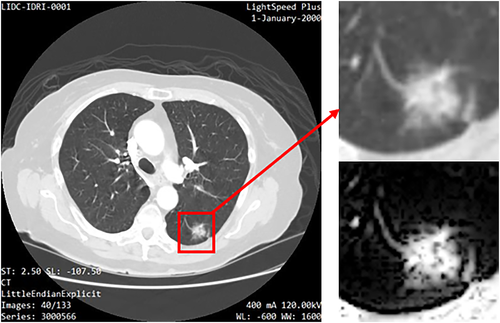
Figure 9 shows the result of the pulmonary nodular region of lung CT image processed by proposed algorithms. The proposed algorithm not only improves the sharpness of details in visual but also greatly restrains the complex noise, eliminates the noise mask, and improves the clarity of the boundary and the overall contrast.
4.2 Objective evaluation
Because medical image enhancement is an inverse problem, the solutions are not unique, so the full reference image quality evaluation indexes PSNR and SSIM as objective evaluation parameters cannot accurately evaluate the actual image quality. Thus, this paper selected contrast and sharpness among the non-reference image quality evaluation parameters as objective evaluation indexes of the processing results of different algorithms.34
Mostly, the larger contrast denotes the richer image level and the more legible visual perception. The larger sharpness indicates the lower blur, with the larger weight ratio of the high-frequency component of the image. All evaluation indicators reflect the clarity of the image.
As shown in Table 1, GAAHE algorithm improves contrast by 100 percent nearly, which performs well than IDE and WTHE in contrast enhancement. But the proposed algorithm improves the image contrast by an average of 9.906% than GAAHE algorithm, while performs superior in visual. Hence, the performance of the proposed algorithm is better than existing algorithms.
As shown in Table 2, the sharpness of IDE methods is equal to the original image almost, which means IDE has no improvement in image sharpness. GAAHE methods have the highest value in sharpness, whereas extra details can lead to misunderstanding for clinical diagnosis. The proposed algorithm performs better than WTHE methods and IDE from the value and visual effect.
All experiments show that the proposed algorithm can not only enhance the foreground image but also handle with local image well. Therefore, the proposed method has excellent robustness.
5 CONCLUSION
Considering the defined complex noise model, CT image is decomposed into low-frequency structure layer with poor contrast and high-frequency detail layer with complex noise by using the total variational framework from the perspective of image decomposition.
Aiming at the high-frequency detail layer of the image, the wavelet transform is used to transform it to the wavelet domain. The wavelet coefficient of noise is processed and reconstructed to obtain the enhancement detail layer. In view of the poor contrast of low-frequency structure layer, by means of analyzing the histogram characteristic, the adaptive polarization histogram adjustment and Gaussian filter are carried out to obtain the contrast-enhanced structure layer. Finally, the enhanced detail layer and structure layer are fused and sharpened to obtain the enhanced image.
In view of the characteristics of lung CT images such as poor contrast, complex noise, and artificial artifacts, this paper proposes an image enhancement algorithm that combines variation framework with wavelet transform, which performance is superior to existing algorithms in objective indicators and subjective visual effect. Experiments verify the effectiveness and robustness of the proposed algorithm on lung CT images.
6 DISCUSSION
The experimental results show that the proposed framework suppresses the complex noise, eliminates the artificial artifacts, and improves the contrast and detail clarity. In detail, this framework has considered complex noise model compared with GAAHE and WTHE methods. In fact, the methods GAAHE and WTHE perform poorly on details due to histogram equalization. Compared with the proposed framework, IDE algorithm considers noise model insufficiently, and sharpness is not enough, which verified on visual effect. Overall, the proposed framework has improved the accuracy of lesion identification for clinical imaging workers and lays a good foundation for further analysis or segmentation of lesions.
The proposed framework takes a complex noise model into account, while the compared experiments have given a positive answer. Because medical image processing is an inverse question, it is difficult to measure the difference accurately between the enhanced image and the true image, thus rigorous analysis about image model is still required, and future work shall consider this more carefully.
ACKNOWLEDGMENT
Hongfei Wang did conceptualization, software, formal analysis, data curation, visualization, and wrote the original draft. Ping Yang did review, supervision, funding acquisition, and formal analysis. Lei Min did conceptualization, validation, and methodology. Chuan Xu did investigation and validation. Shuai Wang and Bing Xu did supervision and review. The authors would like to thank the editor and reviewers for their comments and suggestions, which helped to improve the quality of the paper significantly.
Open Research
DATA AVAILABILITY STATEMENT
The data that support the findings of this study are openly available in LIDC-IDRI at https://wiki.cancerimagingarchive.net/display/Public/LIDCIDRI.


