PD-L1 promotes OCT4 and Nanog expression in breast cancer stem cells by sustaining PI3K/AKT pathway activation
Significance: We have demonstrated that PD-L1 has a direct effect on sustaining the subpopulation that can resist therapy and reinitiate tumor. Our findings suggest targeting PD-L1 would affect the pool of breast CSCs and have an important consequence on the efficacy of breast cancer therapy.
Abstract
The expression of PD-L1 in breast cancer is associated with estrogen receptor negativity, chemoresistance and epithelial-to-mesenchymal transition (EMT), all of which are common features of a highly tumorigenic subpopulation of cancer cells termed cancer stem cells (CSCs). Hitherto, the expression and intrinsic role of PD-L1 in the dynamics of breast CSCs has not been investigated. To address this issue, we used transcriptomic datasets, proteomics and several in vitro and in vivo assays. Expression profiling of a large breast cancer dataset (530 patients) showed statistically significant correlation (p < 0.0001, r = 0.36) between PD-L1 expression and stemness score of breast cancer. Specific knockdown of PD-L1 using ShRNA revealed its critical role in the expression of the embryonic stem cell transcriptional factors: OCT-4A, Nanog and the stemness factor, BMI1. Conversely, these factors could be induced upon PD-L1 ectopic expression in cells that are normally PD-L1 negative. Global proteomic analysis hinted for the central role of AKT in the biology of PD-L1 expressing cells. Indeed, PD-L1 positive effect on OCT-4A and Nanog was dependent on AKT activation. Most importantly, downregulation of PD-L1 compromised the self-renewal capability of breast CSCs in vitro and in vivo as shown by tumorsphere formation assay and extreme limiting dilution assay, respectively. This study demonstrates a novel role for PD-L1 in sustaining stemness of breast cancer cells and identifies the subpopulation and its associated molecular pathways that would be targeted upon anti-PD-L1 therapy.
Abstract
What's new?
Cancer cells that express the T-cell inhibitory molecule programmed death-ligand 1 (PD-L1) readily escape immune attack. In addition, PD-L1 expression contributes to chemoresistance and is associated with epithelial-to-mesenchymal transition, a process that generates cancer stem cells (CSCs). This study shows that in breast cancer, PD-L1 expression further plays a direct part in maintaining CSC stemness. In breast cancer cells, PD-L1 expression sustained stemness factors OCT-4A and Nanog, via a PI3K/AKT-dependent pathway, and promoted expression of the stemness controlling factor BMI1, independent of PI3K/AKT. Targeting PD-L1 could help advance breast cancer therapy, owing to impacts on the pool of breast CSCs.
Abbreviations
-
- PD-L1
-
- programmed death ligand-1
-
- CSCs
-
- cancer stem cells
Background
Breast cancer is the most common malignant disease and the second leading cancer-related death in women around the world.1 Despite the ongoing advances in the treatment and diagnosis of breast cancer, many patients suffer from tumor recurrence even after responding to initial treatment.2 The tumor recurrence in breast cancer is attributed to an intratumor heterogeneity and the existence of a subpopulation that can resist therapy and reinitiate tumor with all its heterogeneity.3 This subpopulation of cells is commonly called cancer stem cells (CSCs) due to their acquisition of some traits of normal stem cells including self-renewal ability.4
The role of immune system in clearing cancer cells has recently been appreciated after the major success in the blockade of the T-cell inhibitory molecule, PD-L1, which is expressed by cancer cells to evade immune response.5 However, beside its established role in the immune response, PD-L1 expression has intrinsic effect on cancer cells themselves (reviewed by Ritprajak et al.6) where it works as a “molecular shield” to protect cancer cells from cytolysis.7, 8
We have previously shown that PD-L1 is mainly expressed in a subset of hormone negative breast cancer patients and its expression correlates with bad prognostic markers.9 In following studies, we have shown that this molecule is associated with highly proliferating cells and contributes to chemoresistance.8, 10 More recently, we have found a reciprocal effect between PD-L1 expression and epithelial to mesenchymal transition (EMT), an oncogenic process, well known to generate CSCs.11
All these previous observations have intrigued us to investigate the role of PD-L1 in regulating CSC properties. In this study, we have shown that PD-L1 maintains breast CSCs by sustaining BMI1, and activating PI3K/AKT pathway.
Methods and Materials
Expression analysis of human breast tumor microarray datasets
The dataset consisted of mRNA expression profiling of breast invasive carcinoma samples (n = 530) performed using Agilent G450A_07 arrays from The Cancer Genome Atlas (TCGA) project (http://cancergenome.nih.gov),12 and TCGA level 3. The most highly processed data were downloaded in accordance with TCGA Data Access Policies (https://tcga-data.nci.nih.gov/tcga/) and used for analysis. Stemness score was calculated as the average expression of consensus stemness gene expression signature (CSR gene signature markers from ref. 13) for each sample. Correlations between continuous data were estimated by Pearson's correlation coefficient (r) and associated p values were computed by transforming the correlation to create a t statistic. All statistical analyses were performed with the MATLAB software packages (Mathworks, Natick, MA, USA), PARTEK Genomics Suite (Partek Inc., St. Lois, MO, USA) and SAS 9.4 (Statistical Analysis System, SAS Institute Inc., Cary, NC, USA). A p value of <0.05 was considered significant.
Cell culture and treatments
MDA-MB-231 and T-47D cells were maintained in DMEM supplemented with 7% fetal bovine serum (FBS, Invitrogen, USA). SUM-159 cells were cultured in DMEM/12 medium supplemented with 10% FBS, 1 µg/mL hydrocortisone and 5 µg/mL insulin (both from Sigma, USA). Cell lines were used within 6 months of purchase from ATCC, otherwise, they were authenticated using STR analysis (Promega).
Protein expression analysis was done on exponentially growing cells at 40–60% confluence unless otherwise stated. For the inhibition of PI3K/AKT/mTOR pathway, 50 µM LY294002 plus 100 nM of Rapamycin was used (all from EMD Calbiochem-Millipore, USA) as previously described.14
PD-L1 was downregulated using specific ShRNA to PD-L1 from OriGene, USA (RS vector, TR314098) as previously described.11 Among provided plasmids, #TI356387 was the most efficient in downregulating PD-L1 in MDA-MB-231 cells. Cells were cloned to obtain a stable PD-L1 knockdown. At least two clones were selected from the Sh-RNA line, their knockdown effect was confirmed (Supporting Information, Fig. 1) and they were designated as Sh-PD-L1 (a) and Sh-PD-L1 (b). To ensure that our findings are not due to off-target effects, we used another ShRNA (lentiviral GIPZ-GFP commercially available vector from openbiosystems, plasmid V2LHS_53668 and hence designated as (GIPZ-Sh-PDL1)). The statistical analysis for this part of the study was done using Student's t test as calculated by Excel.
Immunofluorescence
Immunofluorescence labeling was done as per the antibody provider (cell signaling) instructions with minor modifications. Briefly, cytospin-attached cells were air-dried. Cells were washed in PBS then fixed in 4% paraformaldehyde for 20 min at room temperature. Nonspecific binding was minimized using blocking/permeabilization solution (0.3% triton-X in 5% goat serum in PBS) for 1 hr. Primary antibody was added overnight at 4°C at the indicated dilution (Supporting Information, Table 1) in antibody buffer (1% BSA and 0.3% Triton X in PBS. After PBS washing, an appropriate Alexa conjugated secondary antibody was added at 1:400 dilution in addition to DAPI (both secondary antibody and DAPI are from Molecular probes, USA) for 60 min. Cells were mounted (VectaMount, Vector labs, USA) before analysis using Zeiss Axioimager Z2 (Zeiss, Germany) for image capture and BD pathway 855 image analyzer (Becton Dickenson, USA) for immunofluorescence quantitation.
Quantification of fluorescence intensity was done using BD pathway 855 system and a 20× objective (Olympus, NA 0.75) according to instrument standard protocols. Briefly, predefined analysis protocols (macros) were formed using a montage of 3 × 3, which had at least 2000 cells per montaged image. Data were analyzed in BD Image Data Explorer and the images from at least 4 different experiments were used to calculate the expression level of each studied protein. In addition, the number of cells considered positive for each studied protein was quantified using an arbitrary MFI cutoff that selects for around 50% of total analyzed cells for each experiment. All data were further normalized on the untreated control (Sh-Cont).
Proteomic analysis: Protein in-solution digestion and protein identification by mass spectrometry: LC-MSE
Prior to expression proteomics analysis, total whole cell lysate protein extracts (100 μg) derived from MDA-MB-231 human breast cancer cell lines were subjected to in-solution tryptic digestion as previously described.15 The protein identification was done using one-dimensional Nano Acquity liquid chromatography coupled with tandem mass spectrometry on Synapt G2 HDMS (Waters, Manchester, UK). The sample analysis was done on a Triazaic Nano source (Waters, Manchester, UK) and ionization in the positive ion mobility mode nanoESI as previously described.15, 16 The Progenesis QI for Proteomics version 2.0.5387 (Nonlinear Dynamics/Waters, Manchester, UK) was used for all automated data processing and database searching using the Uniprot database (www.uniprot.org) for protein identification. The data were filtered to show only unambiguous protein identification using multiple parameters including expected molecular mass, percentage coverage, peptides count, unique peptides and confidence scores.
Western blotting
Western blotting was done as previously reported.17 Briefly, cellular proteins were extracted using RIPA lysis buffer or SDS lysis buffer. Proteins were denatured and separated using SDS-PAGE and transferred to PDVF membrane. Membranes were incubated with primary antibodies diluted in PBST (in the presence or absence of 5% BSA) as per antibody data sheet. After using an appropriate secondary antibody, the signal could be developed using SuperSignal kit and visualized by ImageQuant LAS4010 Biomolecular Imager (GE Healthcare, Pittsburgh, PA, USA). Cytoplasmic and nuclear protein extracts were prepared as previously published.18
Flow cytometry and cell sorting
Cells were prepared for flow cytometry, acquired using LSRII and analyzed using DIVA software as previously described.19 Cells were sorted using FACS Aria as previously described.19
Mouse xenotransplantation studies
Animal work, including anesthesia and euthanasia, was done in accordance to protocols approved by the institution animal care committee and the Research Advisory Council (RAC# 2140–001) of King Faisal Specialist Hospital and Research Centre (KFSH&RC). MDA-MB-231 cells transfected with ShRNA that specifically targets PD-L1 (Sh-PDL1 (a)) or a scrambled control Sh-Cont were injected orthotopically in 6–8-weeks-old female NOD.Cg-Prkdcscid IL2rgtm1Wjl/SzJ, also known as NOD/SCID/IL-2Rneg/neg (NSG, obtained from Jackson Laboratories) or NU/J also known as nude mice. Mice were monitored weekly for tumor formation or any sign of illness/weakness and they were sacrificed after 11 and 12 weeks of injection of NSG and nude mice, respectively. The frequency of CSCs was calculated as reported previously20 using publically available website (http://bioinf.wehi.edu.au/software/elda/). Tumor volume was calculated as ½ (length × width2). Kaplan–Meier survival analysis curve was done using GraphPad Prism 5.
Results
PD-L1 expression in breast cancer is associated with stem-like features
To examine the relationship between PD-L1 expression and CSCs, we used publically available breast cancer patients' gene expression data (TCGA, n = 530 patients). To this end, PD-L1 expression was evaluated in relation to stemness score, which was calculated from a panel of stem cell gene signature as reported previously by Shats et al.13 In this large dataset, there was a statistically highly significant correlation (Pearson's r = 0.36, p = 1.7 × 10⁻¹⁷) between PD-L1 expression and stemness score (Fig. 1), strongly suggesting a positive association between PD-L1 expression and stem-like cells in breast cancer. Furthermore, we looked at the association between PD-L1 and other common stemness associated genes that were not part of the gene list used by Shat et al. There was also significant correlation between PD-L1 expression and these additional stem cell-associated genes as listed in Supporting Information, Table 2. Altogether, we concluded that there is a statistically highly significant correlation between PD-L1 expression in breast cancer and the expression of stemness-associated genes.
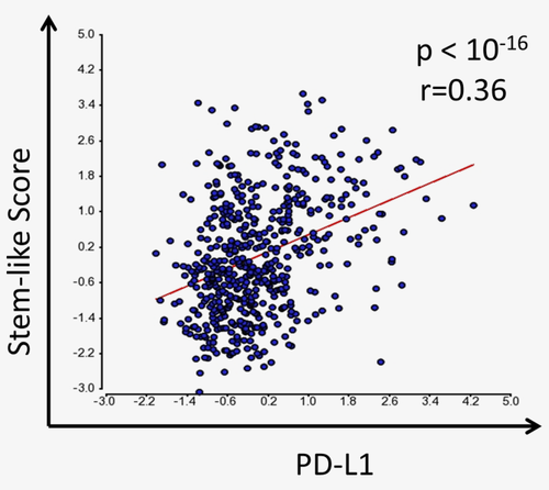
PD-L1 expression in breast cancer is significantly associated with stemness score. Scatter plot of PD-L1 mRNA expression level against the stem-like score, which was calculated based on the expression of 100 stem-cell-associated genes as described in methods, in the TCGA breast cancer gene expression dataset (n = 530). Pearson correlation coefficients (r) and associated p values (p) for the correlation test is shown. [Color figure can be viewed at wileyonlinelibrary.com]
PD-L1 expression is important for the maintenance of stemness factors
To investigate if PD-L1 expression has a direct role in the stemness of cancer cells we used PD-L1 specific ShRNA to knockdown PD-L1 expression in the breast cancer cell line MDA-MB-231 cells. These cells normally express high level of PD-L1 compared to other breast cancer cell lines.11 Downregulation of PD-L1 was confirmed using western blot, flow cytometry and immunofluorescence in two different clones of PD-L1 ShRNA (Sh-PD-L1 (a) and Sh-PD-L1 (b), Supporting Information, Fig. 1a,b).
Owing to the critical role of embryonic antigens on the self-renewal ability of CSCs, we used the two PD-L1 knockdown clones to check for the effect of PD-L1 knockdown on the expression of OCT-4A, Nanog and SOX-2 embryonic stem cell transcriptional factors. Provided that there is a previously reported effect of cell density on stemness of cancer cells,21 we carried out experiments on cells harvested from exponential or confluent conditions. Downregulation of PD-L1 significantly reduced OCT-4A expression in both exponentially growing as well as confluent cells as shown by immunofluorescence (Fig. 2a,b and Supporting Information, Fig. 2). Similarly, although to a lesser extent, Nanog was also downregulated upon PD-L1 loss but only in one clone (Sh-PDL1 (b) in exponentially growing cells. Interestingly, the effect of PD-L1 on Nanog expression became significant when tested on confluent cells (Fig. 2a,b and Supporting Information, Fig. 2). PD-L1 loss had minimal effect, if any, on SOX-2 expression (Supporting Information, Fig. 3).
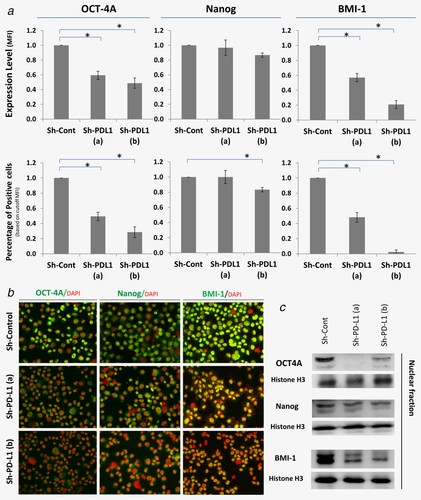
PD-L1 expression is important for the expression of stemness factors. The expression level of OCT-4A, Nanog and BMI-1 is shown in PD-L1-knockdown clones (a & b) of MDA-MB-231 cells compared with the control cells (Sh-Cont). (a) Bar graph showing the expression level (measured as mean fluorescence intensity, MFI) of nuclear OCT-4A, Nanog and BMI1 (top) and the percentage of cells expressing them (bottom). Data is normalized on the control (Sh-Cont) and displayed as mean of 4 independent experiments ± SEM as measured by immunofluorescence and quantified using BD pathway 855 system. (b) Representative immunofluorescent images of the PD-L1-knockdown clones and the control cells (at 200× magnification) from one of the 4 experiments. (c) Representative western blot images showing downregulation of nuclear OCT-4A, Nanog and BMI1 in PD-L1 knockdown cells. * indicates statistical significance (p < 0.05).
We checked another stemness controlling protein, called B lymphoma Mo-MLV insertion region 1 homolog (BMI1), which is well known to control stemness of CSCs.22 Immunofluorescence shows a dramatic decrease in the BMI1 expression upon PD-L1 knockdown in exponential growing cells (Fig. 2a,b). This significant inhibition of BMI1 expression upon PD-L1 knockdown was lost when tested on confluent cells (Supporting Information, Fig. 2). Thereafter, all later experiments were done using exponentially growing cells. PD-L1 knockdown mediated downregulation of OCT-4A, Nanog and BMI-1 was further confirmed using western blot (Fig. 2c). Altogether, PD-L1 is important to maintain the expression of the embryonic antigens OCT-4A and Nanog and the stemness factor BMI-1.
PD-L1 knockdown inhibits AKT phosphorylation
To gain insight into the mechanism of how PD-L1 can affect the expression of embryonic antigens, we used liquid chromatography coupled with tandem mass spectrometry (LC/MS) to analyze global protein expression in breast cancer cells upon PD-L1 knockdown. Approximately >2000 proteins were identified in total between Sh-Cont and both Sh-PD-L1 (a) and (b). We identified significant changes (ANOVA, p values <0.05) with at least fivefold in the expression of a total of 214 different proteins upon PD-L1 knockdown. To summarize all data and understand the main pathway responsible for most of these changes, data were subjected to ingenuity pathway analysis (IPA). The network analysis showed AKT1 as a central node that is related, directly or indirectly, to many of the protein expression changes detected upon PD-L1 knockdown (Supporting Information, Fig. 4).
To validate this observation, we used immunofluorescence to measure the phosphorylation (S473) of AKT in our PD-L1 knockdown clones. There was a dramatic decrease in the phosphorylation of AKT that was prominent in the nucleus (Fig. 3a,b). To further confirm these results, we measured phospho-AKT using western blot. PD-L1 knockdown cells had significantly lower level of phosphorylated AKT confirming that PD-L1 expression is important to maintain phosphorylated AKT (Fig. 3c). To further confirm the effect of PDL1 on AKT activity, we measured the activation of mTOR, which is very well-known to be under the control of PI3K/AKT activity. PD-L1 knockdown resulted in a dramatic decrease in the mTOR activity as shown by the reduced phosphorylation (at S235/236 site) of its downstream target, the ribosomal S6 (Fig. 3b).

PD-L1 knockdown impaired the phosphorylation of AKT. (a) Bar graph showing the expression level (measured as MFI) of phospho (S473)-AKT or phospho (S235/236)-S6 in PD-L1-knockdown clones (a & b) of MDA-MB-231 cells compared with the control cells (Sh-Cont). Data are normalized to the control (Sh-Cont) nuclear MFI of phospho AKT (left) or the control cytoplasmic phospho S6 (right) and displayed as mean of 4 independent experiments ± SEM as measured by immunofluorescence and quantified using BD pathway 855 system. (b) Representative immunofluorescent images of the PD-L1-knockdown clones and the control cells (at 200× magnification) from one of the experiments. (c) Western blot showing phospho AKT expression following PD-L1 knockdown in MDA-MB-231 cells. (d) Bar graph showing the expression level of phospho-AKT, OCT-4A and Nanog in PD-L1 knockdown clones and control ± PI3K/AKT/mTOR inhibitors as compared with untreated cells as mean of 10 independent experiments ± SEM. (e) Representative immunofluorescent images showing staining of phospho (S473)-AKT, OCT-4A, Nanog (at 200× magnification) from one of the 10 experiments in D for clone Sh-PD-L1 (b). * indicates statistical significance (p < 0.05).
We then used pathway inhibitors to test whether the effect of PD-L1 knockdown on the OCT-4A and Nanog was due to the inhibition of AKT activation. Given our data (Fig. 3b) and the well-known feedback loops of PI3K/AKT and mTOR pathways,23, 24 we inhibited the entire PI3K/AKT/mTOR pathway to examine whether the effect of PD-L1 on OCT-4 A and Nanog expression is PI3K/AKT pathway dependent. PI3K/AKT/mTOR inhibition significantly reduced the phosphorylation of AKT (Fig. 3d, top). PD-L1 knockdown did not have further inhibition on OCT-4A when PI3K/AKT/mTOR pathway was inhibited (Fig. 3d,e). Similarly, the PD-L1 (b) clone that showed significant effect on Nanog expression did not show further effect when the PI3K/AKT/mTOR pathway was inhibited (Fig. 3d,e). These data suggest that the effect of PD-L1 on OCT-4A and Nanog is PI3K/AKT-pathway dependent.
Inhibiting PI3K/AKT/mTOR pathway did not have an effect on BMI1 expression (data not shown), suggesting that the effect of PDL1 on BMI expression is PI3K/AKT-independent.
To test the universality of our findings to other types of breast cancer cells, we knocked down PD-L1 in another breast cancer cell line (SUM-159), which normally expresses abundant PD-L1 in a similar fashion to MDA-MB-231 cells. PD-L1 knockdown in SUM-159 cells validated the decrease in OCT-4A, Nanog and BMI1 that we obtained with PD-L1 knocked in MDA-MB-231 cells (Supporting Information, Fig. 5). Furthermore, we transfected PD-L1 ORF in T-47D breast cancer cells, which are normally PD-L1 negative, to test whether we could replicate the PD-L1 mediated effects on stemness factors in this cell line. Upon ectopic PD-L1 expression, OCT-4A, Nanog and BMI1 were upregulated as shown by immunofluorescence (Supporting Information, Fig. 6a) and western blot (Supporting Information, Fig. 6b).
The above findings depended on PD-L1 knockdown using one sequence of PD-L1 Sh-RNA. Therefore, to ensure that these observed changes are not due to an off-target effect, we transfected MDA-MB-231 cells with another PD-L1 ShRNA that utilizes different sequence and inserted on a different vector. Results with the second Sh-RNA (GIPZ-Sh-PDL1) were consistent with the previous one confirming the downregulation of OCT-4A, Nanog and BMI1 we observed in these cells are specifically due to PD-L1 knockdown (Supporting Information, Fig. 7).
Altogether, we have demonstrated that PD-L1 expression sustains AKT phosphorylation, which in turn is important for the maintenance of OCT-4A and Nanog expression. On the other hand, we have shown that PD-L1 regulate BMI1 expression in a PI3K/AKT pathway-independent manner.
PD-L- mediated PI3K/AKT pathway activation promotes the phosphorylation of (T235) OCT-4
Lin et al. has previously shown that AKT maintains OCT-4 expression by sustaining its phosphorylation at threonine 235 site in both embryonic stem cells and embryonic carcinoma cells.25 This AKT-mediated phosphorylation of OCT-4 promotes its stabilization and nuclear localization. Therefore, we checked the effect of PD-L1 expression on the phosphorylation (T235) of OCT-4. Indeed, PDL1 knockdown significantly decreased the phosphorylation of OCT-4 in both clones (Fig. 4a,b). More importantly, inhibition of PI3K/AKT/mTOR pathway decreased the phosphorylation of OCT-4 in a similar fashion to knocking down PDL1. The effect of PD-L1 knockdown on the phosphorylation of OCT-4 and its dependence on PI3K/AKT/mTOR pathway was confirmed by western blot (Fig. 4c). Altogether, PD-L1 maintains OCT-4 phosphorylation and increases its total protein expression in a PI3K/AKT pathway-dependent fashion.
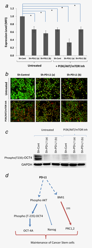
PD-L1-mediated PI3K/AKT/mTOR activity promotes phosphorylation of OCT-4 (T235). (a) Bar graph showing the expression level of phospho (T235) OCT-4 in PD-L1-knockdown clones (a & b) of MDA-MB-231 cells compared with the control cells (Sh-Cont). Data are normalized on the nuclear MFI of the control (Sh-Cont) and displayed as mean of 3 independent experiments ± SEM as measured by immunofluorescence and quantified using BD pathway 855 system. (b) Representative immunofluorescent images of the PD-L1-knockdown clones (a & b) and the control cells (at 200× magnification) from one of the experiments. (c) Western blot showing expression of phospho OCT4 following PD-L1 knockdown in MDA-MB-231 cells in the presence or absence of PI3K/AKT/mTOR inhibitors. (d) Schematic diagram showing the effect of PD-L1 on stemness of breast cancer cells via a PI3K/AKT-dependent and independent pathways. Solid blue lines indicate a demonstrated direct effect (thick = strong, thin = mild). Dashed lines indicate a demonstrated effect (direct or indirect). Solid red line indicates a demonstrated effect by other studies. * indicates statistical significance (p < 0.05).
Based on the above data presented we propose that PD-L1 regulate breast cancer stemness via modulating OCT-4A and Nanog expression, which is PI3K/AKT pathway-dependent and through promoting BMI1 expression, in a PI3K/AKT-independent fashion (Fig. 4d).
PD-L1 is critical for the self-renewal of CSCs
The effect of PD-L1 knockdown on the expression of stem-related molecules suggests a direct role for this molecule in CSC maintenance. To functionally test the role of PDL1 on maintaining CSCs, we examined the ability of PD-L1 knockdown cells to grow in an anchorage-independent manner (tumorsphere formation ability). PD-L1 knockdown significantly decreased the ability of breast cancer cells to form tumorspheres as compared with the scrambled control (Fig. 5a). PD-L1-mediated effect on tumorsphere formation was consistent in three subsequent passages supporting for the role of PD-L1 in breast cancer stemness.
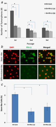
(a) PD-L1 is overexpressed by CSCs and is important for its self-renewal of potential. (a) PD-L1 knockdown abrogates the self-renewal of CSCs in the tumorsphere assay. Bar graph showing the number of tumorspheres formed from 1000 cells (mean± SD, n = 3) for three consecutive passages. (b) Representative immunofluorescent images showing PD-L1 expression in sorted CSCs (Ep-CAM+/CD44high/CD24low). (c) Bar graph showing percentage of CSCs (Ep-CAM+/CD44high/CD24low) in PD-L1 knockdown cells compared with the control as measured by flow cytometry (mean± SEM, n = 6). * indicates statistical significance (p < 0.05).
We then sorted MDA-MB-231 cells stem-like cells (CSCs enriched population) using standard Ep-CAM+/CD44high/CD24low cell surface markers and their differentiated counterparts (Ep-CAMlow/neg/CD44low/CD24high) and compared their level of PD-L1 expression. Immunofluorescence results confirmed the higher level of PD-L1 expression in CSCs compared with the more-differentiated breast cancer cells. Interestingly, in addition to membranous PD-L1 there was a nuclear fraction PD-L1 in CSCs (Fig. 5b). Quantitation of stem-like cells in PD-L1 knockdown using flow cytometry showed a decrease in the percentage of these cells compared with the control (Fig. 5c and Supporting Information, Fig. 8).
We validated our work in vivo by performing an extreme limiting dilution assay (ELDA), the golden standard of CSCs frequency estimation in vivo. PD-L1-knockdown MDA-MB-231 breast cancer cells were injected at different inoculation densities (10, 100 and 1000 cells/mice) in 6 mice per group with similar setting for control cells. We have chosen the most immunocompromised strain of mice, which is NOD/SCID/IL-2R−/− (NSG), to neutralize the effect of PD-L1 on the immune system. In this strain of mice the incidence of tumor formation was extremely high, which is consistent with previous reports.26 Even with this high level of tumorigenicity, knockdown of PD-L1 resulted in ninefold decrease the frequency of CSCs from 1:1 in the control cells to 1:9 in the PD-L1 knockdown cells (Table 1). Importantly, the survival of mice injected with PD-L1 knockdown cells were significantly (p < 0.001) longer than mice injected with control cells (median survival were 77 days for mice injected with PD-L1 knockdown cells compared with 53 days for mice injected with control cells (Supporting Information, Fig. 9). Necropsy on dead mice revealed massive metastatic nodules suggesting this as the cause of death. To further ensure that such an effect is reproducible in other type of immunocompromised mice, we repeated the experiment in Nude mice. We obtained similar results; although the tumor incidence was much lower (Table 1). Interestingly, in nude mice, the survival was much better and therefore we could compare the tumor size in each group. Sh-Cont cells formed significantly larger tumors (average of 600 mm2) compared with Sh-PD-L1 cells (average 170 mm2).
| Cells seeded | 1000 | 100 | 10 | Frequency of CSCsa | p valueb |
|---|---|---|---|---|---|
| NOD/SCID/IL-2neg/neg (NSG) | |||||
| Sh-Cont | 6/6 | 6/6 | 6/6 | 1 in 1 cells | 0.075c |
| Sh-PD-L1 | 6/6 | 6/6 | 4/6 | 1 in 9 cells | |
| Nude/Nude (Nude) | |||||
| Sh-Cont | 4/6 | 5/6 | 2/6 | 1 in 316 cells | 0.006 |
| Sh-PD-L1 | 1/6 | 2/6 | 1/6 | 1 in 1500 cells | |
- a Estimated as per ELDA calculating website.
- b Confidence choice entered was 0.95.
- c Overall test for differences in stem cell frequencies between the two groups.
Altogether our data in vivo and in vitro demonstrate that PD-L1 is critical for the maintenance of breast CSCs.
Discussion
There is accumulating evidence that cancer is originated and sustained by a small population of cells called “cancer stem cells (CSCs).” These cells are associated with common features including distinct expression of adhesion molecules, resistance to chemotherapy and signs of EMT. We previously have shown an association between these CSC features and PD-L1 expression,8, 11 triggering us to investigate the direct role of PD-L1 in the function of CSCs. Here, we have demonstrated that PD-L1 has a direct effect on sustaining the stemness of CSCs mainly through regulating OCT-4A and Nanog expression in a PI3K/AKT-dependent and by promoting BMI1 expression through PI3K/AKT-independent fashion.
The ability of cancer cells to reinitiate tumors depends on self-renewal capacity, ability to proliferate continuously and resist genotoxic stress such as chemotherapy. The self-renewal capacity is a feature obtained through the orchestration of multiple important factors including embryonic antigens (OCT-4A, Nanog and SOX-2), the activation of Notch, WNT or Hedgehog self-renewal pathways and facilitation of chromatin modulators. Our presented data have shown that PD-L1 can sustain OCT-4A and Nanog expression through PI3K/AKT pathway activation. Furthermore, we have shown that PD-L1 maintains the expression of BMI1, a well-known oncoprotein that can remodulate chromatin to promote stemness (reviewed in ref. 22). We have further shown that PD-L1 is important to sustain mTOR pathway activation as demonstrated by the decrease in the phosphorylation of its target, ribosomal S6 in PD-L1 knockdown cells. There is an established role of mTOR pathway in regulating cell proliferation,27 which may explain the significantly larger tumor size formed by PD-L1 expressing compared with PD-L1 knockdown cells.
The increase in the expression of PD-L1 in the nucleus specifically in CSCs is in agreement with our previously reported translocation of PD-L1 to the nucleus upon chemotherapy and its interacting effect with PI3K/AKT in the nucleus leading to chemoresistance.8 Very recently, this phenomenon has been appreciated by Satelli et al.28 as they have found a significant correlation between nuclear PD-L1 in breast cancer and poor prognosis. Furthermore, the effect of PD-L1 specifically on the nuclear fraction of AKT in particular, which has an important role in the tumorigenesis of cancer cells (reviewed in ref. 29), supports for an exclusive nuclear interactive pathway (Fig. 4d).
Phosphorylation is an important posttranslational regulatory mechanism for many proteins. Several phosphorylation sites have been reported for OCT-4.30 Subsequent study demonstrated that phosphorylation of OCT-4 at threonine 235 leads to its stabilization and nuclear localization.25 Further study has confirmed the importance of OCT4 phosphorylation at this site (T235) for the stemness of cancer stem cells.31 In agreement, our findings in this study demonstrated the importance of PI3K/AKT pathway for PD-L1 ability to maintain the phosphorylation (T235) of OCT-4, in line with PD-L1 promoting effect on nuclear OCT4A expression and enhanced stemness of breast cancer cells.
PD-L1 is a T-cell inhibitory molecule and its immunomodulatory effect is well established. However, we and others have previously shown that PD-L1 has roles in cancer cell biology beyond its effect on the immune system.7, 8, 11 In this work, we have used the most immunocompromised mouse model available NOD/SCID/IL-2Rneg/neg (NSG) to examine the role of PD-L1 on CSCs. Results have clearly shown PD-L1 expression in cancer cells is important to maintain frequency of CSCs, even in this strain of mice, supporting for PD-L1 role in controlling breast cancer cells stemness independent of its immune modulating function.
Conclusions
In conclusion, we have observed close association between PD-L1 expression and breast cancer stemness in the TCGA invasive breast cancer gene expression dataset. Our work confirmed this association and directly demonstrated the critical role of PD-L1 in maintaining breast cancer stemness in vivo. We have further shown that PD-L1 role in CSCs is mediated through PI3K/AKT activation, which in turn is important to maintain OCT-4A and Nanog. This is in addition to PI3K/AKT-independent effect of PD-L1 on BMI-1 expression. Our findings suggest that targeting PD-L1 would affect the pool of breast CSCs and have an important consequence on the efficacy of breast cancer treatment.
Declarations
Ethics approval and consent to participate
This work was under an institutionally approved King Faisal Specialist Hospital and Research Centre project (RAC# 2140–001). The institutional ethics committee has approved this work.
Consent for publication
All authors read and approved the manuscript. All contributing authors approved the submission of this version of the manuscript and asserted that the document represent valid work. All contributing authors had no disclosures to make.
Availability of data and materials
The datasets generated during and/or analyzed during this study are included in this published article (and its Supporting Information files), otherwise available from the corresponding author on reasonable request.
Competing Interests
All authors declare no conflict of interest.
Funding
Funding bodies did not have any role in the design of the study or the collection, analysis and interpretation of data or in writing the manuscript.
Authors' Contributions
SA: collected and analyzed/interpreted data (immunofluorescence, protein fractionation, western blot, cell culture). DC: supervised and performed all bioinformatics analyses, interpretation. FM: collected and analyzed/interpreted data (immunofluorescence, protein fractionation and cell culture). AA: collected and analyzed/interpreted data (proteomics and IPA analysis). OA: performed some data collection and mining. AQ: performed data analysis and interpretation (miR analysis). FA (veterinarian): collection and analyzed/interpreted data (animal work). MA: performed data interpretation and edited the manuscript. HG (the principal investigator): conceived and designed the study, collected and analyzed/interpreted data (immunofluorescence), performed data analysis and interpretation and wrote the manuscript.
Acknowledgements
We would like to thank Mr Amer Al-Mazrou for his help in cell sorting of CSCs.


