Female with peritonitis and abdominal distention
1 PATIENT PRESENTATION
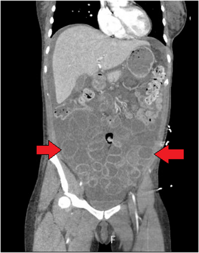
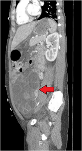
A G0 35-year-old female with a body mass index (BMI) of 21 and history of antibiotic allergies presented to the emergency department with diffuse abdominal pain. Two days prior, she had a transvaginal ultrasound-guided oocyte retrieval for mature oocyte cryopreservation and 36 hours before the procedure received a human chorionic gonadotropin (hCG) “trigger shot.” She was tachycardic, but otherwise hemodynamically stable. On abdominal examination, she had a distended abdomen with diffuse tenderness to percussion and rebound tenderness. A point-of-care ultrasound demonstrated free fluid and a computed tomography (CT) of the abdomen/pelvis with intravenous contrast was obtained after consultation with surgery and obstetrics (Figures 1-3).


2 DIAGNOSIS
2.1 Severe Ovarian Hyperstimulation Syndrome
CT revealed bilateral enlarged ovaries with follicles, the largest at 17 × 15 × 10 cm, with ascites and bilateral pleural effusions. No free air or active hemorrhage was identified. She underwent paracentesis of 2.5 L with administration of albumin and anticoagulation.
Ovarian hyperstimulation syndrome (OHSS) is an iatrogenic complication of assisted reproduction technology, described as cystic enlargement of the ovaries and fluid shifts from the intravascular space to the third space.1, 2 It can occur in 1%–5% of cycles with risk factors of low BMI, polycystic ovarian syndrome, allergy history, high doses of hCG, and large number of follicles.2, 3 Peritonitis can occur because of the release of inflammatory mediators or rupture of follicular cysts.3 Severe OHSS is characterized by ascites, pleural effusions, and hemoconcentration. In addition to OHSS, complications of oocyte retrieval, albeit rare, must also be considered as intraperitoneal hemorrhage, bowel perforation, or infection.4


