Sex differences on peak oxygen uptake in heart failure
Abstract
Women represent nearly half of the adult heart failure (HF) population and they remain underrepresented in HF studies. We aimed to evaluate the evidence about peak oxygen uptake (peak VO2) for clinical stratification in women with HF. This narrative review summarizes (i) the evidence endorsing the value of cardiopulmonary exercise testing for clinical stratification and phenotyping HF population; (ii) the determinants of a person's functional aerobic capacity to understand predicted values for patients with chronic HF; and (iii) sex differences on peak VO2 data in different forms of HF. Lastly, based on existing data in patients with HF, we provide a perspective on how to improve existing gaps about the utility of peak VO2 in clinical stratification in women. Peak VO2 provides prognosis information in patients with HF; however, its use has been limited for a reduced number of patients excluding women, elderly, and HF patients with preserved ejection fraction. Further studies will help to fill the wide gender gap about the utility of cardiopulmonary exercise testing in the risk assessment and management in women with HF.
Introduction
Heart failure (HF) prevalence is increasing over the years in western countries.1 Despite women represent nearly half of the adult HF population,1 they have been classically underrepresented in HF studies.2 Likewise, this underrepresentation of women is even more obvious when we explore cardiopulmonary exercise testing (CPET) literature in HF.3-6 This last gap in knowledge limits our understanding in women HF pathophysiology, risk stratification, recommendations for physical therapy, and advanced HF intervention. Given the limited data in CPET parameters for clinical stratification in women with HF, teasing out the information from previous studies could be an excellent start point for further research lines.
In this narrative review, first, we highlight the valuable role of CPET for clinical stratification and phenotyping HF population. Second, we describe the main determinants of maximal aerobic capacity in healthy individuals and physiological sex differences. Third, we explore sex differences on peak oxygen uptake (peak VO2) data in different forms of HF. Lastly, we discuss existing gaps about the utility of CPET in the risk assessment in women with HF, and we provide a perspective on how to improve the utility of peak VO2 in clinical stratification in women.
Methods
This narrative review was conducted to identify all studies evaluating the role of peak VO2 for clinical stratification in HF. All studies included meeting the search criteria in PubMed (MEDLINE) up to December 2018. Search terms used were the combinations of the terms ‘prognosis’, ‘cardiopulmonary exercise test’ ‘heart failure’, ‘mortality’, ‘morbidity’, ‘rehospitalizations’, ‘maximal aerobic capacity’, and ‘peak oxygen uptake’.
Studies included in this review were required to fulfil the current diagnosis criteria for HF with preserved ejection fraction (HFpEF) and HF with reduced ejection fraction (HFrEF) of European Society of Cardiology.7 Furthermore, studies evaluated were required to include both men and women. We excluded studies including patients with ‘borderline’, ‘non-reduced’, ‘mildly’, or ‘intermediate’ ejection fraction to avoid heterogeneity. To this end, we qualitatively summarized nine HFrEF and one HFpEF eligible studies in order to extract useful information despite the fact that these studies were heterogeneous in outcomes measures, sample size, and stress test protocol.
Cardiopulmonary exercise testing in heart failure
Cardiopulmonary exercise testing (CPET) with measurement of peak VO2 has become the mainstay of assessing functional capacity and predict outcomes in patients with HF, particularly in patients with HFrEF.3, 8, 9 During a CPET, we assess respiratory oxygen uptake, carbon dioxide production, ventilatory response to exercise (ventilatory efficiency or VE/VCO2), and chronotropic response to exercise, among other classical physiological variables. This non-invasive technique provides complete information about (i) pathophysiological causes of exercise intolerance (cardiac, pulmonary, and/or peripheral); (ii) functional capacity and severity of the functional impairment; (iii) recommendations for physical treatment; and (iv) prognostic information. Consequently, the CPET data may proportionate additional advantages in HF patients for a better understanding of the pathophysiology and characterization of the different phenotypes of this complex syndrome.
Determinants of maximal aerobic capacity in healthy subjects
Measured maximal oxygen uptake (VO2max) during a maximal symptom-limited CPET constitutes the most objective method to assess aerobic capacity in healthy individuals9 and it defines the limits of the cardiopulmonary system. According to Fick equation, VO2max is equal to the product of cardiac output and arteriovenous oxygen difference at maximal exercise. The achievement of VO2max requires a plateau in oxygen uptake despite of increasing workload during a specified period. Patients with pulmonary or cardiovascular disease rarely achieve VO2max during a maximal symptom-limited CPET and that is the reason why we use the term peak VO2 instead of VO2max to describe maximal aerobic capacity in patients with HF.
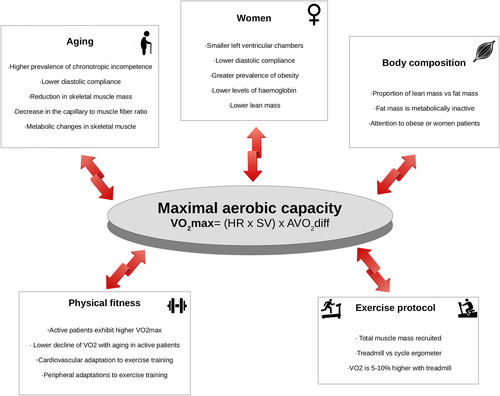
Interpretation of the results of peak VO2 in a chronic HF patient requires information of the normal values of VO2max of a healthy individual.10 VO2max in healthy subjects varies with sex, age, body size, physical fitness, and exercise modality as showed in Figure 1.

Sex
Previous investigators11, 12 have reported that VO2max is significantly lower in women than in men. Several anatomical and physiological sex disparities between women and men could explain these differences. Among them are as follows: (i) women have minor left ventricular chambers and consequently, lower stroke volumes13; (ii) women exhibit lower diastolic compliance14; (iii) women show greater prevalence of obesity13; (iv) women have lower levels of haemoglobin than men and they are more likely to suffer from iron deficiency15; and (v) women have lesser lean mass than men.16
Age
VO2max physiologically declines with age.17 Numerous central and peripheral physiological modifications associated with aging process reduce functional capacity,18 among them are as follows: (i) more prevalence of chronotropic incompetence; (ii) lower diastolic compliance; (iii) reduction in skeletal muscle mass; (iv) decrease in the capillary to muscle fibre ratio; and (v) metabolic changes in skeletal muscle.
Body composition
Absolute VO2max (mL/min) value is traditionally corrected for total body weight and is reported in millilitres per kilogram per minute (mL/kg/min). Corrected value of VO2max (mL/kg/min) underestimates functional capacity in obese healthy individuals19 and HF patients20 because it does not take into account body composition (proportion of lean mass vs. fat mass). Along this line, it is important to note that body fat represents metabolically inactive mass and could be increased in obese, women, and HF patients.20 Thus, Wasserman et al.10 recommends calculation of predicted VO2max using different equations based on estimated normal weight compared with real weight for avoiding misleading results and conclusions.
Physical fitness
Previous studies have showed that the level of ordinary activity is positively associated with VO2max obtained during a CPET.21, 22 Moreover, the decline of VO2 with aging is inversely associated with physical fitness.21 The physiological potential mechanisms proposed to explain better functional capacity in active individuals are central (increase of stroke volume, decrease of diastolic dysfunction, etc.) and peripheral (increase skeletal muscle mass, capillary to muscle fibre ratio, and metabolic efficiency, among others) adaptations to exercise.23
Exercise test protocol
The CPET protocol (cycle ergometer vs. treadmill) used to asses functional capacity is an important determinant of VO2max.10 In fact, during a treadmill exercise testing, we are recruiting a larger muscle mass (upper and lower limbs) and working against gravity. Therefore, on average, VO2max is 5–11% higher with treadmill than on cycle ergometer. Furthermore, the selection of specific exercise protocols (ramped vs. stepped) should be tailored for a sufficient effort (assessed as a respiratory exchange ratio ≥1) and exercise duration of 8–12 min with a progressive and individualized increase of workload.9
The combination of the aforementioned variables that determines the normal values of maximal aerobic capacity indicates that women exhibit lower absolute (mL/min) or corrected (mL/kg/min) values of VO2max than men in healthy subjects. Therefore, it is obvious that we should not use similar cut-off of absolute or corrected VO2max for both sexes. Along this line, in patients with HF, expressing peak VO2 as a percentage of predicted VO2max (pp-peak VO2) adjusted for sex, age, exercise protocol, weight, and height could be a more accurate approach.
Sex differences on peak oxygen uptake data in different heart failure populations
Based on previous evidence about the role of CPET in HF patients, current position paper3 advocates for three prognostic ‘cut-point’ values (>18, 18-10, or <10 mL/kg/min) of peak VO2 for risk stratification in male patients with HFrEF, with still no enough evidence for stratify other subgroups of patients (women, elderly, atrial fibrillation, patients with co-morbidities, HFpEF, etc.). Regarding women, the current data are scarce and inconsistent. We aimed to evaluate and tease out evidence in patients with HFrEF and HFpEF to date in order to get recommendations for further research lines.
Peak oxygen uptake data in HF with reduced ejection fraction patients
Observational studies
In a recent position paper, Corrà et al.3 accurately reviewed six observational studies24-29 endorsing the utility of CPET parameters for risk stratification in women with HFrEF. The mean age of included women in published data was slightly lower than men, and the main justification of female underrepresentation in HFrEF studies was the large proportion of older aged women in HFrEF patients, and the policy of non-inclusion of the elderly. Based on the data of corrected peak VO2 (mL/kg/min) values, the authors showed that women have nearly 2 mL/kg/min lower peak VO2 than men; however, women exhibited better prognosis. Only two studies included in this review24, 29 exhibited both the values of corrected peak VO2 and the pp-peak VO2, and surprisingly, these last ones were higher in women (women exhibited nearly 10% higher pp-peak VO2 than men). In the same way, other subsequent observational study that included 1085 patients (33% women) published by Ehrman et al.6 showed that prognostic peak VO2 might be considered separately for men and women with HFrEF. However, pp-peak VO2 threshold values corresponding to 1- and 3-year survival rates were similar between both sexes.
Controlled studies
Regarding randomized trials in HFrEF patients, the HF-ACTION examined the prognostic value of CPET parameters in terms of mortality in 2100 patients (29% women).4 Women included were slightly younger than men and exhibited better clinical profile. Once again, baseline characteristics showed that corrected peak VO2 was lower [13.4 (7.7–21.0) vs. 15.2 (8.7–24.3) mL/kg/min] and pp-peak VO2 was higher [68 (39–95) vs. 57 (32–86) %] for women compared with men. The results showed that pp-peak VO2 and exercise duration were the strongest predictors of survival in this population.
Peak oxygen uptake data in HF with preserved ejection fraction patients
The evidence endorsing the prognostic role of CPET parameters in HFpEF is even scarcer, and once again, women have been underrepresented despite being the most common gender in this syndrome.30 To gather some insight and hope in addressing this lack of knowledge, our group has recently published the results of a prospective study30 where we evaluated the prognostic utility of CPET parameters for predicting recurrent hospitalizations in a cohort of 74 older (72.5 ± 9.1 years), predominantly women (53%) and highly symptomatic patients with HFpEF. We found pp-peak VO2 was independent and linearly associated with recurrent admissions in this sample. In fact, a 10% reduction in pp-peak VO2 was associated with a 32% increased risk of recurrent hospitalization. In a further analysis, we evaluated the distribution of corrected peak VO2 and pp-peak VO2 in both genders. Women were slightly older than men, and after a median duration of follow-up of 2.3 years, they exhibited better prognosis in terms of mortality and all-cause readmissions (20.5% vs. 45.7% and vs. 41.6% vs. 58.4%, respectively). Corrected peak VO2 was significantly higher in men [10.9 ± 2.9 vs. 9.1 ± 2.2 mL/kg/min (P < 0.01)]; however, in agreement with the data in HFrEF, pp-peak VO2 was significantly higher in women [60.8 ± 13.3% vs. 53.3 ± 13.5% (0.02)]. Once again, these results suggest that pp-peak VO2 describes the degree of functional impairment in women more accurately than corrected peak VO2.
General approaches
It is important to remark that exercise test protocol selection of each study evaluated in this review was vastly heterogeneous. Each study chose different protocols (ergometer or treadmill) with different workload increments (Naughton protocol, conservative ramped protocols, or individualized stepped protocol). Likewise, all the studies included in this review reported a respiratory exchange ratio at peak exercise higher than 1; however, only seven studies reported differences of respiratory exchange ratio at peak exercise4, 6, 24-26, 28, 29 between men and women, and it was significantly lower in women.
Clinical implications and future directions
CPET in HF patients provides valuable information of exercise responses and has shown many clinical applications8; however, its use has been limited for a reduced number of HFrEF patients excluding women, elderly, and HFpEF patients. We strongly believe that the incorporation of CPET assessment in the routine work out of whole spectrum of patients with HF may proportionate additional advantages such as (i) a better understanding of the pathophysiology of different subgroups; (ii) characterization of the different phenotypes of the syndrome; (iii) filling gender gaps in HF; and (iv) evaluation of the effect of new therapeutic options.
Although establishing three prognostic ‘cut-point’ values (>18, 18–10, or <10 mL/kg/min) of peak VO2 for risk stratification in male patients with HFrEF simplifies the algorithm,3 it could lead us to inaccuracy and misconception of objective peak VO2. Along this line, the question is whether we can equalize the risk of adverse events between two male HF patients with the same ‘cut-point’ or corrected value of peak VO2 (i.e. 18 mL/kg/min) but different age (30 vs. 59 years old), body mass index (22 vs. 32 kg/m2), and body composition (percentage of fat: 21% vs. 27%). The answer is probably not, but unfortunately with the current knowledge, we cannot unravel this question. Furthermore, women have been excluded from this algorithm because they showed lower corrected values of peak VO2 but without enough evidence to design a specific algorithm. Consequently, it is reasonable that we should recommend expressing peak VO2 as a pp-peak VO2 (percentage of sex-predicted, age-predicted, exercise protocol-predicted, weight-predicted, and height-predicted VO2max) in women in order to more accurate risk stratification, while we hope further studies will help to fill the wide gender gap about the utility of CPET in the risk assessment and management in women with HF.
Conflict of interest
None declared.
Funding
The authors have no funding.


