Regional and age-related differences in GAD67 expression of parvalbumin- and calbindin-expressing neurons in the rhesus macaque auditory midbrain and brainstem
ABSTRACT
Neurons expressing the calcium binding proteins (CaBPs) parvalbumin (PV) and calbindin (CB) have shown age-related density changes throughout the ascending auditory system of both rodents and macaque monkeys. In the cerebral cortex, neurons expressing these CaBPs express markers of γ-aminobutyric acidergic neurotransmission, such as GAD67, and have well-understood physiological response properties. Recent evidence suggests that, in the rodent auditory brainstem, CaBP-containing cells do not express GAD67. It is unknown whether PV- and CB-containing cells in subcortical auditory structures of macaques similarly do not express GAD67, and a better understanding of the neurotransmission of neurons expressing these proteins is necessary for understanding the age-related changes in their density throughout the macaque auditory system. This was investigated with immunofluorescent double-labeling techniques to coregister PV- and CB-expressing neurons with GAD67 in the superior olivary complex and the inferior colliculus of young and aged rhesus macaques. The proportions of GAD67-expressing PV- and CB-positive neurons were computed with unbiased sampling techniques. Our results indicate that between 42% and 62% of PV- and CB-positive neurons in the auditory brainstem and midbrain express GAD67, which is significantly less than in the cerebrum. In general, fewer PV+ neurons and more CB+ neurons expressed GAD67 as a function of age. These results demonstrate that the inhibitory molecular profile of PV- and CB-expressing neurons can change with age in subcortical auditory structures and that these neurons are distinct from the well-described inhibitory interneurons that express these proteins in the cerebral cortex. J. Comp. Neurol. 522:4074–4084, 2014. © 2014 Wiley Periodicals, Inc.
The primate auditory system is characterized by two parallel, chemically defined processing pathways that can be segregated based on immunoreactivity to either parvalbumin (PV) or calbindin (CB). The immunoreactivity to these calcium binding proteins (CaBPs) is sufficient to differentiate specific subdivisions of auditory nuclei, especially in midbrain, thalamic, and cortical structures (Jones, 2003; Parvizi and Damasio, 2003; Engle et al., 2014). The relative densities of neurons expressing these CaBPs and other calcium-dependent molecules change with age in auditory structures throughout the rodent auditory system (O'Neill et al., 1997; Zettel et al., 1997; Idrizbegovic 2003, 2004, 2006; Ouda et al., 2008, 2012), and similar observations have recently been made in the macaque monkey (Gray et al., 2013, 2014a; Engle et al., 2014). These density changes occur in parallel with age-related cochlear pathologies that lead to decreased excitatory output from the sensory periphery (Schuknecht, 1955, 1964; Schuknecht and Gacek, 1993; Nelson and Hinojosa, 2006; Fetoni et al., 2011; Engle et al., 2013) and changes in the balance of excitation and inhibition in auditory neurons that lead to overexcitable central auditory structures in aged animals (Caspary et al., 1995, 1999, 2005, 2006, 2008; Juarez-Salinas et al., 2010). Currently, the functional purpose of these density changes remains unknown.
PV- and CB-expressing neurons are chemically and physiologically well characterized in the cerebral cortex, striatum, and hippocampus, where they have been shown to release γ-aminobutyric acid (GABA; Klausberger and Somogyi, 2008; Rudy et al., 2011; Bartos and Elgueta, 2012) and have fast spiking response properties that produce brief, nonadapting action potentials (Kawaguchi et al., 1987; Cauli et al., 1997; Kawaguchi and Kubota, 1997; Gibson et al., 1999; Ascoli et al., 2008; Xu and Callaway, 2009). Together, these characteristics make CaBP-expressing interneurons powerful inhibitors in these cerebral structures. If these neurons behave analogously in the subcortical structures of the auditory system, one hypothesis explaining the function of their age-related density changes is as an inhibitory compensatory mechanism combatting the hyperexcitability of the aged auditory system.
Few studies, however, have investigated the physiological properties of subcortical neurons expressing PV and CB, and those that have suggest that these neurons are not always inhibitory as in higher brain regions. For example, in the superior olivary complex (SOC) and cochlear nucleus of rodents, PV+ and CB+ neurons do not express markers of GABAergic neurotransmission, such as glutamic acid decarboxylase (GAD); however, these markers are present in the majority of CaBP-expressing neurons in the inferior colliculus (IC; Fredrich and Reisch, 2009). This work has not been replicated in primates, so it is unknown whether CaBP-expressing neurons in the macaque auditory brainstem and midbrain are inhibitory (as in the cerebral cortex), not inhibitory (as in the rodent auditory brainstem), or a combination of the two.
Whether PV- and CB-expressing neurons are GABAergic in the macaque auditory system is a piece of information critical for understanding the functional consequences of the density changes of neurons expressing these proteins during natural aging. We therefore characterized the GABAergic molecular profiles of PV+ and CB+ neurons in the macaque SOC and IC using double-labeling immunofluorescent protocols with GAD67, an isoform that has been found previously in subcortical auditory structures (Burianova et al., 2009). We calculated the proportion of PV+ and CB+ neurons expressing GAD67 and compared these percentages between the auditory brainstem (SOC) and the auditory midbrain (IC). Finally, given the age-related changes in the density of PV+ and CB+ neurons in these auditory structures (Gray et al., 2014a; Engle et al., 2014), we segregated the animals into young and old age groups and compared the proportions between age groups within these two auditory structures.
MATERIALS AND METHODS
The IC and SOC from eight rhesus macaques ranging in age from 12 to 28 years (corresponding to roughly 36–84 human years; Davis and Leathers, 1985) were quantified for GAD67 expression in PV+ and CB+ neurons. Table 1 gives the demographic information for all animals used in this analysis. No animal had a history of loud noise exposure, ear trauma, or ototoxic drug treatment. Other than one monkey (327 month old), none of the animals had received auditory training prior to analysis. All procedures adhered to the National Institutes of Health guidelines and were approved by the UC Davis Institutional Animal Care and Use Committee.
| Age (months) | Human age (years) | Gender | Thickness (µm) | Age croup |
|---|---|---|---|---|
| 146 | 36.5 | Male | 25 | Middle-aged |
| 148 | 37 | Male | 30 | Middle-aged |
| 161 | 40.25 | Female | 30 | Middle-Aged |
| 180 | 45 | Female | 25 | Middle-Aged |
| 291 | 72.75 | Male | 25 | Old |
| 302 | 75.5 | Male | 25 | Old |
| 327 | 81.75 | Male | 30 | Old |
| 331 | 82.75 | Female | 30 | Old |
Histological processing and antibody characterization
Animals were euthanized with an overdose of sodium pentobarbital (60 mg/kg, i.v.) and transcardially perfused with a solution of 4% paraformaldehyde in 0.01% phosphate-buffered saline (PBS; pH 7.4), followed by a mixture of 4% paraformaldehyde and 10% sucrose. After perfusion, the brains were extracted and placed in a solution of 4% paraformaldehyde and 30% sucrose for cryoprotection. All brains were cut transversely at either 25 or 30 μm. Sections were stained for PV or CB immunohistochemistry along with GAD67 immunohistocemistry (see below). In total, at least eight and up to 20 individual sections showing colocalization of both PV and CB with GAD67 were analyzed per animal, and this range depended on the size of the region studied. Adjacent sections were always processed for different combinations of CaBP and GAD67 or with other markers not described in the current article, which allowed for a minimum spacing of 100–120 μm between sections processed for a given combination. This spacing was sufficient in sampling neurons from the majority of the rostrocaudal axis of both the IC and the SOC in all animals.
Brain sections were blocked for 24 hours in 3% normal horse serum (Vector, Burlingame, CA; S-2000) and 0.25% Triton X-100 (Sigma-Aldrich, St. Louis, MO), followed by a 24-hour incubation in a solution containing a mixture of either the primary PV antibody (anti-PV in mouse, 1:2,000; Sigma-Aldrich; p3088; RRID: AB_477329) or primary CB antibody (anti-CB in mouse, 1:2,000; Sigma-Aldrich; c9848; RRID: AB_476894) along with the primary GAD67 antibody (anti-GAD1 in rabbit; 1:1,000; Sigma-Aldrich; SAB4300642; RRID: AB_11129033). After this incubation, the sections underwent three 15-minute washes in 0.01% PBS. The sections were then incubated for 48 hours in a solution containing two secondary fluorescent antibodies of different emission wavelengths, one specific to mouse IgG (Alexa Fluor 594; 1:250; Life Technologies, Grand Island, NY; A21203; RRID: AB_10563558) and the other specific to rabbit IgG (Alexa Fluor 488; 1:250; Life Technologies; A21206; RRID: AB_10049650). After three more 15-minute rinses in PBS, the sections were mounted with a Vectashield fluorescent mounting medium containing 4',6-diamidino-2-phenylindole (DAPI; Vector; H1200) and coverslipped. The information for all antibodies and chemicals used in these procedures is summarized in Table 2.
| Antibody/chemical | Working concentration (% by volume) | Immunogen structure/sequence | Manufacturer/lot No./other informatiion |
|---|---|---|---|
| Anti-parvalbumin | 0.05 | PARV-19 hybridoma | Sigma-Aldrich, P3088; monoclonal; RRIDAB_477329l raised in mouse |
| Anti-Calbindin | 0.05 | CB-955 | Sigma-Aldrich, C9848; monoclonal; RRID: AB_476894; raised in mouse |
| Anti-GAD1 | 0.1 | aa 17–21(A-D-P-N-T) | Sigma-Aldrich, SAB4300642; RRID: AB_11129033; raised in rabbit |
| Alexa Fluor 488 donkey anti-rabbit IgG | 0.4 | Rabbit IgG | Life Technologies, A-21203; RRID: AB_105635586; raised in donkey |
| Alexa Fluor 594 donkey anti-mouse IgG | 0.4 | Mouse IgG | Life Technologies, A-21206; RRID: AB_100496503; raised in donkey |
| Normal horse serum | 3 | N/A | Vector, S-2000 |
Several tests were used to confirm the isotype specificity of all antibodies used in the study. The Sigma ImmunoType Kit (product code ISO-1) and a double diffusion immunoassay using mouse monoclonal antibody isotyping reagents (product code ISO-2) confirmed isotype specificity of the anti-PV antibody as frog leg PV and the anti-CB antibody as purified bovine kidney CB-D-28k. Immunoblotting of the anti-GAD antibody against extracts from rat and mouse brain yielded a single band, confirming specificity to the GAD67 protein. Furthermore, exclusion of the primary antibody yielded only weak background signal in all animals and no punctuate cellular labeling, indicating that positive signals resulted from the antibodies used and not extraneous label.
Identification of the IC and SOC
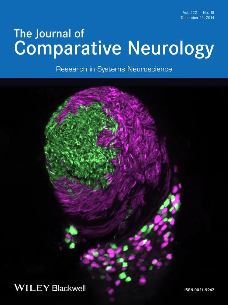
The different subregions of the IC (central and external nucleus) and the SOC (medial, lateral, and trapezoid regions) were defined by using the DAPI counterstain on all CaBP-stained sections. In accordance with previous investigations, the IC was defined as being dorsal to the cuneiform nucleus and the nucleus of the lateral lemniscus, lateral to the periaquaductual gray, ventral and caudal to the superior colliculus, and caudal to the central tegmental field (Oliver, 2005). We differentiated the central nucleus (ICc) from the peripheral cortex (ICx) based on their relative immunoreactivity to PV and CB, because the ICc and ICx of primates have previously been segregated based on the immunoreactivity to these molecules, with the ICc being PV rich and the ICx being CB rich (Jones, 2003; Parviz and Damasio, 2003; Engle et al., 2014; Fig. 1A,C). The three principle subdivisions of the SOC, the medial nucleus of the trapezoid body (MNTB), medial superior olive (MSO), and lateral superior olive (LSO), were segregated based on the conventions of Strominger and Hurwitz (1976) and Irving and Harrison (1967). Briefly, the MSO was characterized as a vertically oriented cell-rich region in the caudal pontine tegmentum surrounded by heavy staining of the neuropil. The LSO was characterized by a globular cell-rich region located laterally and slightly dorsally to the MSO, and the MNTB was identified as a cell rich region with large perikarya lying medially and ventrally to the MSO (Fig. 1B; see also Gray et al., 2014a).

Transverse sections of the auditory midbrain and brainstem. A: PV (magenta) and GAD67 (green) colabeled section of the inferior colliculus. The two principle subdivisions, the core (ICc) and cortex region (ICx), are labeled. B: PV (magenta) and GAD67 (green) colabeled section of the superior olivary complex. The medial superior olive (MSO), lateral superior olive (LSO), and medial nucleus of the trapezoid body (MNTB) are labeled. C: The CB (magenta)-rich ICx region colabeled with GAD67 (green). Scale bars = 500 µm in A,B; 250 µm in C.
Data analysis
Fluorescence microscopy was used to coregister CaBP immunoreactivity (PV or CB) with GAD67 immunoreactivity. Individual neurons were sampled by taking ×20 or ×40 images of the different subdivisions of the IC and SOC using an appropriate set of fluorescent filters. All images were line averaged to reduce background noise and imported into Adobe Photoshop CS5, in which they were coregistered. Images of the counting fields were taken away from the anatomical borders of the regions of interest to ensure that all sampled neurons belonged to the appropriate subdivisions.

Statistical analysis
Two statistical tests were performed, first at the level of individual neurons and second at the level of individual animals. In the first analysis, neurons from all animals were grouped and categorized as olivary/collicular for the between-region analysis and young/old for the aging analysis. A Mann-Whitney U test for nonparametric populations was used to analyze whether the proportions of neurons that coexpressed GAD67 and PV/CB were significantly different between the IC and the SOC or between age groups. The age cutoff was 21.66 years (roughly 65 human years). A relatively stringent significance criterion of P < 0.01 was used in these analyses. The total number of neurons sampled in each comparison ranged between 272 and 498 neurons per group for the regional analysis and between 51 to 202 neurons per group for the aging analysis. This grouping increased the sample size because each neuron was considered a data point that either did or did not express GAD67. This analysis provided more power to detect changes (given the relatively small number of animals) than the multiple-way ANOVA described below, which uses each individual animal as a data point.
In the second analysis, we sought to understand the data at the level of individual animals by using a two-way ANOVA with region (IC vs. SOC) and age as factors. A significance criterion of P < 0.05 was used in this analysis because protection against the effects of multiple comparisons is built into the ANOVA model. Overall, this is a more stringent analysis than the Mann-Whitney U test, especially given the much smaller sample size (n = 8 animals). Both of these analyses together are useful for understanding the degree of intersubject variability (reflected strongly in the ANOVA) for both region- and age-related differences (detected through the less stringent Mann-Whitney U test).
Finally, to determine age-related density changes in PV+ and CB+ neurons (as opposed to the proportions described above), the density values from young and old animals underwent an unpaired t-test with a significance criterion of P < 0.01.
RESULTS
Characterization of the IC and SOC
We examined the IC and SOC in transverse histological sections from eight rhesus macaques ranging in age from 12 through 28 years, which roughly corresponds to 36 through 84 human years (Davis and Leathers, 1985). Neurons from these sections were labeled with GAD67 antibodies along with either PV or CB antibodies (see Materials and Methods). All animals stained positively for all antibodies used, so both combinations of PV-GAD67 and CB-GAD67 were observed and analyzed. Borders of the IC and SOC (Fig. 1A,B) were determined with the help of a DAPI counterstain (see Materials and Methods).
The proportion of PV+ and CB+ neurons expressing GAD67 differed in the IC and SOC
In rodents, most PV+ and CB+ neurons in the IC express GAD67, whereas for the SOC this is not the case (Fredrich and Reisch, 2009). Because these observations have not been made in the macaque, at the first level of analysis we compared the proportion of sampled PV+ and CB+ neurons that expressed GAD67 in the IC and SOC (Fig. 2A,B). We observed PV+ and CB+ neurons expressing GAD67 in both the IC and the SOC, although in different proportions. In the IC, 57% of PV+ neurons sampled were also GAD67+, whereas only 42% expressed GAD67 in the SOC (Mann-Whitney U, P < 0.01; Fig. 2D). Conversely, CB+ neurons in the IC and SOC did not differ in the proportion of the neurons expressing GAD67 (61% and 62%, respectively; Mann-Whitney U, P > 0.05; Fig. 2D). These data suggest that at the level of individual neurons there are regional differences in the expression of GAD67 for populations of PV neurons but not CB neurons. These observations held true at the level of individual animals (two-way ANOVA; PV: P = 0.03; CB: P > 0.05), suggesting that there is little variability in these observations between monkeys.

Proportion of sampled PV-positive and CB-positive neurons expressing GAD67 in the IC and SOC. Neurons containing a significant amount of both PV (magenta) and GAD67 (green) resulted in white label in the coregistered images, and these neurons were considered GAD67-expressing PV-positive neurons. The same analysis was carried out for CB-positive neurons (data not shown). Arrowheads indicate PV neurons that did not contain sufficient GAD67 label to be considered GAD67 positive. In the SOC (A), IC (B), and striatum (C), there appeared to be differences in the proportions of PV- and CB-expressing neurons that expressed GAD67. Quantifications revealed that the proportion of GAD67-immunoreactive PV- and CB-positive neurons in the superior olivary complex (SOC), inferior colliculus (IC), and striatum indeed differed. D: In the SOC, 38% of PV-positive neurons expressed GAD67, whereas 52% were GAD67 positive in the IC. Both of these percentages were significantly less than the 82% of PV-positive neurons expressing GAD67 in the striatum. Conversely, the GAD67 expression of CB-positive neurons did not differ between the SOC and the IC, but both were significantly less than the 93% GAD67 expression of CB-positive neurons in the striatum. *P < 0.05. Scale bar = 40 microns.
As a control for the GAD67 specificity observed in the IC and SOC, we also sampled neurons from the striatum (Fig. 2C), because the molecular profiles of PV+ and CB+ neurons in this region are well known (Carder et al., 1996; Parent et al., 1996; Prensa et al., 1998; Cicchetti et al., 2000; Rudy et al., 2011) and make up separate chemically defined classes of GABAergic interneurons. Our results are consistent with these previous findings in that PV+ and CB+ neurons in the striatum expressed GAD67 in most neurons sampled. Quantification of these observation revealed that PV+ neurons expressed GAD67 in 83% of neurons sampled, and CB+ neurons expressed GAD67 in 92% of the neurons sampled (Fig. 2, light gray bars). These proportions are significantly different from the proportion of PV+ and CB+ neurons expressing GAD67+ in the IC and SOC (Mann-Whitney U, P < 0.01; Fig. 2D, asterisks). Again, a two-way ANOVA yielded identical results (both PV and CB P < 0.05), indicating that these differences are also seen at the level of individual animals. Thus, our methodology yielded results consistent with those found previously for the striatum, and the percentage of PV+ and CB+ neurons expressing GAD67 in the macaque SOC and IC is significantly different from that in the striatum. Together these data suggest that there exist subclasses of CaBP-expressing neurons in the auditory brainstem and midbrain that differ chemically and physiologically from cerebral neurons expressing the same CaBPs and that these observations are consistent across animals.
Age-related changes in PV+ and CB+ neurons expression
To determine whether there is an age-related change in the density of neurons expressing these CaBPs, as reported previously (Gray et al., 2013, 2014a,b; Engle et al., 2014), we quantified the age-related density changes of PV+ and CB+ neurons by counting the number of PV+ or CB+ neurons per section analyzed and compared these values across age groups. The results of the current study were consistent with prior studies in that PV expression was significantly increased in the MSO, LSO, and ICc with age (unpaired t-test, P < 0.01; Fig. 3A), but not in the MNTB or ICx. Age-related increases in CB expression were noted only in the MSO (unpaired t-test, P < 0.01; Fig. 3B), and to our knowledge this is the first report of age-related changes in the density of CaBP-expressing neurons in the macaque auditory system.

Age-related changes in PV- and CB-expressing neurons. The average number of cells expressing PV (A) and CB (B) in the young animals (solid bars) differed compared with old animals (open bars). Statistically significant differences in PV+ neurons were not seen in the medial nucleus of the trapezoid body (MNTB) or cortex region of the inferior colliculus (ICx) but were observed in the medial superior olive (MSO), lateral superior olive (LSO), and core region of the inferior colliculus (ICc). For CB+ neurons, only the MSO showed an age-related difference. *P < 0.05.
Age-related changes in the proportion of GAD67-expressing PV+ and CB+ neurons in the IC and SOC
Given that we observed age-related changes in the density of PV+ and CB+ neurons, the next level of analysis was to determine whether there was also a difference in the percentage of these neurons that also expressed GAD67. The quantification of the proportion of these cells within the IC and SOC revealed changes in some, but not all, subdivisions of these structures. PV and GAD67 showed no significant changes in coexpression in the ICc as a function of age (Mann-Whitney U; P > 0.05) but an increase with age in the ICx (Mann-Whitney U; P < 0.01; Fig. 4A). The proportion of CB+ neurons in both the ICx and the ICc expressed GAD67 equally with age (Mann-Whitney U; P > 0.05; Fig. 4B). Changes were also noted in the SOC, although they differed from the changes seen in the IC. The proportion of PV+ neurons expressing GAD67 significantly decreased with age in the LSO (Mann-Whitney U; P < 0.01; Fig. 4A) and showed a trend to decrease in the MSO (Mann-Whitney U; P = 0.06). The changes in CB and GAD67 coexpression with age were confined to the MSO, where significant increases were noted (Mann-Whitney U; P < 0.01; Fig. 4B).

Age-related changes in the proportion of GAD67-expressing PV- and CB-positive neurons in the inferior colliculus (IC) and superior olivary complex (SOC). A: The proportion of GAD67-expressing PV-positive neurons remained constant with age in the ICc but increased in the ICx. In the SOC, only the lateral superior olive (LSO) showed significant decreases in the proportion of PV-positive neurons expressing GAD67. B: The proportion of GAD67-expressing CB-positive neurons remained constant with age in the IC and decreased only in the medial superior olive (MSO) of the SOC. C: When the subdivisions of the IC and SOC were combined and compared across age groups, the proportion GAD67-expressing PV- and CB-positive neurons remained constant, whereas significantly more PV-positive neurons and fewer CB-positive neurons expressed GAD67 in the SOC. D: Scatterplots of age against the proportion of GAD67-expressing cells shows the considerable variability across animals in these age-related changes. *P < 0.05.
Because one or more subdivisions showed either significant differences or trends toward increases or decreases in the coexpression of GAD67 and PV+/CB+ neurons as a function of age, we combined the values of all IC and SOC subdivisions. This analysis revealed that as a population there was no change in the coexpression of GAD67 with either PV+ or CB+ neurons in the IC with age; however, in the SOC, PV+ neurons express GAD67 less often and CB+ neurons express GAD67 more often with age (Mann-Whitney U, P < 0.01; Fig. 4C). Together these data suggest that, at the level of individual neurons, age-related changes in the inhibitory molecular profile in populations of PV+ and CB+ neurons exist in the SOC, but not the IC, and are different among CaBPs. In general, the populations of PV+ neurons became less inhibitory with age, whereas populations of CB+ neurons become more inhibitory with age, but these observations are not absolute (PV+ neurons in the ICx) nor present in all auditory subdivisions. When these same data underwent a two-way ANOVA, no statistically significant changes were observed, indicating that there is considerable inter-animal variation in these age effects that, given the small sample size, masks any age-related effects at the level of individual animals (Fig. 4D). Together these data suggest that populations of PV- and CB-expressing neurons in the auditory brainstem change their expression of GAD67, but these changes do not necessarily occur in all aged animals.
DISCUSSION
Age-related changes in PV and CB neuron density
Neurons of the central auditory system containing CaBPs have repeatedly been shown to change in density during the natural aging process of both rodents (O'Neill et al., 1997; Zettel et al., 1997; Idrizbegovic 2003, 2004, 2006; Ouda et al., 2008, 2012) and nonhuman primates (Gray et al., 2013, 2014a, b; Engle et al., 2014; Fig. 3). The present study is the second report of age-related density changes in PV-expressing neurons within the auditory system of the macaque and the first report of age-related changes in CB-expressing neurons. Although the density changes of CB-expressing neurons were confined to the MSO, unlike the more widespread density changes of PV-expressing neurons, it is clear that the number of neurons expressing both of these CaBPs can change in aged auditory nuclei. Notably, all reported age-related changes in the density of CaBP-expressing neurons have resulted in increases, whereas both increases and decreases have been shown in rodents. These observations suggest that there are cross-species differences in the expression of these neurons and in their density changes with age. The root of these inconsistencies may arise from cross-species differences in sound processing or from differences in the distribution of inhibitory neurons throughout the auditory pathway. For example, the auditory thalamus of rodents does not contain inhibitory neurons, but the same structure in primates and felines does (Winer and LaRue, 1996; Bartlett and Smith, 1999). Also, the specifics of binaural sound processing can differ significantly between species with different hearing ranges (for reviews see Moore, 1991; Pollack et al., 2002; Grothe and Koch, 2011). Regardless of the exact cross-species differences, the results from rodent models cannot be completely generalized to primates, and vice versa.
What remains unclear, however, is the role of these changes in the aging process. This gap in understanding arises partially from our minimal knowledge of the physiological response properties of CaBP-expressing neurons of subcortical structures. Most studies investigating the physiology of these neurons have used the cerebral cortex, striatum, or hippocampal formation, where most are GABAergic inhibitory neurons; therefore, the first goal of the present study was to uncover the GABAergic profiles of populations of PV+ and CB+ neurons in the auditory midbrain and brainstem.
Subcortical PV+ and CB+ cells differ from cerebral PV+ and CB+ neurons
The results of the current study reveal two important findings. First, as a population, PV+ neurons of the IC express GAD67 more often than in the SOC, a difference that is not seen with populations of CB+ neurons. Therefore, populations of CaBP-expressing neurons can differ in their net GAD67 expression between the midbrain (IC) and the brainstem (SOC), but this is not the case with all classes of CaBP-expressing neurons. Second, as a population, subcortical PV+ and CB+ neurons expressed GAD67 in significantly fewer cells than in populations of chemically analogous (with respect to CaBPs) neurons in the cerebrum (Fig. 2). This observation suggests that the presence of PV and CB in these subcortical structures does not relate to a single specific physiological profile of neurons as it does in the cerebrum but rather PV and CB are likely expressed in neurons performing different cellular processes, some of which are likely affected during the aging process.
Together these data suggest that CaBP-expressing neurons of the subcortical auditory system cannot, as a population, be considered inhibitory through GABAergic neurotransmission. Caution should be taken, however, when interpreting these results. It is possible that GAD67 expression is simply lower in SOC neurons compared with IC neurons, bringing them below detection levels. Also, we cannot definitively rule out that these non-GAD67-expressing neurons are not inhibitory in these areas of the brain through a different neurotransmitter system. As an example, many subcortical auditory neurons use glycine as an inhibitory neurotransmitter instead of GABA. To investigate these possibilities, it would be informative for future studies to colabel these CaBP-expressing neurons with other markers of GABA neurotransmission, such as the GABA vesicular transporter, GAT, or markers for other neurotransmitters. Alternatively, some neurons may express a different isoform of GAD, such as GAD65, which would elude the present analysis.
Despite the possibilities described above, substantial proportions of both PV+ and CB+ neurons do express GAD67 in both the SOC and the IC. These large values suggest that subclasses of CaBP-expressing neurons contain GAD67 and can be differentiated from one another based on independent morphological or chemical characteristics. The distribution of these subclasses of neurons may differ between regions of the auditory system, which would explain the IC–SOC differences in PV+ inhibitory cells. It is unknown what morphological cell types comprise the chemically defined neurons described in the current study. A better understanding of the morphological and chemical properties characterizing these CaBP-containing, GAD67-expressing neurons would be useful in elucidating their different expression patterns observed in the IC and SOC.
Functional implications
Aged neurons of the auditory system of both rodents and nonhuman primates have increased spontaneous and driven firing rates in both subcortical and cortical structures (Caspary et al., 2005, 2006; Juarez-Salinas et al., 2010; Engle and Recanzone, 2012). These increases result in broader spectral and spatial tuning of auditory neurons, both deficits that correlate with decreases in psychophysical metrics of auditory perception (Engle and Recanzone, 2012). Aside from disrupting auditory perception, these age-related increases in neural activity put auditory neurons at risk for excitotoxicity. This toxicity occurs when relatively high levels of glutamate cause greater depolarizations and more frequent action potentials, increasing the influx of calcium, which activates intracellular enzymes that in turn damage vital cellular components (Manev et al., 1989). Given the increased physiological activity in aged auditory neurons, mechanisms to prevent such excitotoxicity are likely necessary for normal cell maintenance. An effective way to compensate for the increased intracellular calcium would be to increase the expression of CaBPs, which act as calcium buffers.
The current data suggest that in the macaque, as in rodents (Fredrich and Reisch, 2009), the presence of CaBP within subcortical auditory neurons does not necessarily relate to the neurotransmitters used by these neurons. Rather, the CaBPs likely play different, nonelectrophysiological roles in the aging process of the auditory system. One hypothesis explaining the age-related increases in these neurons is that they function as a homeostatic response combatting the toxic effects of excitotoxicity. This idea is applicable to both rodent and nonhuman primate models because both experience age-related hyperexcitability in the auditory system (Caspary et al., 2006; Juarez-Salinas et al., 2010), both possess subcortical PV- and CB-expressing neurons that do not relate to a single class of neurotransmitter, and the basic intracellular physiological mechanisms (metabolic homeostasis, control of Ca2+ levels, etc.) are very similar. Whether this is true remains to be seen; however, these observations give credibility to such a preventive function for these cells.
SUMMARY AND CONCLUSIONS
In the brainstem, the proportion of GAD67-expressing PV and CB cells changed in aged animals such that PV+ neuron populations were less likely to express GAD67 and CB+ populations were more likely to express GAD67, and this effect was not noted in the IC. Therefore, if the speculated compensatory effort of the auditory system to combat the hyperexcitability of neurons that occurs with age is true, neurons expressing CB are more likely to be responsible for this because the aged population of these cells expressed GAD67 more often. Conversely, PV+ neuron populations become less inhibitory with age, but these cells still increase in density, suggesting that the Ca2+ buffering capacity of PV may be of importance in combatting the excitotoxic effects from an intracellular level. Whether either of these two hypotheses are true cannot be determined from the present data. Regardless, this study suggests that the neurons of the IC and SOC are not equivalently affected by the aging process, which supports the hypothesis noted above that different morphological classes of neurons express PV or CB and are distributed differently throughout the ascending auditory system. Taken together, the current data demonstrate that densities of GAD67-expressing PV+ and CB+ neurons change in the brainstem with age, which would clearly influence local and global circuit and network processing of auditory information between young and old animals. The functional consequences of these age-related changes at the brainstem and midbrain levels and how they relate to age-related hearing deficits defined at the perceptual level remain to be seen.
ACKNOWLEDGMENTS
The authors thank Carol A. Barnes for her generous contribution of tissue to the current project. We also thank Mary Baldwin, Xochi Navarro, and Leah Krubitzer for their contributions of expertise and resources to this project.
CONFLICT OF INTEREST STATEMENT
The authors declare no competing financial interests.
ROLE OF AUTHORS
All authors had full access to all the data in the study and take responsibility for the integrity of the data and the accuracy of the data analysis. Study concept and design: DTG, JRE, GHR. Acquisition of data: DTG, MLR, JRE. Analysis and interpretation of data: DTG, JRE, GHR. Drafting of the manuscript: DTG, GHR. Critical revision of the manuscript for important intellectual content: DTG, JRE, GHR. Statistical analysis: DTG. Obtained funding: GHR. Administrative, technical, and material support: DTG, JRE, MLR. Study supervision: GHR.