NAT10 regulates tumor progression and immune microenvironment in pancreatic ductal adenocarcinoma via the N4-acetylated LAMB3-mediated FAK/ERK pathway
Enhong Chen, Qin Wang, and Leisheng Wang contributed equally to this work.
Abstract
Background
N-acetyltransferase 10 (NAT10) was reported to be associated with the immune microenvironment in several cancers. However, it is not known in pancreatic ductal adenocarcinoma (PDAC). This study aimed to elucidate the roles and mechanisms of NAT10 in tumor malignancy and the tumor microenvironment (TME) in PDAC.
Methods
NAT10 expression and its role in tumor progression and clinical prognosis were analyzed using bioinformatics and functional assays. Downstream genes regulated by NAT10 and their underlying mechanisms were explored using acetylated RNA immunoprecipitation, quantitative polymerase chain reaction, RNA immunoprecipitation, and Western blotting. The role and mechanism of NAT10 in the PDAC TME were further explored using bioinformatics, single-cell RNA sequencing, multiplexed immunofluorescence, and flow cytometry. The association between NAT10 and immunotherapeutic response was investigated in a mouse model by inhibiting the programmed cell death 1/programmed cell death ligand 1(PD-1/PD-L1) axis with a PD-1/PD-L1 binding inhibitor, Naamidine J.
Results
NAT10 was upregulated in PDAC tissues and cell lines, and was associated with poor progression-free survival of PDAC patients. NAT10 promoted tumor progression by enhancing the mRNA stability of laminin β3 (LAMB3) via N4-acetylation modification, thereby activating the focal adhesion kinase (FAK)/extracellular regulated protein kinases (ERK) pathway. NAT10 promoted subcutaneous tumor growth, increased the proportion of exhausted CD8+ T cells (CD8+ Tex), especially the intermediate CD8+ Tex subset, and decreased the proportion of cytotoxic CD8+ T cell (CD8+ Tc) subset in the PDAC TME. Naamidine J treatment significantly enhanced the proportion of CD8+ Tc subset and reduced the proportion of intermediate CD8+ Tex subset in mice bearing subcutaneous tumors with high NAT10 expression. Regarding the regulatory mechanism, NAT10 increased PD-L1 expression and abundance in tumor cells by activating the LAMB3/FAK/ERK pathway, thereby reducing the cytotoxicity of CD8+ T cells. Inhibition of the PD-1/PD-L1 axis with Naamidine J retrieved CD8+ T cell cytotoxicity.
Conclusions
This study proposes a regulatory role of NAT10 in tumor progression and immune microenvironment via the LAMB3/FAK/ERK pathway in PDAC. These findings may favor the selection of candidates who may benefit from immunotherapy, optimize current therapeutic strategies, and improve the clinical prognosis of PDAC patients.
Abbreviations
-
- ac4C
-
- N4-acetylcytine
-
- acRIP
-
- Acetylated RNA immunoprecipitation
-
- Act_CD4+ T
-
- Activated CD4+ T cell
-
- Act_B
-
- Activated B cell
-
- Bcl2
-
- B-cell-lymphoma 2
-
- CAR-T
-
- Chimeric antigen receptor-T
-
- CD8+ Tex
-
- Exhausted CD8+ T
-
- CD8+ Tn
-
- Naïve CD8+ T
-
- CD8+ Tc
-
- Cytotoxic CD8+ T
-
- CTLA-4
-
- Ctlantigen-4
-
- Casp3
-
- Caspase-3
-
- Cdk1
-
- Cyclin-dependent kinase 1
-
- PFS
-
- Progression-free survival
-
- DAPI
-
- 4',6-Diamidino-2-phenylindole
-
- DC
-
- Dendritic cell
-
- DEGs
-
- Differential expressed genes
-
- γδ T
-
- Gamma-delta T cells
-
- ECM
-
- Extracellular matrix
-
- ERK1/2
-
- Extracellular regulated kinases 1/2
-
- FBS
-
- Fetal bovine serum
-
- FAK
-
- Focal adhesion kinase
-
- GEO
-
- Gene Expression Omnibus
-
- GSE
-
- Gene Expression Omnibus Series
-
- GEPIA2
-
- Gene Expression Profiling Interactive Analysis 2
-
- GTEx
-
- Genotype-Tissue Expression
-
- GSEA
-
- Gene Set Enrichment Analysis
-
- GAPDH
-
- Glyceraldehyde 3-phosphate dehydrogenase
-
- Gzmb
-
- Granzyme B
-
- HPNE
-
- Human pancreatic ductal immortalized cells
-
- iDC
-
- Inducible dendritic cell
-
- ICIs
-
- Immune checkpoint inhibitors
-
- IRS
-
- Immunoreactive score
-
- IFNg
-
- Interferon gamma
-
- KD
-
- Knockdown
-
- LAMB3
-
- Laminin β3
-
- Lag3
-
- Lymphocyte-activation gene 3
-
- m6A
-
- N6-methyladenosine
-
- m1A
-
- N1-methyladenosine
-
- m5C
-
- 5-methylcytosine
-
- m7G
-
- N7-methylguanosine
-
- NAT10
-
- N-acetyltransferase 10
-
- NKT
-
- Natural killer T cell
-
- NK
-
- Natural killer cell
-
- NC
-
- Negative control
-
- OS
-
- Overall survival
-
- OE
-
- Overexpression
-
- PBS
-
- Phosphate-buffered saline
-
- PDAC
-
- Pancreatic ductal adenocarcinoma
-
- PD-1
-
- Programmed cell death 1
-
- Pdcd1
-
- Programmed cell death 1
-
- PD-L1
-
- Programmed cell death ligand 1
-
- PFA
-
- Paraformaldehyde
-
- p-FAK
-
- Phosphorylated focal adhesion kinase
-
- p-Src
-
- Phosphorylated sarcoma
-
- p-ERK1/2
-
- Phosphorylated extracellular regulated protein kinases1/2
-
- Prf1
-
- Perforin 1
-
- PCA
-
- Principal component analysis
-
- qPCR
-
- Quantitative polymerase chain reaction
-
- RIP
-
- RNA immunoprecipitation
-
- RNA-seq
-
- RNA sequencing
-
- Src
-
- Sarcoma
-
- scRNA-seq
-
- Single-cell RNA sequencing
-
- TCGA
-
- The Cancer Genome Atlas
-
- TISIDB
-
- The Tumor-Immune System Interactions Database
-
- TIMER
-
- Tumor Immune Estimation Resource
-
- TME
-
- Tumor microenvironment
-
- Tem_CD8+ T
-
- Effector memory CD8+ T cell
-
- Tem_CD4+ T
-
- Effector memory CD4+ T cell
-
- Treg
-
- Regulatory CD4+ T cell
-
- Tfh
-
- Follicular helper T cell
-
- Th1
-
- T helper cell type I
-
- T_NK cells
-
- T cell and NK cell
-
- Tim-3
-
- Mucin-3/T-cell immunoglobulin
-
- Tgfb1
-
- Transforming growth factor beta 1
-
- UMI
-
- Unique molecular identifier
-
- UMAP
-
- Uniform Manifold Approximation and Projection
1 BACKGROUND
Pancreatic ductal adenocarcinoma (PDAC) is one of the most lethal malignancies globally. It is characterized by late diagnosis and poor 5-year survival (∼9%) due to recurrence and distant metastasis [1, 2]. Chemotherapeutics with moderate benefits remain the cornerstone of advanced PDAC treatments [3]. There is an urgent need to improve and optimize current therapeutic strategies.
The recent breakthroughs of immunotherapy through immune checkpoint inhibitors (ICIs) have substantially changed therapeutic paradigms in several types of cancers with considerable survival benefits [4-13]. However, PDAC has been largely refractory to immunotherapeutic approaches, mainly because of its immune-cold phenotype [14-22]. Therefore, an in-depth understanding of the tumor progression mechanisms and the characterization of the tumor microenvironment (TME) will provide insights into the development of novel and targeted therapeutic strategies for PDAC management.
Epigenetic modifications, such as RNA modifications, play a crucial role in regulating molecular function, including N6-methyladenosine (m6A), N1-methyladenosine (m1A), 5-methylcytosine (m5C), N7-methylguanosine (m7G), and N4-acetylcytidine (ac4C) [23-32]. RNA modifications mainly modulate RNA stability and translational efficacy, and are associated with malignant biological properties and carcinogenesis in several types of cancer [33]. Pathways modulated by RNA modifications could be potential targets for cancer therapy [34, 35]. For instance, methyltransferase-like 3 promoted tumor progression in PDAC via the m6A-mediated LncRNA 00662/general transcription factor IIB/integrin alpha 1/focal adhesion kinase (FAK) pathway. Targeting this pathway is a potential strategy to inhibit the growth of PDAC tumors in vitro and in vivo [36].
In addition, ac4C plays vital roles in malignant tumor progression, including gastric cancer [37], hepatocellular carcinoma [38], acute myeloid leukemia [39], colorectal cancer [40], and bladder cancer [41]. N-acetyltransferase 10 (NAT10) is recognized as the only known ac4C “writer” protein, containing both an N-acetyltransferase domain and a nucleotide-binding region, and acts as an acetyltransferase [42-45]. In PDAC, NAT10-mediated ac4C modifications on receptor tyrosine kinases promote tumorigenesis and metastasis [46]. Moreover, several recently published studies have indicated that NAT10 regulates programmed cell death ligand 1 (PD-L1) expression and is associated with the immune microenvironment in several types of cancer, including breast [47], melanoma [47], colorectal [48], and cervical cancers [49]. Targeting NAT10 or combining it with ICIs represents a promising strategy to improve therapeutic efficacy in tumors [47]. However, for immune-cold tumors, such as PDAC, the exact functions and mechanisms of NAT10 in tumor progression and the TME remain largely unknown.
In the current study, we focused on the roles and regulatory mechanisms of NAT10 in tumor properties and the TME of PDAC. These findings may facilitate the identification of molecular targets and predictive markers for immunotherapeutic response, thus optimizing current therapeutic strategies, especially immunotherapy, and improving the clinical prognosis of PDAC patients.
2 MATERIALS AND METHODS
2.1 Tissue microarray
Tissue microarray (TFPanc-02) of 108 pairs of PDAC tissues and adjacent normal tissues (collected from December 2011 to December 2015) was obtained commercially (TUFEIBIO, Shanghai, P. R. China). All the patients were followed up regularly until September 2022. This study was approved by the Ethics Committee of the Affiliated Hospital of Jiangnan University (No. LS2024065), and written informed consent was obtained from all participants.
2.2 Immunohistochemistry
Paraffin-embedded sections were dewaxed in xylene and rehydrated in a graded alcohol series. Heat-activated antigen retrieval was performed using a sodium citrate buffer. The sections were heated for 20 minutes using a microwave. Endogenous peroxidase activity was suppressed by exposure to 3% hydrogen peroxide for 10 minutes. The sections were then blocked with 5% bovine serum albumin and incubated with primary antibody at 37°C for 1 hour, followed by incubation with horseradish peroxidase-conjugated secondary antibody at 37°C for 20 minutes. The sections were visualized using diaminobenzidine and counterstained with hematoxylin for microscopic examination. The immunoreactive score (IRS, IRS = staining intensity × positive cell proportion) was used to assess protein expression. Staining intensity was graded as 0, 1, 2, and 3 (no staining, weak, moderate, and strong staining, respectively). The proportion of positive cells was scored as 1, 2, and 3 (<10%, 10%-50%, and >50%, respectively).
2.3 Bioinformatic analysis
Expression patterns of NAT10 and laminin β3 (LAMB3) were analyzed based on the data from two Gene Expression Omnibus (GEO) datasets (GSE15471: 39 pairs of PDAC tissues and adjacent normal tissues, and GSE16515: 26 pairs of PDAC tissues and adjacent normal tissues). The expression pattern of LAMB3 in PDAC and normal tissues was analyzed using Gene Expression Profiling Interactive Analysis 2 (GEPIA2, http://gepia2.cancer-pku.cn) [50], with matched normal tissue data from The Cancer Genome Atlas (TCGA, project ID: TCGA-PAAD) and Genotype-Tissue Expression (GTEx) databases. Survival analysis and the correlation between NAT10 and CD274 expression were also performed using GEPIA2 with the data from TCGA-PAAD. The correlations between NAT10 and LAMB3/ITGA2/CEACAM6 expressions were analyzed using an online tool named TNMplot (http://TNMplot.com/analysis/; PDAC tissues: n = 248; correlation method: Spearman) [51]. The correlations between NAT10 expression and the clinicopathologic features were analyzed using an online tool named The Tumor-Immune System Interactions Database (TISIDB, http://cis.hku.hk/; PDAC tissues: n = 179; correlation method: Spearman). The correlations between NAT10 expression and immune cell infiltration were analyzed using two online tools: TISIDB (PDAC tissues: n = 179; correlation method: Spearman) [52] and Tumor Immune Estimation Resource (TIMER, http://timer.cistrome.org/; PDAC tissues: n = 179; correlation method: Spearman) [53].
2.4 Cells and cell culture
Four human PDAC cell lines (BxPC-3, MIA-PaCa2, CoLo357, and PANC-1), a mouse PDAC cell line (PanO2), and a human pancreatic ductal immortalized cell (HPNE) were purchased from Shanghai Zhong Qiao Xin Zhou Biotechnology Co., Ltd. (Shanghai, P. R. China). All the cells were cultured in high-glucose DMEM (Wisent, Nanjing, Jiangsu, P. R. China) supplemented with 10% fetal bovine serum (FBS, Wisent), 100 IU/mL penicillin, and 100 µg/mL streptomycin (Thermo Fisher Scientific, MA, USA) at 37°C in a humidified atmosphere containing 5% CO2.
2.5 RNA extraction and real‑time quantitative polymerase chain reaction (qPCR)
Total RNA was extracted using TRIzol reagent (Invitrogen, CA, USA). cDNA was synthesized using a reverse transcription kit (ProteinBio, Nanjing, Jiangsu, P. R. China) according to the manufacturer's instructions. SYBR Green qPCR Supermix (ProteinBio) was used for qPCR analysis in an ABI 7900 real-time PCR Detection System (Applied Biosystems, CA, USA). β-actin was used to normalize the RNA levels using the 2−ΔΔCt method. All qPCR assays were performed in triplicate. The primer sequences are listed in Supplementary Table S1.
2.6 Stable cell line construction
Lentiviruses encoding small hairpin RNA (shRNA) targeting human NAT10 and mouse Nat10, as well as a lentivirus encoding human NAT10, were constructed by GeneChem (Shanghai, P. R. China). Four human PDAC cell lines (BxPC-3, MIA-PaCa2, CoLo357, and PANC-1) and a mouse PDAC cell line (PanO2) were seeded in a 24-well plate at a density of 2 × 10⁴ cells per well. After a 24-hour culture, the cells were infected with lentiviruses at a multiplicity of infection of 50. The plasmid encoding mouse Nat10 was constructed by Miaoling Biotechnology (Wuhan, Hubei, P. R. China). PanO2 cells (2 × 10⁴ cells/well) were seeded in a 24-well plate. After a 24-hour culture, the cells were transfected with 5 µg of plasmids using 2.5 µL Lipofectamine 3000 (Thermo Fisher Scientific). Stable cell clones were selected for 1 week using 0.5 µg/mL puromycin (MedChemExpress, NJ, USA). The efficiency of NAT10/Nat10 overexpression and knockdown was analyzed using qPCR. The primer sequences and target sequences of shRNAs are listed in Supplementary Table S1 and Supplementary Table S2, respectively.
2.7 RNA sequencing (RNA-seq) and data processing
Total RNA was extracted from the Nat10 overexpression and the negative control PanO2 cells for RNA-seq by Beijing Tsingke Biotech Co., Ltd. (Beijing, P. R. China). RNA-seq was performed on the Illumina HiSeq platform. After quality control, the clean reads were aligned to the mouse reference genome GRCm39 (https://ftp.ensembl.org/pub/release109/fasta/mus_musculus/dna/Mus_musculus.GRCm39.dna.toplevel.fa.gz) with Hisat2 software (v2.0.4) [54]. The read counts were adjusted by the edgeR package [55] through one scaling normalized factor. Differential expression analysis of two samples was performed using the EBSeq R package [56].
2.8 RNA interference and plasmid transfection
Cells (6 × 10⁵/well) were seeded in a 6-well plate and were subjected to RNA interference and plasmid transfection upon reaching ∼80% confluence after a 24-hour culture. For RNA interference, a mixture of 50 nmol/L siRNA and 7.5 µL of Lipofectamine 3000 was incubated at room temperature for 20 minutes and then added to the cells. For plasmid transfection, a mixture of 15 µg of plasmids and 7.5 µL of Lipofectamine 3000 was incubated at room temperature for 20 minutes and then added to the cells. The transfected cells were collected after a 48-hour culture. The siRNAs were listed as follows: siLAMB3-sense: 5'-CCCUGGUGUGUAGCUAGUAAGTT-3'; siLAMB3-antisense: 5'-CUUACUAGCUACACACCAGGGTT-3'.
2.9 Western blotting
Total protein was extracted from the cells using radioimmunoprecipitation assay lysis buffer (Beyotime, Suzhou, P. R. China). The protein extracts were separated on 10% gels via sodium dodecylsulfate-polyacrylamide gel electrophoresis and then transferred to 0.45 µm polyvinylidene difluoride membranes (Millipore, MA, USA). The proteins were probed with specific antibodies after the blot was blocked with a blocking buffer (Beyotime). Protein expression was visualized using Super ECL Chemiluminescent Substrate Kit (Millipore). The antibodies used in this process are listed in Supplementary Table S3.
2.10 Immunofluorescence
Cells were fixed in 4% paraformaldehyde (PFA) for 10 minutes. After blocking, the cells were incubated with the primary antibody overnight at 4°C, followed by incubation with secondary antibodies for 1 hour at room temperature. The cells were counterstained with 4',6-diamidino-2-phenylindole (DAPI; Beyotime) and YF488-Phalloidin (Proteinbio) if needed. Stained cells were stored in the dark at 4°C until further investigation. The antibodies used in this process are listed in Supplementary Table S3.
2.11 Clone formation assay
Cells were seeded in a 6-well plate (500 cells/well) and incubated in complete medium at 37°C with 5% CO2. The medium was replaced every 3 to 4 days. After 2 weeks, the colonies were fixed in 4% PFA for 20 minutes and stained with 0.1% crystal violet solution for 30 minutes.
2.12 Wound healing assay
Cells were seeded in a 6-well plate and cultured until 100% confluence. A wound was inflicted using a 200 µL pipette tip. The cells were washed with phosphate-buffered saline (PBS) to remove the detached cells and cultured in a serum-free medium. Microscopic images were captured at 0 and 24 hours after the wound was generated. The area of the wound was measured using the ImageJ software (version 1.53, National Institutes of Health, MD, USA).
2.13 Cell invasion assay
The upper chambers (Corning, NY, USA) were coated with Matrigel (Becton, Dickinson and Company, NJ, USA). The cells (30,000 cells/well) suspended in serum-free medium were placed into the upper chamber, and the culture medium supplemented with 10% FBS was added to the lower chamber. The plates were incubated at 37°C with 5% CO2 for 24 hours. The cells in the top chamber were removed using a cotton swab. The cells on the lower membrane surface were fixed in 4% PFA for 20 minutes and stained with 0.1% crystal violet for 30 minutes. The membrane was washed and dried. The cells in three to four random fields of view were counted using a microscope.
2.14 RNA decay assays
Cells were treated with 5 µg/mL Dactinomycin (Sigma, MO, USA) and harvested at different time points to extract RNA using TRIzol reagent (Invitrogen) for qPCR. The primer sequences are listed in Supplementary Table S1.
2.15 RNA immunoprecipitation (RIP)-qPCR
RIP was performed using a Magna RIP RNA-Binding Protein Immunoprecipitation Kit (Millipore) according to the manufacturer's instructions. Cells were collected and lysed with RIP lysis buffer. Magnetic beads were incubated with primary antibodies for 1 hour at room temperature. IgG was used as a negative control. After washing, the beads were incubated with cell lysis at 4°C for 3 hours. The beads were then collected and washed, and the RNA complexes were isolated by phenol-chloroform extraction. Enriched RNA fragments were subjected to qPCR analysis. The input RNA served as an internal control for normalization.
2.16 Acetylated RNA immunoprecipitation (acRIP)-qPCR
Total RNA was subjected to immunoprecipitation using aGenSeq®ac4C-IP Kit (GenSeq, Shanghai, P. R. China) according to the manufacturer's instructions. RNA was randomly fragmented into pieces of ∼200 nt with RNA fragmentation reagents. Protein A/G beads were coupled to the anti-ac4C antibody or IgG by incubation with rotation at room temperature for 1 hour. The RNA fragments were incubated with the antibody-conjugated beads and incubated with rotation at 4°C for 4 hours. The RNA/antibody complexes were then digested with Proteinase K, and the eluted RNA was purified by phenol/chloroform extraction. Based on the typical ac4C motifs (CXXCXXCXXCXX, X refers to A, C, G, U) [57], the primers were designed around the ac4C motif. The enrichment of RNA fragments bound to the ac4C antibody was quantified via qPCR. The input RNA served as an internal control for normalization.
2.17 Xenograft model
Four-week-old female C57BL/6 mice (18 to 20 g) were housed in a specific pathogen-free facility with a 12-hour light/12-hour dark cycle and free access to food and water. The animals were terminated if the body weight loss exceeded 20%-25% or the tumor weight accounted for more than 10% of the body weight. All animals were euthanized. For tumor growth evaluation, the flanks of mice were subcutaneously injected with PanO2 cells (3 × 10⁶ cells) suspended in 0.1 mL of PBS. After four weeks, the xenograft tumors were harvested for analysis. Tumor volumes were calculated as 0.5 × length × width2. To examine the role of Naamidine J (MedChemExpress) in tumor growth, mice were subcutaneously injected in the flanks with PanO2 cells (3 × 10⁶ cells) suspended in 0.1 mL of PBS. After four weeks, Naamidine J was intraperitoneally injected into mice (30 mg/kg, once every 3 days), and the tumors were harvested on day 56 after injection for analysis. The animal study was approved by the Laboratory Animal Welfare and Ethics Committee of Jiangnan University (IACUC Issue No: JN.No202312 30c0520505[626]).
2.18 Preparation of single-cell suspension
The subcutaneous tumors were excised, cut into small pieces, and digested in a dissociation medium containing 1 mg/mL collagenase IV (Sigma) and 0.2 mg/mL DNase I (Sigma). The tissue pieces were then incubated for 30 minutes at 37°C with vortexing manually every 10 minutes. After digestion, the cells were resuspended in 1 mL of ACK lysis buffer and incubated for 5 minutes to remove the red cells. Then, the cells were passed through 70 µm cell strainers to make a single-cell suspension. Finally, the cell suspension was centrifuged at 300 ×g for 5 minutes at 4°C and resuspended in PBS without calcium or magnesium.
2.19 Single-cell RNA sequencing (scRNA-seq) and data preprocessing
Single-cell suspensions were converted into uniquely barcoded RNA libraries using the MobiNova-100 Library (Mobidrop, Jiaxing, Zhejiang, P. R. China). The libraries were sequenced on a DNBSEQ-T7 (Beijing Genomics Institute, Shenzhen, P. R. China). The output raw data was mapped to the mouse reference genome (version mm10) using MobiVision software (version 3.0, Mobidrop) with unique molecular identifier (UMI) counts summarized for each barcode. The UMI count matrix was analyzed using the Seurat (version 4.0.0) R package [58]. To remove low-quality cells and likely multiplet captures, cells were filtered based on the following criteria: (1) gene number < 200, (2) UMI number < 1,000, (3) UMI per gene < 0.7, (4) proportion of UMIs mapped to mitochondrial genes > 10%, and (5) proportion of UMIs mapped to hemoglobin genes > 5%. Subsequently, the DoubletFinder [59] package (version 2.0.3) was used to identify potential doublets and multiplets. To obtain the normalized gene expression data, library size normalization was performed using the “NormalizeData” function of the Seurat R package. Specifically, the global-scaling normalization method “LogNormalize” normalized the gene expression measurements for each cell by the total expression, multiplied by a scaling factor (10,000 by default), and log-transformed the results.
Principal-component analysis (PCA) was performed to reduce dimensionality using the “RunPCA” function of the Seurat R package. Graph-based clustering was performed to cluster the cells according to their gene expression profiles using the “FindClusters” function of the Seurat R package. Cells were visualized using a two-dimensional Uniform Manifold Approximation and Projection (UMAP) algorithm with the “RunUMAP” function of the Seurat R package. The “FindAllMarkers” function (test.use = presto) was used to identify marker genes in each cluster. Differentially expressed genes (DEGs) were selected using “FindMarkers” function (test.use = presto). P value < 0.05 and |log2 (fold change)| > 0.58 was set as the threshold for significantly differential expression. KEGG pathway enrichment analyses of DEGs were performed using the clusterProfiler (version 3.18.1) R package [60].
Pseudotime trajectory analysis was determined with the Monocle2 (version 2.9.0) R package [61] to reconstruct the trajectory of CD8+ T cell differentiation from the naïve state to the exhausted state. The raw count was first converted from a Seurat object into a CellDataSet object with the “importCDS” function in Monocle2. The “differentialGeneTest” function of the Monocle2 R package was used to select ordering genes (q value < 0.01) that were likely to be informative in the ordering of cells along the pseudotime trajectory. The dimensional reduction clustering analysis was performed with the “reduceDimension” function, followed by trajectory inference with the “orderCells” function using default parameters. Gene expression was plotted with the “plot_genes_in_pseudotime” function to track changes over pseudotime. Sequencing and bioinformatic analyses were performed by OE Biotech Co., Ltd. (Shanghai, P. R. China).
2.20 Flow cytometry
The phenotypes of CD8+ T cells obtained from subcutaneous tumors in mice were evaluated using flow cytometry. Cells were first incubated with TruStainFcX™ PLUS (anti-mouse CD16/32) antibody (BioLegend, CA, USA) for 10 minutes on ice for blocking. Cell viability (Zombie Aqua™ Fixable Viability Kit) and cell surface markers (CD45, CD3, CD8, and CD279) were stained for 30 minutes at 4°C followed by the fixation and permeabilization using a Permeabilization/Fixation Kit (MultiSciences, Hangzhou, Zhejiang, P. R. China), according to the manufacturer's instructions. After washing, intracellular cytokines (IFNg and Prf1) were stained for 30 minutes at 4°C. All cells were acquired using Cytoflex S (Beckman, CA, USA) and analyzed using CytoExpert software (version 2.4, Beckman). The reagents used in this process are listed in Supplementary Table S3.
2.21 Multiplexed immunofluorescence staining
Multiplexed immunofluorescence staining was performed using a multiplexed immunofluorescence kit (RecordBio, Shanghai, P. R. China) according to the manufacturer's instructions. Paraffin-embedded sections were dewaxed and rehydrated. Heat-activated antigen retrieval was performed using a sodium citrate buffer. The sections were then heated for 20 minutes using a microwave. Endogenous peroxidase activity was suppressed by exposure to 3% hydrogen peroxide for 10 minutes. After blocking with 3% bovine serum albumin, the sections were incubated with multiple primary antibodies, followed by incubation with horseradish peroxidase-conjugated secondary antibodies and tyramide signal amplification. The sections were subjected to microwave heat treatment after each tyramide signal amplification. Nuclei were stained with DAPI after all antigens were labeled. Stained sections were imaged using the Pannoramic MIDI Digital Pathology System (3DHISTECH, Budapest, Hungary). The antibodies used in this process are listed in Supplementary Table S3.
2.22 PD-L1 and programmed cell death 1 (PD-1) binding assay
The interactions between PD-1 and PD-L1 were also assessed. The cells were fixed in 4% PFA at room temperature for 15 minutes and incubated with recombinant human/mouse PD-1 Fc chimeric protein (MedChemExpress) at room temperature for 1 hour, followed by incubation with Alexa FluorTM 647-conjugated secondary antibodies (Invitrogen) at room temperature for 1 hour. Nuclei were stained with DAPI. The cells were visualized using a confocal microscope (LSM700, Carl Zeiss, Oberkochen, Germany), and fluorescence intensity was measured using ImageJ software (version 1.53). The antibodies used in this process are listed in Supplementary Table S3.
2.23 T cell-mediated tumor cell killing assay
Human peripheral blood CD8+ T cells (Cellcook, Guangzhou, Guangdong, P. R. China) were cultured in ImmunoCult-XF T cell expansion medium (STEMCELL, Vancouver, Canada) with ImmunoCult Human CD3/CD28/CD2 T cell activator (STEMCELL) and 10 ng/mL interleukin-2 (IL-2; PeproTech, NJ, USA) for one week to obtain activated CD8+ T cells. Cancer cells were seeded into a 96-well plate, incubated overnight, and then co-cultured with activated T cells at a ratio of 1:5 at 37°C for 24 hours in the presence or absence of Naamidine J. The experiments were performed in triplicate in DMEM/F12 medium with 100 ng/mL anti-CD3 antibody (eBioscience, CA, USA) and 10 ng/mL IL-2. T cells and cell debris were removed after washing. The living cancer cells were stained with 0.1% crystal violet and quantified using a spectrometer (Multiskan GO, Thermo Fisher Scientific) at an optical density of 570 nm.
2.24 Statistical analysis
Statistical analyses were performed using GraphPad Prism software (version 6.0; GraphPad Software, CA, USA). Quantitative data are expressed as mean ± standard deviation. Differences between two groups were analyzed using the Student's t-test. Categorical data were analyzed using the χ2-test or Fisher's exact test. Statistical significance was set at P < 0.05.
3 RESULTS
3.1 NAT10 was associated with disease progression and poor prognosis in PDAC
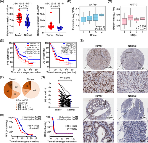
NAT10 expression in PDAC tissues was analyzed using two GEO datasets. In both GSE15471 and GSE16515 datasets, NAT10 expression was upregulated in PDAC tumor tissues compared to adjacent normal tissues (P < 0.001 and P = 0.005, respectively; Figure 1A). Based on the analysis in TISIDB, NAT10 expression was positively associated with the pathological grade (r = 0.187, P = 0.013) and clinical stage (r = 0.158, P = 0.036) of PDAC patients (Figure 1B-C). Survival analysis based on TCGA-PAAD data indicated that high NAT10 expression was significantly associated with poor progression-free survival (PFS; HR = 1.600, P = 0.032), although a similar trend was observed for overall survival (OS; HR = 1.500, P = 0.076), the association did not reach statistical significance (Figure 1D). To further confirm the results, we analyzed NAT10 expression in tissue microarray using immunohistochemistry. Of 108 pairs of PDAC tissues, 103 were enrolled for analysis (three pairs of tissues detached during staining and two pairs of tissues without complete information were excluded). NAT10 expression was detectable in 68.93% (71/103) of PDAC tissues, which was higher than that in the adjacent normal tissues (36.89%, 38/103; P < 0.001). In addition, the IRS of NAT10 expression was also significantly higher in PDAC tissues than in adjacent normal tissues (P < 0.001; Figure 1E-G, Supplementary Figure S1). Based on the IRS of NAT10 expression, 103 tissues were classified into NAT10 high/medium group (IRS = 6-12 (+++)/2-4 (++); n = 49) and NAT10 low/negative group (IRS = 1 (+)/negative (-); n = 54). No significant differences were found between the NAT10 high/medium group and the NAT10 low/negative group regarding each clinicopathological characteristic of PDAC (Table 1). Kaplan-Meier survival analysis showed that high/medium NAT10 expression was significantly associated with poor PFS (HR = 1.628, P = 0.030), whereas NAT10 expression was not associated with OS (HR = 1.317, P = 0.209) of PDAC (Figure 1H). The results indicate that NAT10 is associated with disease progression and poor prognosis in PDAC.
| Clinicopathological characteristics |
High/mediuma (n = 49) |
Low/negativeb (n = 54) |
t/χ2 | P |
|---|---|---|---|---|
| Sex, male/female, n | 33/16 | 29/25 | 1.467 | 0.226 |
| Age, mean ± standard deviations, year | 64.39 ± 9.18 | 62.15 ± 11.77 | 1.001 | 0.319 |
| Tumor size, cm | 3.25 ± 1.26 | 3.13 ± 1.18 | 0.281 | 0.780 |
| Tumor location, n | ||||
| Head | 21 | 23 | 4.556 | 0.472 |
| Neck | 1 | 0 | ||
| Body | 6 | 3 | ||
| Tail | 20 | 24 | ||
| Ampulla | 1 | 2 | ||
| Body and tail | 0 | 2 | ||
| Pathological grade, n | ||||
| Low-median | 21 | 21 | 0.043 | 0.835 |
| Median-high | 28 | 33 | ||
| Capsular invasion, n | ||||
| Yes | 28 | 32 | 0.034 | 0.854 |
| No | 21 | 22 | ||
| Neurological invasion, n | ||||
| Yes | 30 | 24 | 2.266 | 0.132 |
| No | 19 | 30 | ||
| Vascular invasion, n | ||||
| Yes | 39 | 40 | 0.183 | 0.669 |
| No | 10 | 14 | ||
| T stage, n | ||||
| T1-T2 | 33 | 33 | 0.205 | 0.650 |
| T3-T4 | 16 | 21 | ||
| Lymph node metastasis, n | ||||
| N0 | 23 | 29 | 0.239 | 0.625 |
| N1-N2 | 26 | 25 | ||
| Clinical stage, n | ||||
| I | 19 | 21 | 0.036 | 0.849 |
| II-IV | 30 | 33 | ||
- Abbreviations: IRS, immunoreactive score; NAT10, N-acetyltransferase 10; PDAC, pancreatic ductal adenocarcinoma.
- a High/medium NAT10 expression: patients with NAT10 IRS of 6-12 (+++) or 2-4 (++);
- b Low/negative NAT10 expression: patients with NAT10 IRS of 1 (+) or negative (-);
3.2 NAT10 regulated LAMB3 mRNA stability by ac4C modification in PDAC cells
To investigate the potential downstream genes regulated by NAT10 in PDAC, we searched the articles. Two articles [62, 63] reported DEGs that were commonly identified across multiple GEO datasets comparing PDAC and normal tissues. Another article [64] reported the top 50 overlapping DEGs (25 upregulated and 25 downregulated) identified from three GEO datasets comparing PDAC and normal tissues (Supplementary Table S4). The overlapping DEGs are mainly related to extracellular matrix (ECM)-receptor interaction [62-64], cell adhesion [63, 64], and focal adhesion [63, 64]. Among these overlapping DEGs, three were simultaneously reported in the three articles, including laminin subunit beta 3 (LAMB3), integrin subunit alpha 2 (ITGA2), and carcinoembryonic antigen-related cell adhesion molecule 6 (CEACAM6) (Supplementary Figure S2A). The three genes were all upregulated in PDAC tissues compared to normal tissues. Our further analysis in TNMplot revealed a positive correlation between LAMB3 and NAT10 mRNA in human PDAC tissues (r = 0.180, P < 0.001; Supplementary Figure S2B). However, no correlations were observed between ITGA2 and NAT10 mRNA (r = 0.010, P = 0.850, Supplementary Figure S2C), and CEACAM6 and NAT10 mRNA (r = 0.090, P = 0.150, Supplementary Figure S2D) in human PDAC tissues. Survival analysis indicated that LAMB3 expression was negatively associated with OS (HR = 1.900, P = 0.002) and PFS (HR = 1.700, P = 0.023) in patients with PDAC (Supplementary Figure S2E). Then, LAMB3 was chosen for further study. LAMB3 is a component of laminin-332 (a main ECM protein) [65]. Laminin-332 (encoded by LAMA3, LAMB3, and LAMC2) is a key element of the basement membrane barrier unique to epithelial cells, which plays a key role in cell adhesion and migration [66, 67] and is associated with invasiveness in various types of malignant tumors [68].
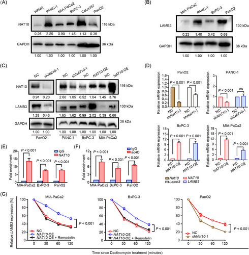
After examining the expression levels of NAT10 and LAMB3 among the PDAC cell lines, we selected three human PDAC cell lines (PANC-1, MIA-PaCa2, and BxPC-3) and one mouse PDAC cell line (PanO2) to examine the effect of NAT10/Nat10 expression on LAMB3 expression (Figure 2A-B). Immunofluorescence showed that NAT10 predominantly localized in the cell nucleus, while LAMB3 predominantly localized in the cytoplasm (Supplementary Figure S2F-G). The knockdown of Nat10 reduced LAMB3 expression in mouse PanO2 cells, while the knockdown of NAT10 had no effect in human PANC-1 cells. In contrast, NAT10 overexpression increased the expression of LAMB3 in both BxPC-3 and MIA-PaCa2 cells (Figure 2C-D, Supplementary Figure S2H-I). Thus, MIA-PaCa2, BxPC-3, and PanO2 cells were used to verify the effect of NAT10-mediated ac4C modification on LAMB3/Lamb3. RIP-qPCR confirmed that LAMB3/Lamb3 mRNA was enriched in the NAT10 group but not in the IgG group, and acRIP-qPCR confirmed that LAMB3/Lamb3 mRNA was enriched in the ac4C group but not in the IgG group (Figure 2E-F). Furthermore, RNA decay assays indicated that NAT10 overexpression prolonged the half-life of LAMB3 mRNA, and vice versa. Treatment with Remodelin (an inhibitor of NAT10) reversed the effect of NAT10 overexpression on the half-life of LAMB3 mRNA (Figure 2G). These results indicate that NAT10 improves LAMB3/Lamb3 mRNA stability via an N4-acetylated modification in PDAC cells.
3.3 NAT10 promoted tumor malignant progression by the LAMB3-mediated FAK/extracellular regulated protein kinases (ERK) signaling pathway
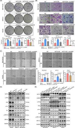
To verify the roles and regulatory mechanisms of NAT10 in malignant tumor progression in PDAC cells, a series of functional assays were performed, including clone formation, wound healing, and Transwell assays. The results showed that NAT10 overexpression promoted cell proliferation, invasion, and migration, while the knockdown of LAMB3 reversed the effects of NAT10 overexpression in MIA-PaCa2 and BxPC-3 cells (Figure 3A-C). In contrast, the knockdown of Nat10 inhibited cell proliferation, invasion, and migration, while the overexpression of Lamb3 rescued the inhibitory effects of Nat10 knockdown on these malignant behaviors in PanO2 cells (Figure 3A-C). Given that NAT10 regulates LAMB3/Lamb3 mRNA stability in PDAC cells and LAMB3 is a subunit of laminin-332 (an important component of ECM) [65], NAT10 may be related to TME by ECM signaling (a process in which ECM ligands bind to cell surface receptors to transmit signaling [69]). The focal adhesion pathway is an important ECM signaling pathway, which can promote cell attachment and migration [70]. To verify it, we examined the expression of several molecules involved in the focal adhesion pathway, including FAK, sarcoma (Src), and ERK. We found that Nat10 knockdown decreased the levels of phosphorylated-FAK (p-FAK), phosphorylated-Src (p-Src) and phosphorylated-ERK1/2 (p-ERK1/2) in PanO2 cells, whereas NAT10 overexpression increased the levels of p-FAK, p-Src and p-ERK1/2 in BxPC-3 and MIA-PaCa2 cells (Figure 3D, Supplementary Figure S3A). Furthermore, we found that LAMB3 knockdown or Remodelin treatment reversed the activation of the focal adhesion pathway in NAT10 overexpression cells, as evidenced by reduced levels of p-FAK, p-Src, and p-ERK1/2 in MIA-PaCa2 and BxPC-3 cells. Overexpression of Lamb3 resulted in the activation of the focal adhesion pathway in Nat10 knockdown cells, as evidenced by the up-regulated levels of p-FAK, p-Src, and p-ERK1/2 in PanO2 cells (Figure 3E, Supplementary Figure S3B). These results indicate that NAT10 promotes malignant tumor progression by activating the LAMB3-mediated FAK/ERK pathway.
3.4 NAT10 was associated with the abundance of infiltrating immune cells in the PDAC TME
To verify the relationship between NAT10 and the TME in PDAC, we analyzed the effects of NAT10 expression on the abundance of infiltrating immune cells using two public online databases (TIMER and TISIDB). NAT10 expression was positively associated with the infiltrating abundance of B cells, CD8+ T cells, macrophages, neutrophils, and dendritic cells, while negatively associated with the infiltrating abundance of CD4+ T cells in human PDAC tissues based on TIMER (Figure 4A). According to TISIDB, NAT10 expression was negatively associated with the abundance of infiltrating immune cells, including mast cells, eosinophil cells, regulatory T cells (Treg), follicular helper T cells (Tfh), T helper cells type 1 (Th1), macrophages, inducible dendritic cells (iDC), activated B cells (Act_B), natural killer cells (NK), natural killer T cells (NKT), effector memory CD4+ T cells (Tem_CD4+ T), and effector memory CD8+ T cells (Tem_CD8+ T), while positively associated with the abundance of infiltrating activated CD4+ T cells (Act_CD4+ T) in human PDAC tissues (Figure 4B, Supplementary Table S5).
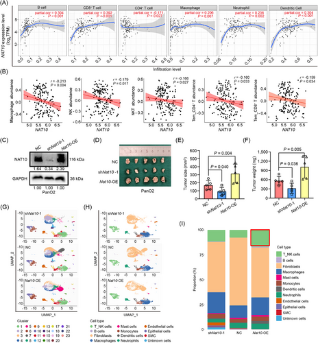
We constructed Nat10 overexpression and Nat10 knockdown PanO2 cells (Figure 4C). Immunocompetent C57BL/6 mice were inoculated with PanO2 cells of Nat10 overexpression, Nat10 knockdown, and a negative control. The tumors were harvested four weeks after inoculation. Nat10 knockdown significantly reduced tumor size and weight, whereas Nat10 overexpression significantly increased them (Figure 4D-F).
scRNA-seq was performed on 10 fresh tumor tissues from 10 mice (three from the Nat10 overexpression group, three from the negative control group, and four from the Nat10 knockdown group). After multiple quality controls, single-cell transcriptomes were acquired from 37,623 cells (11,395 cells from the Nat10 knockdown group, 14,228 cells from the negative control group, and 12,000 cells from the Nat10 overexpression group, respectively) for downstream analyses. These cells were classified into 23 clusters using the UMAP dimensionality reduction algorithm (Figure 4G) and further annotated to 13 major cell types by well-recognized marker genes (Figure 4H), including T_NK cells (T cells and NK cells), B cells, fibroblasts, macrophages, mast cells, monocytes, dendritic cells, neutrophils, endothelial cells, epithelial cells, smooth muscle cells (SMC), and unknown cells. Notably, fibroblasts, macrophages, and T_NK cells were the top three dominant cell types in the PDAC TME, and T_NK cells were predominant in the Nat10 overexpression group compared to the negative control group (Figure 4I). These results indicate that NAT10 is associated with the abundance of infiltrating immune cells in the PDAC TME.
3.5 NAT10 overexpression was associated with CD8+ T cell exhaustion in the PDAC TME
Notably, T_NK cells are two major immune-killing cell types, which were predominant in the Nat10 overexpression group compared to the negative control group (Figure 4I). However, the tumor size and weight did not decrease, but increased significantly in the Nat10 overexpression group compared to the negative control group (Figure 4D-F). The seemingly contradictory phenomena triggered our interest. We further identified 10 distinct subsets of T_NK cells using the UMAP dimensionality reduction algorithm (1,323 T_NK cells from the Nat10 knockdown group, 1,072 T_NK cells from the negative control group, and 2,006 T_NK cells from the Nat10 overexpression group; Figure 5A-B) with marker genes and functional signatures listed in Figure 5C. The 10 subsets included regulatory T cells (Treg), exhausted CD8+ T cells (CD8+ Tex) type I, cytotoxic CD8+ T cells (CD8+ Tc), naïve CD4+ T cells (CD4+ Tn) type I, NK, naïve CD8+ T cells (CD8+ Tn), CD4+ Tn (type II), exhausted CD4+ T cells (CD4+ Tex), Gamma-delta T cells (γδ T), and CD8+ Tex (type II). The top 10 marker genes of each subset are listed in Supplementary Figure S4. CD8+ Tex (type I) was the predominant subset in the Nat10 overexpression group, whereas Treg was the predominant subset in the Nat10 knockdown and negative control groups, indicating the immune exhaustion property (CD8+ Tex subset) in the Nat10 overexpression group, whereas immune suppression property (Treg subset) in the Nat10 knockdown and negative control groups.
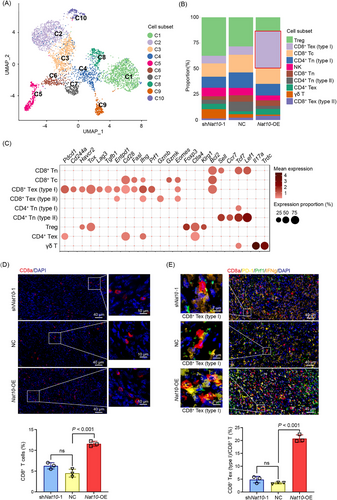
We then focused on the CD8+ T cell subset because of its vital role in tumor immune-killing capacity [71]. CD8+ Tex (type I) subset was characterized by the expressions of both exhausted and inhibitory marker genes, such as programmed cell death protein 1 (Pdcd1, encoding protein PD-1), Cd244a, Havcr2 (also known as mucin-3/T-cell immunoglobulin, Tim-3), thymocyte selection-associated high mobility group box (Tox), lymphocyte-activation gene 3 (Lag3), transforming growth factor beta 1 (Tgfb1), and ectonucleoside triphosphate diphosphohydrolase 1 (Entpd1), along with stimulatory and effector molecule genes, such as Cd28, fas ligand (Fasl), interferon gamma (Ifng), and perforin (Prf1). This pattern suggests a potential for reactivation. In contrast, the CD8+ Tex (type II) subset was characterized by the expression of the exhausted marker Entpd1, along with effector molecule genes, such as Ifng and granzyme B (Gzmb), indicating a reduced potential for reactivation (Figure 5C).
Finally, we analyzed the phenotypes of CD8+ T cells in the PDAC TME via multiplexed immunofluorescence staining of CD8a, IFNg, Prf1, and PD-1. In subcutaneous tumors of mice, the proportions of CD8+ T cells and CD8+ Tex (type I) subset were both substantially higher in the Nat10 overexpression group than the negative control group (Figure 5D-E), which was consistent with the result of scRNA-seq. In human PDAC tissues, the proportion of CD8+ Tex (type I) subset was significantly higher in PDAC tissues with high/medium NAT10 expression than those with low/negative NAT10 expression (P = 0.024; Supplementary Figure S5).
These findings suggest that NAT10 overexpression is associated with CD8+ T exhaustion in the PDAC TME.
3.6 CD8+ T differentiation analysis in the PDAC TME
To gain an in-depth understanding of CD8+ T cell exhaustion in the PDAC TME, KEGG pathway enrichment analysis of the top 100 marker genes in each CD8+ T cell subset was performed. The FoxO and Wnt signaling pathways were enriched in the CD8+ Tn subset (Supplementary Figure S6A). The NF-kappa B signaling pathway, chemokine signaling pathway, and leukocyte transendothelial migration were substantially enriched in the CD8+ Tc subset (Supplementary Figure S6B), which may facilitate their migration, proliferation, and cytotoxic function in TME. Antigen processing and presentation, as well as PD-L1 expression and PD-1 checkpoint pathway in cancer, were predominant in the CD8+ Tex (type I) subset (Supplementary Figure S6C), which may facilitate exhaustion and dysfunction in the TME. Notably, several enriched pathways were identified in both CD8+ Tc and CD8+ Tex (type I) subsets, including natural killer cell mediated cytotoxicity, T cell receptor signaling pathway, cytokine-cytokine receptor pathway, and PI3K-Akt signaling pathway, suggesting a close relationship between these two CD8+ T subsets. However, cellular senescence and p53 signaling pathway were enriched in the CD8+ Tex (type II) subset (Supplementary Figure S6D), indicating a terminally dysfunctional state.
Pseudotime trajectory analysis using Monocle2 revealed that CD8+ T cells followed a characteristic trajectory from naive T cells to exhausted T cells along two distinct linear differentiation pathways. CD8+ Tex (type II) subset was located at the end of one linear differentiation pathway, while CD8+ Tex (type I) subset was positioned at the end of another linear differentiation pathway, indicating that CD8+ T cell exhaustion was a gradual progress involving distinct dysfunctional states (Supplementary Figure S6E-F). According to a previous report [72], we then re-designated CD8+ Tex (type I) as intermediate CD8+ Tex and CD8+ Tex (type II) as terminally CD8+ Tex. Thus, two distinct linear differentiation pathways were observed during CD8+ T cell development. One pathway was CD8+ Tn /CD8+ Tc/intermediate CD8+ Tex/terminally CD8+ Tex, while another was CD8+ Tn/CD8+ Tc/intermediate CD8+ Tex.
3.7 NAT10 regulated PD-L1 expression in PDAC cells via the FAK/ERK pathway
As mentioned above, the pathway named PD-L1 expression and PD-1 checkpoint pathway in cancer was enriched in the CD8+ Tex (type I) subset (Supplementary Figure S6C), indicating PD-1/PD-L1 axis may be involved in CD8+ T cell exhaustion. We then examined the role of NAT10 in PD-L1 expression in PDAC cells. RNA-seq identified 392 DEGs (86 downregulated and 306 upregulated) between the Nat10 overexpression and the negative control PanO2 cells under the criteria of P < 0.05 and |log2 (fold change)| > 1, among which Cd274 was significantly upregulated in the Nat10 overexpression group (Figure 6A). qPCR and Western blotting confirmed that NAT10 promoted Cd274 (encoding protein PD-L1) expression at both the mRNA and protein levels in PanO2 cells (Figure 6B-C). We further examined the relation between NAT10 and CD274/PD-L1 expression in human PDAC tissues and cells. Correlation analysis indicated a positive correlation between NAT10 and CD274 mRNA expression in PDAC tissues (Figure 6D). qPCR and Western blotting revealed that NAT10 promoted CD274/PD-L1 expression at both the mRNA and protein levels in MIA-PaCa2 and BxPC-3 cells (Figure 6E-F). Immunofluorescence staining indicated that PD-L1 was localized to both the cytoplasm and cell membrane, and NAT10 upregulated PD-L1 expression in PDAC cells (Figure 6G). Notably, PD-L1 upregulation may not be consistent with its increased abundance on the cell membrane. Flow cytometry confirmed that NAT10 upregulated PD-L1 expression on PDAC cell membranes (Figure 6H).
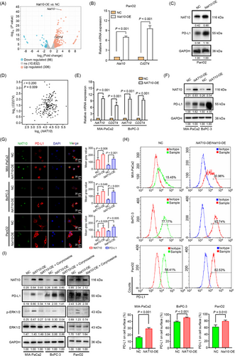
Several signaling pathways on PD-L1 regulation have been reported, including ERK signaling [73]. Considering the regulatory role of NAT10 in LAMB3, which activates the FAK/ERK signaling pathway, we speculated that NAT10 promoted PD-L1 expression via the FAK/ERK pathway. We treated NAT10/Nat10-overexpressed PDAC cells with an ERK inhibitor (Corynoxeine) and found that the inhibition of ERK activity reversed PD-L1 up-regulation (Figure 6I).
The results indicate that NAT10 regulates PD-L1 expression in PDAC cells via the FAK/ERK pathway.
3.8 NAT10 may regulate CD8+ T cell function via the PD-1/PD-L1 axis
Given that NAT10 enhanced PD-L1 expression and abundance in PDAC cells, we speculated that NAT10 may regulate CD8+ T cell function via the PD-1/PD-L1 axis. To verify this, we analyzed the binding capacity of PD-L1 on tumor cells to PD-1 and found that NAT10/Nat10 overexpression enhanced PD-1/PD-L1 binding, while Naamidine J (an inhibitor of PD-1/PD-L1 binding) reduced it (Figure 7A). Further analysis indicated that Naamidine J treatment reduced PD-L1 expression in PDAC cells (Figure 7B). In vitro, the tumor-killing capacity of CD8+ T cells was significantly suppressed when co-cultured with the NAT10 overexpression cells compared to the negative control cells, which was retrieved by treatment with Naamidine J (Figure 7C).
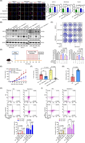
To further verify whether NAT10 regulates CD8+ T cell function via the PD-1/PD-L1 axis in vivo, we treated mice bearing subcutaneous tumors with Naamidine J (Figure 7D) and found that Naamidine J treatment substantially repressed tumor growth in the Nat10 overexpression group, but not in the negative control group (Figure 7E-F). The inhibitory efficacy of Naamidine J treatment on tumor size was significantly higher in the Nat10 overexpression group than in the negative control group (P < 0.001; Figure 7G). The results indicate that mice in the Nat10 overexpression group benefit more from Naamidine J treatment. Then, flow cytometry was performed to analyze the phenotypes of CD8+ T cells in the subcutaneous tumors. We found that Nat10 overexpression substantially increased the proportion of intermediate CD8+ Tex subset (PD-1+IFNg+ or PD-1+Prf1+) and decreased the proportion of CD8+ Tc subset (PD-1−IFNg+) in the subcutaneous tumors (Figure 7H-I, Supplementary Figure S7). In addition, Naamidine J treatment significantly reduced the proportion of intermediate CD8+ Tex subset (PD-1+IFNg+ or PD-1+Prf1+) and increased the proportion of CD8+ Tc subset (PD-1−IFNg+ or PD-1−Prf1+) in the Nat10 overexpression group. However, Naamidine J treatment failed to increase the proportion of CD8+ Tc subset (PD-1−IFNg+ or PD-1−Prf1+) in the negative control group (Figure 7H-I). The results indicate that NAT10 may regulate CD8+ T cell function via the PD-1/PD-L1 axis.
4 DISCUSSION
In the current study, we reported the role and mechanism of NAT10 in tumor progression and the immune microenvironment in PDAC. NAT10 regulates the malignant behaviors of PDAC cells by enhancing ac4C-mediated LAMB3 stability, thereby activating the FAK/ERK pathway. In addition, NAT10 overexpression upregulates PD-L1 expression in tumor cells through the LAMB3/FAK/ERK pathway and is associated with an abundance of intermediate CD8+ Tex subset in the PDAC TME. The inhibition of PD-1/PD-L1 axis with Naamidine J substantially increases the proportion of CD8+ Tc subset and decreases the proportion of intermediate CD8+ Tex subset in the PDAC TME (Figure 8). These findings may favor the selection of candidates who may benefit from immunotherapy, optimize current therapeutic strategies, and improve the clinical prognosis of PDAC patients.
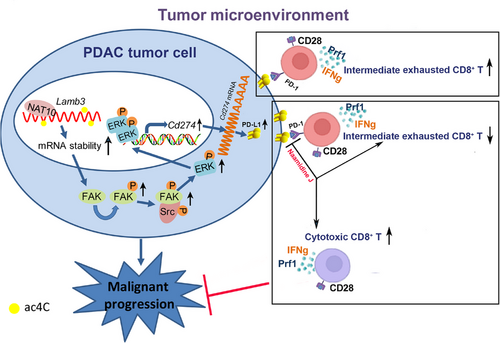
Consistent with a previous study [46], we found that NAT10 was upregulated in PDAC tissues and closely associated with poor clinical prognosis of PDAC. Moreover, we elucidated that NAT10 enhances LAMB3 mRNA stability via ac4C modification, thereby promoting the proliferation, invasion, and metastasis of PDAC cells. Reportedly, LAMB3 was upregulated in PDAC tissues and was associated with shorter PFS and OS in patients with PDAC. The knockdown of LAMB3 inhibited cell proliferation, migration, and invasion, promoted cell apoptosis, and regulated EMT in PDAC cells [63, 74-76]. Our study further revealed that LAMB3 was associated with focal adhesion formation in PDAC. Focal adhesion contains an array of proteins, including FAK and Src [77, 78]. FAK is a highly auto-phosphorylated protein in response to integrin activation, and regulates cytoskeletal dynamics and cell movement [79]. p-FAK and p-Src then form complexes to activate or inhibit multiple downstream signaling pathways, including the PI3K/AKT, P53, and ERK pathways. The FAK/Src/ERK signaling pathway plays an important role in tumorigenesis and metastasis in lung cancer [80, 81] and PDAC [36]. In the current study, we verified that NAT10 enhances LAMB3-mediated focal adhesion formation and revealed that NAT10 regulates the malignant behaviors of PDAC cells by activating the LAMB3-mediated FAK/Src/ERK pathways.
Given that NAT10 enhances LAMB3-mediated focal adhesion formation, we examined the role of NAT10 in the PDAC TME. The results of scRNA-seq indicated that fibroblasts and macrophages were the two most abundant cell types in the TME, which was consistent with previous reports that the PDAC TME was characterized by desmoplastic dense stroma and unique immunosuppression [3, 82-84]. In addition, Nat10 overexpression was associated with CD8+ T cell exhaustion in the PDAC TME, as evidenced by the predominant infiltrating abundance of intermediate CD8+ Tex subset in the Nat10 overexpression group. This may partially explain the preferential tumor growth in this group.
T cell exhaustion is a common event in tumors, and the aberrant expression of immune checkpoint proteins resulting in T cell dysfunction is associated with disease relapse and progression in tumors and can be used to predict clinical outcomes [85]. However, the mechanisms underlying T cell exhaustion are not fully understood. The presence of persistent tumor antigen stimulation induces prolonged and increased expression of inhibitory receptors on the T cell surface, including cytotoxic T lymphocyte associated antigen-4 (CTLA-4), PD-1, Tim-3, Lag-3, and T-cell tyrosine-based inhibitory motif [86, 87]. Upon binding to their cognate ligands on cancer cells, T cells' effector function and proliferation are gradually reduced. The binding between PD-1 on T cells and PD-L1 on tumor cells induces T cell dysfunction and exhaustion, which is recognized as an important mechanism by which tumor cells evade immune surveillance [88-90]. The current study reveals that NAT10 regulates PD-L1 expression via the LAMB3/FAK/ERK pathway in PDAC cells. NAT10 overexpression reduced the CD8+ T-mediated cytotoxicity to tumor cells, which was reversed after blocking of the PD-1/PD-L1 axis, indicating NAT10 may regulate CD8+ T cell function through the PD-1/PD-L1 axis. This may partially explain the predominant abundance of CD8+ Tex subset in the Nat10 overexpression group.
Although PDAC is refractory to immunotherapy, regardless of monotherapy or combination therapy, progress has recently been achieved. There is a potential therapeutic window for the formation of the Tex subset. Tex subset still proliferates and expresses a broad spectrum of effector function-related genes. The effective antitumor immune responses elicited by ICIs may depend on exhausted T cell subset in the tumor, whose function is restored by therapy [72]. Our study revealed that the intermediate CD8+ Tex subset in the PDAC TME expresses the exhausted marker (Pdcd-1), stimulatory marker (Cd28) and cytotoxic markers (Ifng and Prf1), indicating the potential for proliferation and cytotoxicity of this subset. Naamidine J treatment significantly increased the proportion of CD8+ Tc subset in the Nat10 overexpression group but not in the negative control group. This may partially explain why mice in the Nat10 overexpression group benefit more from Naamidine J treatment.
In PDAC, the dense stoma in the TME obstructs the delivery of therapeutics and chimeric antigen receptor-T (CAR-T) cells to the tumor core [3, 91]. In addition, CAR-T cells always develop exhaustion soon after administration, which hinders their clinical efficacy [3, 91]. In view of this, the intermediate CD8+ Tex subset may be an ideal autologous responder for immunotherapy in PDAC. However, whether PDAC patients with high NAT10 expression can benefit from immunotherapy needs clinical validation.
The current study has some limitations. First, the subcutaneous tumor does not reflect all conditions of the TME. Therefore, the final verification should be performed in an orthotopic environment. In addition, PDAC is driven by KRAS and p53 mutations. However, PanO2 is a mouse cell line with wild-type Kras, which does not reflect all human PDAC pathological features. A KPC orthotopic model (with Kras and p53 mutations) is more suitable for further verification. Second, the knockdown of NAT10 displayed a limited effect on LAMB3 expression. Therefore, NAT10 overexpression was used in subsequent studies. Notably, NAT10 overexpression may lead to supraphysiological changes. Third, the PDAC TME is characterized by desmoplastic dense stroma and limited immune cell infiltration; however, single-cell transcriptomics cannot determine the location of the immune cells in the PDAC TME. Spatial transcriptomics combined with single-cell transcriptomics may be useful to resolve these concerns. Finally, an in-depth investigation is needed to analyze and compare the PDAC TME of patients before and after ICI treatment, which can further verify our findings.
5 CONCLUSIONS
This study proposed a regulatory role of NAT10 in tumor progression and immune microenvironment via the LAMB3/FAK/ERK pathway in PDAC. These findings may favor the selection of candidates who may benefit from immunotherapy, optimize current therapeutic strategies, and improve the clinical prognosis of PDAC patients.
AUTHOR CONTRIBUTIONS
Hao Hu, Yong Mao, and Youzhao He conceived and designed the project, supervised and coordinated all aspects of the work. Enhong Chen, Qin Wang, Leisheng Wang, Zebo Huang, Dongjie Yang, Changyong Zhao, Wuqiang Chen, Shuo Zhang, and Shuming Xiong performed the experiments. Enhong Chen, Qin Wang, Leisheng Wang, Zebo Huang, Dongjie Yang, and Changyong Zhao analyzed the data and prepared the figures and tables. Enhong Chen, Qin Wang, Leisheng Wang, Wuqiang Chen, Shuo Zhang, and Shuming Xiong wrote the manuscript, and all of the authors proofed it.
ACKNOWLEDGMENT
We would like to thank Editage (www.editage.cn) for English language editing, and we are indebted to all the authors and our colleagues for the fruitful suggestions and discussions. This work was supported by Jiangsu Natural Science Foundation Project (No. BK20191142), Scientific Research Project of Jiangsu Provincial Health Commission (No. M2024075), High-level talent training project of Wuxi Taihu Talent Plan (No. BJ2020055, BJ2023048), Clinical Medical Research Project of the Affiliated Hospital of Jiangnan University (No. LCYJ202321), Postgraduate Research & Practice Innovation Program of Jiangsu Province (No. KYCX24_2648).
CONFLICT OF INTEREST STATEMENT
The authors declare that they have no competing interests.
ETHIC STATEMENT
This study was approved by the Ethics Committee of the Affiliated Hospital of Jiangnan University (No. LS2024065), and written informed consent was obtained from all participants. The animal studies were conducted in compliance with protocols approved by the Laboratory Animal Welfare and Ethics Committee of Jiangnan University (IACUC Issue No: JN.No202312 30c0520505[626]).
Open Research
DATA AVAILABILITY STATEMENT
Data, analytical methods, and study materials supporting the findings of this study will be available from the corresponding author upon request. The datasets analyzed in this study are publicly available in NCBI under accession numbers (PRJNA1209794, PRJNA1209905, PRJNA1209847, PRJNA1210509, PRJNA1210634, PRJNA1210995, and PRJNA1214184 (https://www.ncbi.nlm.nih.gov/bioproject/) and the Gene Expression Omnibus (GEO) database under accession numbers GSE15471 and GSE16515 (https://www.ncbi.nlm.nih.gov/geo/)


