Anatomy education for medical students in a virtual reality workspace: A pilot study
Abstract
The COVID-19 pandemic has posed a challenge for many medical schools, as they have had to adjust their curricula into an online format. This was particularly problematic for anatomy courses as in person dissections have historically been preferred for providing students with a three-dimensional learning environment. In this study, we aim to share our experience with conducting anatomy lectures for medical student using a virtual reality (VR) workspace. Additionally, we discuss the advantages of using VR and expand on how it may be used to improve students' understanding of anatomy in comparison to various other online lecture formats. To do this, we utilized a post-lecture survey to gain feedback from the medical students that participated in a VR anatomy workspace. We found that many of our participants expressed that having access to their course material from anywhere and anytime via a virtual space, and being able to manipulate anatomical structures by moving and modifying them provided the student with a strong advantage. Although there are still limitations, we hope that our experience will assist other anatomy teachers with improving their lecture methods, especially during the pandemic.
1 INTRODUCTION
Anatomy educators from around the world have been required to modify their curricula due to the COVID-19 pandemic. To reduce transmission of the virus, most classes have been conducted online (Byrnes et al., 2021; Iwanaga et al., 2020). Several anatomy lectures have utilized two-dimensional (2D) online communication tools such as Zoom (Iwanaga et al., 2020). One of the main advantages of such tools over traditional in-person classes is that they enable students from different courses, schools, cities, and even countries to participate at the same time (Iwanaga et al., 2020). However, there are still limitations with 2D lectures, as mastery of anatomy requires a three-dimensional (3D) understanding of the material (Iwanaga, Terada, et al., 2021). Although the pandemic has made it more difficult to access cadaveric materials, there are various other educational tools that can be incorporated into anatomy education, for example, video lectures, 3D printing material, augmented reality, and virtual reality (VR) (Iwanaga, Loukas, et al., 2021; Iwanaga, Terada, et al., 2021). In order to continue providing anatomical content to medical/dental students and other healthcare providers, various other available materials have been utilized during the pandemic (Iwanaga, Kamura, et al., 2021; Iwanaga, Loukas, et al., 2021; Kockro et al., 2015; Ross et al., 2020). Of these, VR has been found to be a potential alternative resource for cadaveric dissection (Saverino, 2020; Serhan, 2020; Solyar et al., 2008). Recently, we suggested a new option for educating professional healthcare providers in clinical anatomy with the use of a VR workspace, that is, VR headset (Oculus, Menlo Park, CA) and VR workspace (Spatial, New York, NY) (Zhao et al., 2020). During the lecture, several 3D anatomical structures were used, and the participants were able to actively participate. As the 3D aspect of anatomy is often the most challenging aspect for students to grasp, we studied the use of VR in anatomy instruction for medical students.
2 ANATOMY LECTURES FOR MEDICAL STUDENTS IN THE VIRTUAL REALITY WORKSPACE (SPATIAL)
- The participant must be a second year or older medical student in Japan.
- The participant must have completed a traditional medical school anatomy class.
The anatomy lectures covered three different topics (cardiovascular system, musculoskeletal system, and nervous system) and took place in a VR workspace (Spatial) using VR headset (Oculus Quest 2, Menlo Park, CA). Thirty medical students from all over Japan attended the meeting.
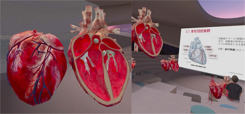
- The participants were allowed to enter anatomical structures such as the heart by magnifying them and seeing, for example, in the heart, the mitral valve and chordae tendineae and this improved their overall three-dimensional understanding of this structure. Having access to multiple 3D models enabled participants to share their knowledge and experience with the other participants in real time (Figure 1).
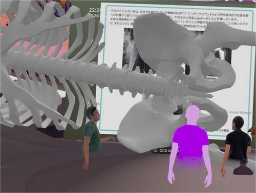
- The use of 3D models of skeletal material and muscles improved visualization of the origins and insertions of various muscles (Figure 2).
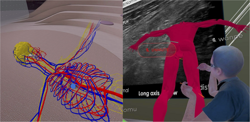
- The participants found it useful to see multiple materials such as real-time images (e.g., ultrasound) and 3D models of the nervous system at the same time (Figure 3).
- In general, lectures in the VR workspace allowed for more interaction between the teacher and students.
- Headsets are required to enter the VR space.
- It might take additional time to learn how to navigate 3D models for beginners.
- Wi-Fi access is necessary for operating in the VR space.
- Motion sickness might be experienced in the VR space, that is, VR sickness.
3 ADVANTAGE OF USE OF TWO-DIMENSIONAL TOOLS (ZOOM) FOR ANATOMY LECTURES
The Zoom application has gained significant popularity throughout the COVID-19 pandemic and has provided educators with a simple video conferencing application to conduct lectures through, without needing extensive training. Zoom allows a host to share their screen and deliver a presentation with a large amount of data to a very large group of participants (Byrnes et al., 2021). In comparison, VR workspaces have smaller limits on the size of the files that can be used and the number of participants allowed joining at one time. Generally, this is not large enough to accommodate an entire medical school class. Additionally, with Zoom, presenters can record the meeting and later upload it for students to rewatch at their convenience. While there is some debate regarding the efficacy of using online platforms to teach anatomy, researchers such as Singh and Min (2017) compared the performance of first year anatomy students in 2013 (classroom lectures) and 2014 (digital lectures). They found that the 2014 cohort performed better on both short essay (47.65 vs. 36.80) and multiple-choice questions (51.19 vs. 49.22) compared with the 2013 cohort (Singh & Min, 2017).
The greatest benefit of Zoom is the ability for any student in the world to join a meeting and participate in a discussion (Iwanaga et al., 2020). By using this to their advantage, institutions can invite speakers from all around the world to share their experiences with their students (Byrnes et al., 2021). Zoom also allows presenters to delegate a moderator for their session who can mediate conversations and control who is able to enter the meeting (Byrnes et al., 2020). Furthermore, Zoom has a special feature that allows the host to separate all of the participants into breakout rooms for smaller group discussions (Rucker et al., 2020). The breakout rooms have become incorporated into several other medical school classes and are now being used to help train medical students in Telemedicine using standardized patients (Rucker et al., 2020).
4 PERSPECTIVE ON FUTURE ANATOMY EDUCATION TO STUDENTS IN THE VR WORKSPACE
One of the greatest advantages of conducting anatomy lectures via a VR workspace is the ability for participants to gain access to the material from anywhere in space, and the functionality of being able to move/magnify structures of interest (Iwanaga, Loukas, et al., 2021). Additionally, the 3D workspace allows students to view multiple models simultaneously and compare them to each other, which is more difficult to do with 2D tools. From the students' perspective, participating in an anatomy lecture through a VR workspace improved their knowledge and three-dimensional comprehension of the anatomical structures that were presented.
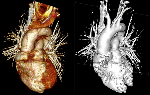
Although there are still limitations to learning in a VR space - quality of 3D models, need for headsets, limit in the number of participants - the VR space is an alternative method for teaching medical students three-dimensional anatomy without dissecting cadavers. While the quality of the 3D models can be improved with the use of computed tomography data or better 3D scanners (Figure 4), the level of detail that can be represented in the VR workspace is limited by the amount of data that can be stored and integrated. The greater the size, detail, and realism of the models, the more space they require. Therefore, it may be more beneficial to utilize simplified 3D models that allow students to observe and manipulate them from various viewpoints. We hope that our experience of providing anatomy lectures using a VR workspace will lead to increased utilization of this technology and potentially give rise to wider applications and newer technologies.
ACKNOWLEDGMENT
The authors sincerely thank those who donated their bodies to science so that anatomical research could be performed. Results from such research can potentially increase mankind's overall knowledge that can then improve patient care. Therefore, these donors and their families deserve our highest gratitude (Iwanaga, Singh, et al., 2021).


