Gastric tube perforation penetrating the pericardium after esophagectomy that needed surgical repair
Abstract
Background
As the prognosis of esophageal cancer surgery has improved, reports on postoperative complications of gastric tubes have increased. Among them, gastric tube ulcer perforation is infrequent but often severe and difficult to treat.
Case Presentation
A 73-year-old man had undergone thoracoscopic subtotal esophagectomy and laparoscopic-assisted gastric tube reconstruction via the retrosternal route for thoracic esophageal cancer 8 years previously. He was transferred to our hospital with a diagnosis of gastric tube ulcer perforation, penetrating the pericardium. Emergency surgery was performed to remove the gastric tube, followed by immediate reconstruction by right colon interposition. The patient was discharged on postoperative day 142.
Conclusion
We report a rare complication of gastric tube ulcer perforation, penetrating the pericardium, after esophagectomy for esophageal cancer. It was successfully treated with appropriate surgical management.
BACKGROUND
In recent years, as the prognosis of esophageal cancer surgery has improved, reports on postoperative complications related to gastric tube reconstruction have increased.1 The incidence of esophageal perforation is 3.1 per million per year.2 Although the incidence of gastric tube ulcers is reported to be 6.6%–19.4% among patients with postoperative esophageal cancer,3, 4 that of perforation is not clear. Considering the incidence of gastroduodenal ulcer perforation in general is 2%–5%, it can be estimated to be 0.1%–0.9% among postoperative patients for esophageal cancer.1 We encountered a case of ulcer in a retrosternal-reconstructed gastric tube, which penetrated the pericardium.
CASE PRESENTATION
A 73-year-old man had undergone thoracoscopic subtotal esophagectomy and laparoscopic-assisted gastric tube reconstruction via the retrosternal route for esophageal cancer 8 years previously. The esophageal cancer was not recurrent. He had been taking vonoprazan fumarate for 7 years because of a recurrent gastric tube ulcer with bleeding. He visited his previous doctor for chest pain that appeared 1 day earlier. The physician diagnosed an abscess in the mediastinum and pericardium due to gastric tube perforation, and the patient was transferred to our hospital.
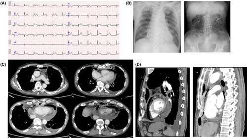
On physical examination at our emergency department, his Glasgow coma scale score was 4, 4, and 6 for eye opening, verbal, and motor responses, respectively; blood pressure was 98/78 mmHg without catecholamine support; heart rate was 68 beats per minute; regular respiration was 22 breaths per minute; oxygen saturation was 95% with 4-L oral mask; and temperature was 36.1°C. Laboratory data at the time of arrival are listed in Table 1. White blood cell count, C-reactive protein, blood urea nitrogen, creatinine, and hepatobiliary enzyme levels were prominently elevated, and arterial blood gas analysis showed elevated lactate levels and severe metabolic acidosis. An electrocardiogram and chest and abdominal radiographs are shown in Figure 1A,B, respectively. Contrast-enhanced computed tomography (CT) showed free air around the gastric tube of the retrosternal route, fluid collection in the pericardial sac that appeared like an abscess, and continuity between the gastric tube and pericardium (Figure 1C,D). Therefore, the patient was diagnosed with gastric tube perforation and penetration to the pericardium.
| Complete blood count data | Biochemistry data | Coagulation data | Arterial blood gas data | ||||
|---|---|---|---|---|---|---|---|
| White blood cells (/μL) | 10,950 | Total bilirubin (mg/dL) | 0.7 | Prothrombin time (%) | 75 | pH | 7.290 |
| Stab cell (%) | 40.5 | Aspartate aminotransferase (IU/L) | 650 | Prothrombin time-international normalized ratio (mg/dL) | 1.17 | Partial pressure of carbon dioxide (mmHg) | 24.3 |
| Segment cell (%) | 36.0 | Alanine aminotransferase (IU/L) | 374 | Activated partial thromboplastin time (seconds) | 27.4 | Partial pressure of oxygen (mmHg) | 118.0 |
| Metamyelocyte cell (%) | 9.5 | Lactate dehydrogenase (IU/L) | 865 | Fibrinogen (mg/dL) | 619 | (mmol/L) | 11.3 |
| Monocyte (%) | 7.0 | γ-Glutamyl transpeptidase (IU/L) | 38 | D-dimer (μg/mL) | 2.5 | Base excess (mmol/L) | −13.7 |
| Lymphocytes (%) | 6.5 | Creatine phosphokinase (IU/L) | 138 | Lactate (mmol/L) | 7.8 | ||
| Red blood cells (×104/μL) | 277 | Blood urea nitrogen (mg/dL) | 72 | ||||
| Hemoglobin (g/dL) | 8.8 | Creatinine (mg/dL) | 3.50 | ||||
| Hematocrit (%) | 27.6 | Sodium (mEq/L) | 136 | ||||
| Platelet (×104/μL) | 18.7 | Chloride (mEq/L) | 106 | ||||
| Potassium (mEq/L) | 6.7 | ||||||
| Calcium (U/L) | 8.1 | ||||||
| Total protein (g/dL) | 5.6 | ||||||
| Albumin (g/dL) | 2.7 | ||||||
| C-reactive protein (mg/dL) | 22.34 | ||||||

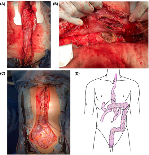
Emergency surgery was performed. First, the cervical esophagus was secured from the cervical incision, then median sternotomy and laparotomy were performed (Figure 2A), and finally the perforated gastric tube was removed. A large perforation was observed in the dorsal side of the gastric tube and pericardium (Figure 2B). The myocardium was discolored, and a large amount of contaminated fluid was observed in the pericardium (Figure 2B). Reconstruction was performed via the retrosternal route using the right ileum and colon according to the formula for colon interposition during esophagectomy. Although the residual cervical esophagus was quite short, anastomosis was performed with the elevated ileocolon (Figure 2C,D). After the lavage of mediastinum and pericardial sac with 7500 mL of saline, six drainage tubes were placed at anterior to the cervical anastomosis, into the posterior sternum via the posterior aspect of the cervical anastomosis, and into the posterior sternum from the abdomen, pericardial sac, Winslow, and under the cysto-rectum, which were managed without continuous perfusion after surgery. A jejunostomy tube was inserted as the feeding route. The operative time was 542 min, blood loss was 1350 mL, and the transfusion consisted of 1960 mL red cell concentrate and 1680 mL fresh frozen plasma.

On postoperative day (POD) 7, CT and gastroscopy revealed leakage at the cervical anastomosis, and a drainage tube was inserted into the neck. The patient was extubated on POD 10 and transferred to the general ward from the intensive care unit. Anastomosis leakage was treated by continuing drain management while administering nutritional supplements through the feeding tube. The leakage was cured on POD 38, and the patient underwent swallowing training. He was discharged on POD 142. The pathological findings of the resected gastric tube were consistent with those of ulcer perforation and no malignancy.
DISCUSSION
We report an uncommon complication of a gastric tube ulcer perforation penetrating the pericardium after esophagectomy for esophageal cancer. It was successfully treated by gastric tube resection and reconstruction using the right colon.
The clinical condition and severity of gastrointestinal perforation vary considerably depending on the reconstruction route after esophageal cancer surgery.1, 5 In general, there are three routes of reconstruction after esophagectomy for esophageal cancer: posterior mediastinal, retrosternal, and antesternal. In the posterior mediastinal route, gastric tube perforation generally occurs into the thoracic cavity, which is serious and sometimes fatal.1, 5-8 By contrast, retrosternal-reconstructed gastric tube perforation is less severe due to the narrow space in the retrosternal cavity.9 However, in the present case, due to the repeated gastric tube ulcer recurrence and chronic inflammation, penetration between the gastric tube and pericardium occurred, resulting in retrosternal and pericardial abscess formation.
The mortality rate in esophageal perforation is 10%–20%, and the success rate in conservative treatment is reported to be 68%.2 Perforation of the gastric tube has a mortality rate of approximately 66%,1 although there are few reports of such cases. There are no reports of successfully managed cases of cardiovascular penetration without surgery,1, 6, 8 which supported the treatment strategy in the present case. While staged reconstruction was considered relatively safe given the contaminated surgical field and patient's septic status, the remaining cervical esophagus was rigid and expected to shorten further. Establishing cervical esophageal fistula requires additional resection of the cervical esophagus in the second-stage reconstruction, which makes future anastomosis quite difficult. Further, anastomosis with the hypopharynx would likely result in loss of swallowing function. Considering the maximum possibility to maintain future swallowing function, immediate reconstruction was performed in the present case.
Gastric tube perforation in the retrosternal route causes pericardial and right ventricular perforation or retrosternal abscess formation.10 Some surgical techniques, including simple suturing, omental filling, external fistulization with a T-tube, partial gastric tube resection, free jejunal reconstruction, and pectoralis major myocutaneous flap, have been reported;1, 5, 7, 10 few cases have achieved successful reconstruction, even with staged surgery.1, 7 To our knowledge, no report exists on immediate colonic reconstruction for gastric tube ulcer perforation.
CONCLUSION
We encountered a rare complication of gastric tube ulcer perforation penetrating the pericardium after esophagectomy for esophageal cancer. It was resolved by appropriate surgical management.
ACKNOWLEDGEMENTS
We thank the patient and Tsuchiura Kyodo General Hospital Intensive Care unit, and Emergency room staff for their help. We also thank Editage (www.editage.com) for English language editing.
CONFLICT OF INTEREST STATEMENT
The authors have no conflict of interest to declare.
ETHICS STATEMENT
Approval of the research protocol: N/A.
Informed consent: Yes.
Registry and the registration no. of the study/trial: N/A.
Animal studies: N/A.
Open Research
DATA AVAILABILITY STATEMENT
All data generated during this study can be accessed through direct communication with the corresponding author and through the agreement of all research team members.


