Size and placement of developing anterior teeth in immature Neanderthal mandibles from Dederiyeh Cave, Syria: Implications for emergence of the modern human chin
ABSTRACT
Evolutionary and functional significance of the human chin has long been explored from various perspectives including masticatory biomechanics, speech, and anterior tooth size. Recent ontogenetic studies have indicated that the spatial position of internally forming anterior teeth partially constrains adult mandibular symphyseal morphology. The present study therefore preliminarily examined the size and placement of developing anterior teeth in immature Neanderthal mandibles of Dederiyeh 1 and 2, compared with similarly-aged modern humans (N = 16) and chimpanzees (N = 7) whose incisors are comparatively small and large among extant hominids, respectively. The Dederiyeh 1 mandible is described as slightly presenting a mental trigone and attendant mental fossa, whereas Dederiyeh 2 completely lacks such chin-associated configurations. Results showed that, despite symphyseal size being within the modern human range, both Dederiyeh mandibles accommodated overall larger anterior dentition and displayed a remarkably wide bicanine space compared to those of modern humans. Dederiyeh 2 had comparatively thicker deciduous incisor roots and more enlarged permanent incisor crypts than Dederiyeh 1, but both Dederiyeh individuals exhibited a total dental size mostly intermediate between modern humans and chimpanzees. These findings potentially imply that the large deciduous/permanent incisors collectively distended the labial alveolar bone, obscuring an incipient mental trigone. It is therefore hypothesized that the appearance of chin-associated features, particularly of the mental trigone and fossa, can be accounted for partly by developmental relationships between the sizes of the available mandibular space and anterior teeth. This hypothesis must be, however, further addressed with more referential samples in future studies. Am J Phys Anthropol 156:482–488, 2015. © 2014 Wiley Periodicals, Inc.
The presence of a chin has generally been recognized as a diagnostic trait discerning Homo sapiens from earlier Homo species and nonhuman primates. The evolutionary and functional significance of the modern human chin has, therefore, been a frequently discussed anthropological issue for over a century (see reviews in Daegling, 1993, 2012; Dobson and Trinkaus, 2002; Gröning et al., 2011). Although the word “chin” can be described by various definitions within particular contexts (Schwartz and Tattersall, 2000), this study refers to “chin configurations” in terms of the labio-basal symphyseal contours with a mental trigone that consists of a central mental protuberance and bilateral mental tubercles and is demarcated by bilateral mental fossa (Weidenreich, 1936). Many attempts have been made to evaluate the functional significance of the chin (in a broad sense) in association with, for instance, masticatory biomechanics (e.g., Daegling, 1993; Dobson and Trinkaus, 2002; Gröning et al., 2011), speech (Ichim et al., 2007; Daegling, 2012; Coquerelle et al., 2013), and sexual selection (Barber, 1995; Thayer and Dobson, 2010). Meanwhile, the chin has also been posited to be a mere residual or byproduct without such primary and active functions, related to overall maxillofacial retraction and extreme size reduction of the anterior dentition (Weidenreich, 1936; Berger, 1969; Riesenfeld, 1969; Marshall et al., 2011). Weidenreich (1936) particularly illustrated the chin emergence and development with interspecies and intraspecies variation in the size of the anterior tooth roots. Such hypotheses cannot alone explain all evolutionary and functional contexts of human chin emergence (Daegling, 1993, 2012) and ultimately are not mutually exclusive to each other. What is certain, however, is that chin configurations did not become clearly definable until the dramatic size reduction of the anterior teeth in the process of human evolution.
It is in this context of great importance to investigate modern humans' close relatives, the Neanderthals. Neanderthal mandibles mostly present no discernible chin, with diagnostically large deciduous and permanent anterior tooth crowns and roots (Smith, 1978; Smith and Paquette, 1989; Hublin, 1998; Le Cabec et al., 2013). These facts may favor the hypothesis that the human chin emerged as a consequence of alveolar recession accompanied by incisor size reduction. However, considerable variation in mental configurations of Neanderthal mandibles is also recognized. Some adult mandibles (e.g., Amud 1, La Quina H9, Vindija 206) are described as bearing an incipient chin and/or a slightly discernible mental trigone (Suzuki and Takai, 1970; Wolpoff, 1975; Lam et al., 1996; Dobson and Trinkaus, 2002). Furtheremore, earlier studies acknowledged the presence of the mental trigone in immature Neanderthal mandibles such as, Pech d'Aze 1, Archi 1, Barakai, La Chaise 13, Gibraltal 2, Hortus II (Ascenzi and Segre, 1971; Tillier, 1981, 1982; Faerman et al., 1994; Mallegni and Trinkaus, 1997). In this light, the chin-associated morphology might not be a genuine synapomorphy of anatomically modern humans but a trait that can be to some extent variably expressed depending on relationships between the sizes of the anterior teeth and available mandibular space, as implied by many previous studies (e.g., Howell, 1960; Day and Leakey, 1973; Wolpoff, 1975; Hublin and Tillier, 1981; Lieberman, 1995; Rosas, 1995; Lam et al., 1996; Bermudez de Castro et al., 2011).
Investigation on the size and placement pattern of the developing teeth should provide further insights into this issue, since recent CT-based ontogenetic studies have increasingly indicated that the spatial context of internal forming anterior teeth partly constrains adult symphyseal morphology (Fukase and Suwa, 2010; Fukase, 2012). Therefore, in the present study, the available infant mandibles of Dederiyeh 1 and 2 were preliminarily examined using CT system (Fig. 1), compared with modern humans and chimpanzees whose deciduous/permanent incisors are comparatively small and large among extant hominids, respectively (Dean and Beynon, 1991). Both Dederiyeh individuals are often described as approximately two-year old Neanderthal infants, but strictly, Dederiyeh 2 exhibits slightly advanced dental development compared with Dederiyeh 1 (Akazawa and Muhesen, 2003; Ponce de Leon et al., 2008). Dodo et al. (2003) and Ishida and Kondo (2003) described that both Dederiyeh mandibles had a wider anterior corpus with more inclined symphysis than those of similarly-aged modern humans, while the symphyseal height and breadth fell within the range of modern humans. However, in terms of mental configurations, Dederiyeh 1 displays faintly visible mental trigone and fossa, whereas there are no such chin-associated features for Dederiyeh 2 (Dodo et al., 2003; Ishida and Kondo, 2003).

Immature mandibles of a chimpanzee, Neanderthals (Dederiyeh 1 and 2), and a modern human. The chimpanzee and modern human mandibles are of ∼2 years of dental age.
Development of the mandibular alveolar bone entirely depends on the presence of the teeth (Cawood and Howell 1988; Soikkonen et al. 1995), and therefore it can be hypothesized that relatively large developing anterior teeth should distend the symphyseal alveolar bone, obscuring the inverted T-shaped relief (or incipient mental trigone/fossa) in an immature mandible. Given such a simple association between relative dental size and chin development, we tested the hypothesis that the Dederiyeh mandibles accommodate relatively larger total size of the anterior dentition than similarly-aged modern human mandibles. Considering the different mental configurations between the two Dederiyeh mandibles, the Dederiyeh 2 symphysis may experience greater spatial requirements for dental accommodation, partly related to the slightly advanced dental development.
MATERIALS AND METHODS
The mandibles used in this study consist of the two Neanderthals, 16 modern humans, and seven chimpanzees. For comparisons with the around two-year-old Dederiyeh specimens, modern human and chimpanzee mandibles with dental ages ranging from one to three years were selected. Dental ages of Dederiyeh 1 and 2 are estimated to range approximately 1.0-2.0 and 1.5-2.5 years, respectively, from previous descriptive and histological reports (Akazawa and Muhesen, 2003) as well as from modern human dental eruption and formation schedule (Ubelaker, 1989; Moorrees et al., 1963; Liversidge and Molleson, 2004). Despite a degree of potential uncertainty in age estimation of fossil and living humans, we chose to employ the rough dental ages of 1.5 and 2.0 years for Dederiyeh 1 and 2, respectively. Dederiyeh 1 and 2 were excavated from Mousterian Layers 11 and 3 of Dederiyeh Cave in Syria, respectively (Akazawa and Muhesen, 2003). Radiocarbon dates for Layer 3 range from ca. 48,000 to 54,000 BP, and Layer 11 is supposed to be earlier. The modern human mandibles were of a pooled sample of prehistoric Jomon (N=9; ca. 5,000 to 2,350 BP) and recent Japanese (N=7; the collections made around 1900) housed in The University Museum, The University of Tokyo. The materials of chimpanzees (Pan troglodytes) are housed in the Primate Research Institute, Kyoto University, and in the Japan Monkey Centre, Inuyama. They derive from both captive and wild-caught individuals. Dental age estimation was conducted by referring to previously reported standards for modern humans (Ubelaker, 1989) and chimpanzees (Dean and Wood, 1981).
CT images of all specimens except the Dederiyeh 2 mandible were acquired using a micro-CT system (Tesco ACTIS) at The University of Tokyo. The slice thickness and interval were 150 μm for the Dederiyeh 1 mandible, and were 180 or 240 μm for the modern humans and chimpanzees, reconstructed in a 512 × 512 matrix with a pixel size equivalent to the slice thickness. CT images of Dederiyeh 2 were obtained with a medical CT scanner (Toshiba Aquilion) at the Kyorin University Hospital. The sequential CT images were taken in both coronal and sagittal directions at the slice thickness and interval were 500 μm, reconstructed in a 512 × 512 matrix with a pixel size of 240 μm. To maintain the measurement accuracy of 240 μm, either of the coronal or sagittal CT imagery was used for analysis of Dederiyeh 2, depending on the measurement direction. The horizontal plane of all reconstructed mandibles was set to coincide with the alveolar plane. CT image processing and analysis were carried out with Analyze 10.0 (Mayo Clinic) and CT-Rugle (Medic Engineering).
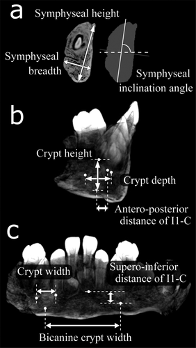
To evaluate the size of mandibular available space for accommodation of the anterior dentition, we first examined symphyseal cross-sectional geometry (Fig. 2a). Symphyseal height was taken as the distance between the most antero-superior and inferior points of the symphysis. Symphyseal breadth was defined as the maximum breadth that was perpendicular to the measurement line of the symphyseal height. Symphyseal inclination was measured as the inclination angle of the principal axis of total subperiosteal area relative to the alveolar plane (Fukase, 2012). The size and placement of developing permanent incisors/canines were then examined with the serial CT images. Maximum vertical height, transverse width, and antero-posterior depth of each crypt were determined (Fig. 2b,c). Crypt volume index was determined as the third power root of the product of the three dimensions. Bicanine crypt width was defined as the transverse distance between the lowest points of the canine crypts. Supero-inferior and antero-posterior distances of I1-C were determined between the lowest points of the central incisor and canine crypts. For evaluation of the root size of the deciduous incisors and canines, the labiolingual cervical diameter was obtained as the distance between the lowest points of the labial and lingual cervical line (Fukase and Suwa, 2008; Suwa et al., 2011). Likewise, the mesiodistal cervical diameter was taken between the highest points of the mesial and distal cervical line. However, it must be noted that these measuring methods for cervical diameter using CT imagery are not necessarily identical with those using a caliper, which cautions against direct and simple comparison between measurement values by the two methods. To evaluate total space occupied by deciduous tooth roots and permanent tooth crypts, we determined the sum of the labiolingual diameter and crypt depth for the pairs of di1-I1, di2-I2, and dc-C.

Metrics used in this study. Symphyseal height, breadth, and inclination angle were taken on the midsagittal cross section (a). In addition to each crypt dimension, antero-posterior and supero-inferior distances between the incisor and canine crypts were measured (b and c). See the text for details.

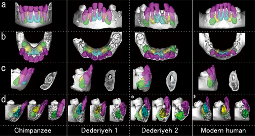
Reconstructed mandibles and teeth in frontal (a), superior (b), and lateral (c and d) views, with midsagittal cross-sections (c, right) and parasagittal cross-sections passing through the root apexes of di1 (d, left), di2 (d, middle), and dc (d, right). *, reflected image. The chimpanzee and modern human mandibles are of approximately two years of dental age.
To compare the measurements among the samples, predicted values at dental ages of 1.5 and 2.0 years were calculated from least-square regressions for modern humans and chimpanzees. Prediction interval was also derived for each measurement of the modern human sample, in order to examine whether the Dederiyeh values fall within the variation ranges of modern humans.
RESULTS
Symphyseal height and breadth of the Dederiyeh and chimpanzee samples mostly fell within the prediction interval of the modern humans (Table 1, and see also Fig. 3). Dederiyeh 1 and 2 had comparable symphyseal inclination angles which were lower than those of any of the modern humans. Dederiyeh mandibles accommodated overall larger anterior tooth crypts than those of the similarly-aged modern humans (Fig. 4a). In particular, the incisor/canine dimensions of Dederiyeh 2 consistently exceeded the prediction interval. In terms of the dental positional relationship, the bicanine crypt width was significantly larger in the Dederiyeh specimens than in the modern humans and chimpanzees. For the supero-inferior I1-C distance, both Dederiyeh values were within the modern human range. Compared with the Dederiyeh and modern human samples, the chimpanzees displayed significantly higher values in the antero-posterior distance of I1-C, indicating that their canines developed more posterior to the central incisors.

Results of the measurements. In the scatter plots (a, c, d), Gray diamond, Dederiyeh 1; gray square, Dederiyeh 2; open circle, modern human; black triangle, chimpanzee. Thick and thin solid lines on dental age (a, c) denote the regression lines of the modern humans and chimpanzees, respectively. Broken lines show the prediction interval of the modern human values. In the box plots (b), the open (right) and darkly colored (left) boxes represent the results of the modern humans and chimpanzees, respectively.
| Chimpanzees | Dederiyeh 1 | Dederiyeh 2 | Modern humans | ||||||
|---|---|---|---|---|---|---|---|---|---|
| N | PV at 1.5ydaa | PV at 2ydaa | N | PV (PI) at l.5ydaa | PV (PI) at 2ydaa | ||||
| Symphyseal height (mm) | 7 | 21.3 | 22.5 | 22.1 | 21.0 | 13 | 19.3(15.5-23.1) | 20.2(16.5-23.9) | |
| Symphyseal breadth (mm) | 7 | 8.9 | 9.5 | 10.4 | 10.3 | 14 | 9.0 (7.5-10.4) | 9.5(8.1-10.9) | |
| Symphyseal inclination (degree) | 7 | 59.3 | 58.7 | 82.3 | 82.6 | 13 | 94.2(81.8-106.6) | 93.8(81.7-105.9) | |
| Crypt height (mm) | |||||||||
| I1 | 7 | 10.0 | 10.9 | 10.2 | 11.5 | 15 | 9.2(7.3-11.0) | 10.1(8.2-11.9) | |
| I2 | 7 | 9.1 | 9.8 | 8.6 | 10.3 | 14 | 8.5 (6.6-10.4) | 9.4(7.5-11.2) | |
| C | 7 | 8.5 | 9.1 | 9.8 | 9.6 | 15 | 8.0(6.5-9.4) | 8.7(7.3-10.2) | |
| Crypt width (mm) | |||||||||
| I1 | 7 | 7.3 | 7.6 | 6.3 | 6.6 | 14 | 5.1 (4.1-6.1) | 5.2 (4.2-6.2) | |
| I2 | 7 | 7.2 | 7.6 | 7.2 | 7.6 | 14 | 6.1 (4.9-7.2) | 6.2(5.1-7.3) | |
| C | 7 | 6.3 | 6.8 | 6.5 | 7.7 | 12 | 5.8(5.1-6.6) | 6.3 (5.5-7.0) | |
| Crypt depth (mm) | |||||||||
| I1 | 7 | 7.7 | 8.1 | 7.1 | 7.7 | 15 | 5.6(4.0-7.1) | 5.8(4.3-7.3) | |
| I2 | 7 | 8.2 | 9.0 | 7.2 | 7.4 | 15 | 5.7(3.8-7.6) | 6.1(4.2-7.9) | |
| C | 7 | 6.6 | 7.4 | 7.1 | 8.8 | 16 | 6.2(4.5-7.9) | 6.8(5.1-8.5) | |
| Crypt volume index | |||||||||
| I1 | 7 | 8.2 | 8.7 | 7.7 | 8.4 | 14 | 6.4(5.3-7.5) | 6.8(5.6-7.9) | |
| I2 | 7 | 8.1 | 8.7 | 7.6 | 8.3 | 13 | 6.7(5.7-7.8) | 7.1(6.1-8.1) | |
| C | 7 | 7.1 | 7.7 | 7.7 | 8.7 | 12 | 6.6(5.5-7.6) | 7.1(6.1-8.2) | |
| Bicanine crypt width (mm) | 7 | 21.2 | 20.1 | 25.4 | 27.4 | 15 | 20.7 (16.7-24.7) | 21.4(17.4-25.3) | |
| Supero-inferior distance of I1-C (mm) | 7 | 1.0 | 1.5 | 4.5 | 2.6 | 15 | 3.9(1.6-6.3) | 3.8(1.5-6.2) | |
| Antero-posterior distance of I1-C (mm) | 7 | 7.3 | 7.7 | 2.9 | 5.1 | 14 | 2.6(0.4-4.9) | 2.8(0.6-5.0) | |
| Labiolingual cervical diameter + crypt depth (mm) | |||||||||
| di1-I1 | 7 | 11.5 | 12.0 | 11.5 | 12.6 | 8 | 9.2(7.1-11.3) | 9.2(7.2-11.3) | |
| di2-I2 | 7 | 13.1 | 13.8 | 12.1 | 12.6 | 8 | 9.8(7.5-12.2) | 10 (7.9-12.2) | |
| dc-C | 7 | 12.3 | 13.2 | 13.21 | 15.1 | 5 | 12.5 (6.7-18.2) | 12.3 (7.2-17.4) | |
- a Predicted value (PV) and prediction interval (PI) were derived at dental age of 1.5 (1.5yda) and 2.0 (2yda) for comparisons with Dederiyeh 1 and 2, respectively.
Both Dederiyeh individuals exhibited larger cervical diameters of the deciduous incisors and canines by approximately 1 mm compared to the modern humans (Table 2, Fig. 4b). The dimensions were consistently slightly greater in Dederiyeh 2 than in Dederiyeh 1. The sum values of LL cervical diameter and crypt depth were overall larger in the Dederiyeh individuals than in the modern humans, both on dental age (Fig. 4c) and symphyseal breadth (Fig. 4d). Specifically, the sum values of di1-I1 of the Dederiyeh specimens fell within the chimpanzee's range.
| Chimpanzees | Dederiyeh 1 | Dederiyeh 2 | Modern humans | |||||
|---|---|---|---|---|---|---|---|---|
| N | Mean | SD | N | Mean | SD | |||
| di1 | 7 | 3.9 | 0.23 | 4.4 | 4.9 | 8 | 3.7 | 0.29 |
| di2 | 7 | 4.8 | 0.37 | 4.9 | 5.2 | 8 | 4.1 | 0.32 |
| dc | 7 | 5.7 | 0.31 | 6.0 | 6.3 | 5 | 5.2 | 0.32 |
| di1 | 7 | 4.7 | 0.22 | 4.0 | 4.5 | 7 | 3.1 | 0.31 |
| di2 | 7 | 4.5 | 0.25 | 4.3 | 4.7 | 8 | 3.3 | 0.30 |
| dc | 7 | 5.2 | 0.22 | 5.2 | 5.6 | 5 | 4.4 | 0.17 |
DISCUSSION
When compared with the modern humans, the two Dederiyeh mandibles exhibited both broader incisors and wider bicanine space, while the chimpanzees had broader incisors and a narrower anterior corpus. Although immature mandibles of chimpanzees usually accommodated considerably anteroposteriorly overlapped dental crypts of incisors and canines (Fukase, 2012), the Dederiyeh anterior teeth were comparatively transversely placed with limited anteroposterior overlap, similar to modern humans (Fig. 3). These observations support the suggestion that inter-taxon differences in the placement patterns of forming anterior teeth should be explained in association with the proportional relationships between the sizes of the available mandibular space and anterior teeth (Fukase, 2012).
The results of this study showed that both Dederiyeh mandibles accommodated overall larger anterior teeth than the modern human mandibles (Fig. 4), supporting the hypothesis that Neanderthal infants without clear chin-like features should experience greater demands for dental accommodation. Furthermore, despite the similar anterior corpus size of the two Dederiyeh infants, Dederiyeh 2 possessed thicker deciduous tooth roots and more expanded permanent tooth crypts than Dederiyeh 1. The crypt size difference partly stems from the slightly different dental developmental stage. It is nonetheless implied that the thick roots and enlarged crypts of Dederiyeh 2 collectively distended the labial alveolar bone (Fig. 3), obscuring the incipient mental trigone. Thus, the results potentially support the hypothesis that a chin, if not with complete mental trigone/fossa, can be individually and variably expressed under certain dento-mandibular spatial conditions, as far as phylogenetically close Neanderthals and modern humans are concerned (e.g. Howell, 1960; Day and Leakey, 1973; Wolpoff, 1975; Hublin and Tillier, 1981; Lieberman, 1995; Rosas, 1995).
In light of ontogenetic constraints on the adult symphyseal morphology (Coquerelle et al., 2010; Fukase, 2011, 2012), it might be further speculated that adult mental configurations are more or less the retention of an immature mental form that is intimately intertwined with dental development (cf. Arensburg et al., 1989). These hypotheses may disagree with the conception of the chin as a strictly species-specific trait of modern humans (Schwartz and Tattersall, 2000), but justifies the presence of an incipient chin in late Neanderthals with comparatively reduced anterior tooth size such as, Amud 1, La Quina H9, Vindija 206 (Suzuki and Takai, 1970; Wolpoff, 1975; Lam et al., 1996). Moreover, an early Upper Paleolithic immature mandible from Lagar Velho (Lagar Velho I, ∼4 years old) displays a retreated symphysis together with a discernible mental trigone, representing a mosaic of Neanderthal and modern human features (Duarte et al., 1999; Trinkaus, 2002). The dental size is also intermediate between late Neanderthals and early modern humans (Duarte et al., 1999; Trinkaus, 2002). The immature mandible of Skhul I lacked definable central keel and mental fossa (Schwartz and Tattersall, 2000), but this may also be associated with the fact that the deciduous anterior teeth of Skhul I are of a comparable size to those of Neanderthals (Smith and Arensburg, 1977; McCown and Keith, 1980). However, despite the large anterior teeth, other immature mandibles of early modern humans (e.g. Skhul X, Irhoud3, Isturitz, 68 and 70) surely exhibit discernible mental trigone (McCown and Keith, 1980; Hublin and Tillier, 1981; Tillier, 1989; Schwartz and Tattersall, 2000), necessitating further investigation on the metric tooth-jaw relationships.
Finally, it must again be noted that this preliminary study investigated only two West Asian Dederiyeh infants as representatives of Neanderthals. The implications presented therefore inevitably require future verification using additional Neanderthal and early modern human fossil samples as well as more modern human samples with varying anterior dental sizes. Investigation of modern humans with congenital anodontia would also provide new insights into the tooth-chin morphological association. Nevertheless, the results obtained in the present study indicate the importance of understanding the dentognathic spatial relationship early during development, for elucidation of mechanisms underlying human mandibular evolution including emergence of the chin.
ACKNOWLEDGMENTS
The authors are grateful to Dr. T. Akazawa and the member of the Japan-Syria joint archaeological project in the Afrin Valley of northeast Syria for permission of the use of the Dederiyeh fossil materials for the present study. Special thanks go to Drs. G. Suwa (The University Museum, The University of Tokyo), M. Takai and T. Nishimura (the Primate Research Institute, Kyoto University), and D. Shimizu (then, the Japan Monkey Centre), for access to the materials used in this study.


