The goniomaxillar length/goniomandibular length ratio in normal newborn infants: A clinical tool for defining chin position abnormalities
Abstract
Retrognathia (recessed chin) and prognathism (prominent chin) often present as signs of an underlying condition. Accurate clinical definitions are important. Yet their definitions were according to “clinical impression”, or to seldom used X-ray criteria. We propose a statistical and anthropometric definition of retrognathia and prognathism based upon the ratio between the goniomaxillar length (distance between the gonion at the mandible angle and the subnasale and the goniomandibular length (distance between the mandible angle and the most anterior point of the bony chin). We assumed that an increase in the ratio indicates retrognathia and a decrease reflects prognathism. We conducted a prospective, observational, anthropometric study in 204 consecutive healthy term infants. Measurements took place on the second day of life, using sliding calipers. Mean ± SD of goniomandibular length (5.1 ± 0.3 cm), goniomaxillar length (5.4 ± 0.3 cm), were calculated. All measurements correlated significantly with gestational age, and with infant birthweight. The mean ± SD goniomaxillar length/goniomandibular length ratio was 1.06 ± 0.05. We defined a normal ratio as being within 2 SD of the mean, that is, between 0.96 and and 1.16. This ratio correlated with neither gestational age nor with birthweight. We conclude that the goniomaxillar length/goniomandibular length ratio can be calculated whenever retro - or prognathism is suspected. A ratio outside of the 95% confidence interval should help in making this diagnosis. An increase in this ratio beyond 2 SD above the mean (1.16) could be interpreted as retrognathia and a decrease beyond 2 SD below the mean (0.96) as prognathism.
1 INTRODUCTION
Prognathism (prominent chin) in a neonate often reflects an underlying medical condition or genetic syndrome. For instance, a protruding jaw, even at birth, can be a prominent feature of Sotos Syndrome (Nalini & Biswas, 2008; OMIM, 2020) or of Autosomal dominant Mandibular Prognathism (OMIM, 2016). Retrognathia (or recessed chin) in the newborn infant may also reflect of a diverse collection of congenital conditions, including Pierre Robin Sequence (Morice et al., 2018), the oculoauriculovertebral spectrum (OMIM, 2019a), or Toriello-Carey Syndrome (OMIM, 2019b; Toriello, Colley, & Bamshad, 2016).
Despite the importance of jaw abnormalities in facilitating the clinical diagnosis of such a wide array of congenital conditions, there exists no known precise clinical definition of retrognathia and prognathism in the neonate. In fact, it is generally relegated to the realm of subjective impression. In her hallmark article on standard terminology for the head and face, Allanson defined prognathism as an “anterior protrusion of the mandibular alveolar ridge beyond the vertical plane of the maxillary alveolar ridge, best appreciated in profile”, but she further specified that this definition was subjective. Similarly, she defined retrognathia as a “posteriorly positioned lower jaw, which is set back from the plane of the face when viewed from the side but not from the front” and also here she specified that the definition remains subjective (Allanson et al., 2009). Thus, until now these two entities have either been described empirically, according to the “clinical impression” of the examiner, or according to X -ray criteria, such as stereophotogrammetry (Cassi et al., 2019), which involves 3 -D reconstructions of several facial images and special hardware and software, and may expose the neonate to unnecessary radiation. van der Haven et al. ( 1997) developed a “jaw index”, which used the tragus of both ears as an anatomic landmark. However, the jaw index is influenced by ear placement (low versus high set ears), and therefore lends itself to misinterpretation.
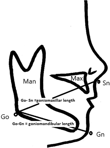
We therefore conducted this prospective, observational, anthropometric study of the chin proportions of normal newborn infants on their second day of life, in order to define normal standards and then to attempt to propose a simple, reproducible, statistical and anthropometric definition of recessed chin and of prominent chin. The availability of definitive clinical standards would facilitate more accurate diagnoses. To accomplish this, we measured the goniomandibular length defined as the distance between the gonion (at the mandible angle) and the gnathion (the most anterior point of the bony chin in the midsagittal plane), and the goniomaxillar length defined as the distance between the gonion and the subnasale at which the nasal septum merges with the upper cutaneous lip in the midsagittal plane). Because the direct measurements are likely to be affected by gestational age and/or birth weight, we also studied the ratio between the goniomaxillar length and goniomandibular length. We hypothesized that an increased ratio indicated retrognathia, while a decreased ratio reflected prognathism.
2 METHODS
2.1 Editorial policies and ethical considerations
The study was approved by the institutional ethics committee in the Rabin Medical Center. All the mothers of the infants participating in the study signed an informed consent.
2.1.1 Patients
We aimed to study approximately 200 healthy, term singleton neonates. We therefore excluded preterm (less than 37 completed weeks of gestation) and post -term (more than 41 completed weeks of gestation) infants. Gestational age was calculated from the first day of the last menstrual period, and verified to be consistent within 1 week of early (first trimester) ultrasonographic assessment. We excluded infants of mothers with pregnancy related complications such as pregnancy-induced hypertension, gestational or pregestational diabetes, maternal chronic disease. We also excluded infants with major congenital malformations, visible craniofacial malformations, low Apgar scores (<7 at 1 and 5 min), and/or infants with unstable vital signs, sepsis, respiratory distress, and facial palsy. These conditions were classified according to Vermont–Oxford criteria (Vermont Oxford Network, 2002). Whenever an infant fulfilling the inclusion criteria was identified his/her mother was asked to sign a consent form.
2.1.2 Measurements
All measurements were performed by a single investigator (GMS) on the second day of life, using a sliding calipers graduated in millimeters (Siber Hegner Maschinen GPM Anthropological Instruments, Switzerland), applying slight pressure on the anatomic landmarks. They were all obtained by one single investigator (GMS), in order to not add to the error of measurement the possible bias of inter -observer variance. As shown on Figure 1, we measured the goniomandibular length defined as the distance between the gonion (Go, at the mandible angle) and the gnathion (Gn, the most anterior point of the bony chin in the midsagittal plane); the goniomaxillar length defined as the distance between the gonion and the subnasale (Sn, the point at which the nasal septum merges with the philtrum in the midsagittal plane). We also measured the intergonion distance, defined as the distance between the two mandible angles. The ratio between goniomaxillar length and goniomandibular length was then calculated, with the assumption that an increase in the ratio meant retrognathia and a decrease in the ratio meant prognathism.

2.1.3 Statistical analyses
Data are reported as mean ± SD or median (range). Statistical analysis was comprised of linear regression. We hypothesized that goniomaxillar length and goniomandibular length correlate with gestational age and birthweight, but that the ratio between them correlates with neither of them.
3 RESULTS
3.1 Patient population
After applying the inclusion and exclusion criteria, 204 mothers remained and consented to participate in the study. Selected clinical characteristics of the infants are summarized in Table 1, including maternal age, infant birthweight and gestational age at delivery.
| Variable | Mean ± SD (range) |
|---|---|
| Maternal age (years) | 29.4 ± 4.8 (18–43) |
| Infant birthweight (kg) | 3.27 ± 0.38 (2.19–4.38) |
| Gestational age (weeks) | 39.3 ± 1.1 (37–41) |
3.2 Neonatal anthropometric measurements
Table 2 depicts the anthropometric measurements (mean ± SD, range and normal range (mean ± 2SD) of the infant mouth and jaw, in terms goniomandibular length, goniomaxillar length, and goniomaxillar length/goniomandibular length ratio. Goniomandibular length and goniomaxillar length correlated significantly with gestational age, and even more with infant birthweight (Table 3). Goniomaxillar length/goniomandibular length ratio correlated neither with gestational age nor with birthweight. By definition, no infants in the study were dysmorphic.
| Variable | Mean ± SD | Range | Normal range |
|---|---|---|---|
| Goniomandibular length (cm) | 5.1 ± 0.3 |
(4.2–6.0) | 4.5–5.7 |
| Goniomaxillar length (cm) | 5.4 ± 0.3 |
(4.6–6.3) | 4.8–6.0 |
| Goniomaxillar length/goniomandibular length ratio | 1.06 ± 0.05 |
(0.85–1.22) | 0.96–1.16 |
| Gestational age | Birthweight | |||
|---|---|---|---|---|
| R2 (%) | p-value | R2 (%) | p-value | |
| Goniomandibular length | 5.0 | .001 | 27.0 | <.001 |
| Goniomaxillar length | 6.2 | <.001 | 31.7 | <.001 |
| Intergonion distance | 5.9 | .001 | 29.8 | <.001 |
4 DISCUSSION
van der Haven et al developed a “jaw index” whose purpose was to define retrognathia in newborns (van der Haven et al., 1997), but since this index uses the tragus of both ears as an anatomic landmark, the jaw index might be affected not only by the chin development, but also by the placement of the ears (abnormally positioned ears). This could lead to significant misinterpretation in conditions where both chin and development and ear shape or placement are affected (such as in hemifacial microsomia) (OMIM, 2019a). Thus to the best of our knowledge, this study is the first to evaluate prospectively and systematically the anthropometric characteristics of the jaws of the healthy neonate, and to establish standards for retro- and prognathism that are unrelated to ear placement.
In this study, we excluded preterm and postterm infants, and infants with major congenital malformations, craniofacial malformations, low Apgar scores (<7 at 1 and 5 min), as well as infants with unstable vital signs, sepsis, respiratory distress, and facial palsy in attempt to eliminate environmental variants which might affect standard measurements. We also excluded pregnancy complications such as pregnancy-induced hypertension, gestational or pregestational diabetes, and maternal chronic diseases with or without chronic intake of medications in an attempt to exclude conditions that may affect growth in general, and facial growth in particular. Thus, we believe that the population herein studied is representative of a general, healthy population of term neonates.
The anthropometric measurements that we obtained on these infants are of importance when dealing with neonatal dysmorphology. Indeed, there are many congenital syndromes in which the infant is quoted to exhibit jaw placement abnormalities. We hereby offer a clinical, statistical definition of retrognathism as a goniomaxillar length /goniomandibular length ratio of ≥1.16 (mean of 1.06 + 2 SD's of 0.05 each) and prognathism as a goniomaxillar length /goniomandibular length ratio of ≤0.96 (mean of 1.06 - 2 SD's of 0.05 each). Values above the upper limit and below the lower limit may still be encountered in 5% of the general population (2.5% at the upper end and 2.5% at the lower one), but should alert the clinician to a possible divergence from normality. Importantly, and contrary to each of the individual components of the ratio, the ratio itself is not affected by either gestational age or birthweight, which enables it to be used without the need for gestational age adjustments.
Since this definition of appropriate jaw position (neither prognathism or retrognathia) is statistical, its main limitation is that it cannot define what is abnormal, it can only define what may be outside the measured range in this single study. Even this might be a matter of definition, whether the second SD (where the risk of abnormality is set arbitrarily at approximately 5%) or the third standard deviation (where the risk of abnormality is set up at approximately 1%) are chosen.
A limitation of this ratio is that it was established in a term Caucasian population, thus these “normal values” might not be applicable to infants of African or Asian ancestry, or in preterm or post term infants. Another limitation is that these measurements are only applicable during the neonatal period. It is likely that in view of the expected growth of the mandible the proportions that we describe might change over time. This measurement, from our experience, could be taught easily but requires a caliper, which is much more precise than a tape measurement. Finally, we have not tested this ratio in real life of dysmorphic patients with diagnosis of retro - or prognathia.
CONFLICT OF INTEREST
The Authors have no conflict of interest.
AUTHOR CONTRIBUTIONS
Galit Mimouni: Collected the data, recruited the patients, reviewed the manuscript.
Paul Melrob: Recruited the patients, reviewed the manuscript.
Francis B. Mimouni: Conceived of the presented idea, wrote the manuscript, analyzed the data.
Alona Bin Nun: Wrote the manuscript, analyzed the data.
Open Research
DATA AVAILABILITY STATEMENT
The data used in this study will be shared upon request after deidentification, including study protocol and statistical analyses, from one author (FBM at the following email address: [email protected]).


