Lipoplexes Could be Alternative to Viral Vectors in Gene Therapy
A computer simulation showed that lipoplexes can deliver DNA fragments into cells, opening the way for more systematic studies
Gene therapy is a promising therapeutic modality that has been studied for more than 4 decades. It involves the introduction of, removal of, or change in the genetic material in the cells of a patient to treat a specifi c disease, and can be used to treat a broad range of disorders, including cancer, genetic syndromes, and infectious diseases. It is possible to employ gene therapy through different types of mechanisms, such as replacement of a disease-causing gene with a healthy copy of the gene, inactivation of a disease-causing gene that is not functioning properly, and introduction of a new or modifi ed gene into the body that could help treat a specifi c disorder.
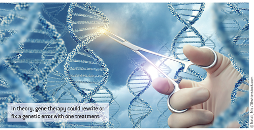
In theory, gene editing could provide a one-time treatment option that rewrites or fi xes errors in the genes. In practice, however, delivering new genetic material to human cells has presented researchers with challenges. Although viral vectors remain a potent gene delivery platform, the immune system has evolved to attack what it perceives as invading pathogens. Immune responses against vector-derived antigens can reduce the effi cacy and stability of in vivo gene transfer and lead to a strong acquired immune response.
There is increasing interest in developing non-viral-based vectors, as they do not trigger a specifi c immune response and may be less expensive. Most such vectors employ cationic lipids or polymers. The drawback to many of these non-viralbased methods is that they have a low rate of transfection in vivo.
The Study
“The models we made were designed to gain insight [into] the molecular detail of lipoplex/membrane fusion,” says study author Bart Bruininks, a PhD student in the group of Siewert-Jan Marrink, Professor of Molecular Dynamics at the University of Groningen, The Netherlands. “Therefore, they aim to accurately simulate such [a] process.”
The detailed computer simulations of the fusion between a lipoplex and model endosomal membranes showed that this is possible, and thus opened the way for more systematic studies using the more advanced lipoplex formulations that are currently available. “We indeed show fusion of lipoplexes with membranes, which is well in line with the experimentally observed phenomena,” says Bruininks. “It is known that for these types of complexes, they can fuse with membranes and liposomes.” He acknowledges, however, that it is unknown how that fusion takes place. “Making optimization of lipoplexes resembles trying to shoot a target in the dark, and we tried to turn the lights on,” he says.
Relating the physico-chemical properties of the vector to transinfection efficiency through experiments has proven very diffi cult. Molecular dynamics simulations, however, can provide an alternative method for studying molecular processes in atomic, or near-atomic, detail. The researchers used a coarsegrain molecular dynamics simulation and were able to visualize the fusion process between the lipoplex and the endosome membrane, along with the subsequent escape of the DNA.
The computational fusion experiments of lipoplexes with endosomal membrane models demonstrated that 2 distinct modes of transfection exist: parallel and perpendicular. In the parallel pathway, it appeared that DNA aligns with the membrane surface and rapidly releases the genetic material soon after the initial fusion pore is formed. Conversely, the release in the perpendicular pathway is slower but also leads to transfection. The investigators' results also demonstrated that the composition and size of the lipoplex have a signifi cant impact on fusion effi ciency in this model, as did the lipid composition of the endosomal membrane.
Building on Results
Bruininks notes that there are ongoing clinical studies using this type of vector. “Instead of reproducing our models, it might be much more interesting to test some of its conclusions,” he says. “We also observed rotation of the lipoplex during fusion, which is something that could be observed with advanced imaging techniques such as FRET microscopy. Such undertakings are far from trivial but could result in a better understanding of the transfection as a whole.”
Commenting on the paper, Kenneth Lundstrom, PhD, CEO of PanTherapeutics, Lausanne, Switzerland, notes that while simulations are useful, they need to be followed up with experiments in animal models. “But studies in animal models, particularly in small rodents, do not refl ect well the situation in humans, which means that delivery effi cacy achieved in mice and rats does not always translate to success in clinical trials in humans,” he says.
Dr. Lundstrom also points out that not all viral vectors are that immunogenic, and many viruses, such as alphaviruses, are not immunogenic in the form of replication-deficient particles, which cannot generate new viral particles in infected cells. “These can be used for repeated injections, especially if encapsulated in liposomes, which is partly to passively target tumors, partly to protect from host immune responses,” he explains. “The other advantage of these RNA viruses is that they have an in-built self-replication machinery, which means that from a single copy of RNA introduced into the cytoplasm, 200,000 copies are made.”
Another issue with DNA delivery for therapeutic use is that even with lipoplexes, it will be diffi cult to achieve high effi cacy in vivo and compete with viral delivery, Dr. Lundstrom explains. “Even if that is the case, I would rather deliver RNA, which allows direct action in the cytoplasm,” he says. DNA vectors have to be delivered to the nucleus and then transcribed to RNA, which then needs to be transported to the cytoplasm again, resulting in loss of time and effi cacy. But for those who wish to use DNA, he recommends using DNA vectors based on alphavirus replicons or self-replicating RNA sequences, “as it has been demonstrated that you need 100— 1000-fold lower concentrations of DNA compared to conventional DNA plasmids to achieve the same immune response in vaccine animal models.”


