Anthropometric findings from birth to adulthood and their relation with karyotpye distribution in Turkish girls with Turner syndrome
Abstract
To evaluate the anthropometric features of girls with Turner syndrome (TS) at birth and presentation and the effect of karyotype on these parameters. Data were collected from 842 patients with TS from 35 different centers, who were followed-up between 1984 and 2014 and whose diagnosis age ranged from birth to 18 years. Of the 842 patients, 122 girls who received growth hormone, estrogen or oxandrolone were excluded, and 720 girls were included in the study. In this cohort, the frequency of small for gestational age (SGA) birth was 33%. The frequency of SGA birth was 4.2% (2/48) in preterm and 36% (174/483) in term neonates (P < 0.001). The mean birth length was 1.3 cm shorter and mean birth weight was 0.36 kg lower than that of the normal population. The mean age at diagnosis was 10.1 ± 4.4 years. Mean height, weight and body mass index standard deviation scores at presentation were −3.1 ± 1.7, −1.4 ± 1.5, and 0.4 ± 1.7, respectively. Patients with isochromosome Xq were significantly heavier than those with other karyotype groups (P = 0.007). Age at presentation was negatively correlated and mid-parental height was positively correlated with height at presentation. Mid-parental height and age at presentation were the only parameters that were associated with height of children with TS. The frequency of SGA birth was found higher in preterm than term neonates but the mechanism could not be clarified. We found no effect of karyotype on height of girls with TS, whereas weight was greater in 46,X,i(Xq) and 45,X/46,X,i(Xq) karyotype groups. © 2016 Wiley Periodicals, Inc.
INTRODUCTION
Turner syndrome (TS), a chromosomal disorder caused by complete or partial X chromosome monosomy, can manifest with various clinical features depending on the karyotype and the genetic background of the affected subjects. It affects approximately 1 in 2500 Live-born females [Davenport et al., 2002]. It is associated with decreased viability in utero, short stature, and gonadal dysgenesis.
Short stature is the most common presenting symptom. Newborns with TS have been reported to be lighter and shorter than their peers [Wisniewski et al., 2007]. However, some studies reported no difference between healthy newborns and children with TS regarding birth length [Davenport et al., 2002]. Although short stature is mild during infancy and early childhood, the height deficit becomes more notable as age progresses. Karyotype, mid-parental height (MPH), hormone replacement with growth hormone or sex steroids and spontaneous onset of puberty may affect the height of girls with TS.
The aim of the present study was to describe the anthropometric features of patients with TS at birth, at presentation, and to evaluate the effect of karyotype on these measurements.
MATERIALS AND METHODS
Patients and Data Collection
Patient recruitment was undertaken by the Turner Syndrome Study Group in Turkey as described previously [Yeşilkaya et al., 2015]. A total of 842 patients were included, with an age of diagnosis ranging from birth to 18 years from a large Turkish patient cohort examined between 1984 and 2014. Patients' data were obtained from 35 different centers throughout Turkey via a web registry. The study was approved by the Ethics Committee of Gulhane Military Medical Academy.
The standard Case Report Form (CRF) developed for this study was uploaded to the online web registry system on the web site “http://www.favorsci.org” The CRF included birth data and data at presentation (age, length/height, weight, body mass index (BMI), MPH and karyotype). Physicians who were working in an outpatient clinic for patients with TS were asked to complete the CRF forms. Data were collected by physicians who were chosen to be responsible for registration from each center. The timeframe for patient enrollment was from September 2013 to January 2014.
Both birth and presenting weight and length/height standard deviation scores (SDS) were calculated, in addition to BMI SDS at presentation. Patients whose medical records could not be found and patients who had received any injections of growth hormone, estrogen or oxandrolone were excluded. Presentation characteristics of the remaining 720 patients were evaluated. Data were compared against healthy age-matched Turkish reference data [Neyzi et al., 2008]. In addition, their parents' mean height SDS values were calculated.
Clinical and Laboratory Evaluation
The karyotype analysis was performed on peripheral blood lymphocytes. Karyotypes of patients were classified by one geneticist (DG) as 45,X, 45,X/46,XX, 46,X,i(Xq), 45,X/46,X,i(Xq), and others.
Patients were classified according to gestational age; term (between 37 and 42 weeks); late preterm (32–36 weeks); very preterm (28–31 weeks) and extremely preterm (<28 weeks) [Moutquin, 2003]. Preterm neonates accounted for 51of the 582 patients. Data of these groups were compared with each other regarding karyotypes and against the healthy population. Patients with a birth weight less than the 10th percentile according to gestational age were classified as small for gestational age (SGA) [Kurtoğlu et al., 2012]. SGA neonates accounted for 176 of the 531 term patients. Frequency of SGA and karyotype distribution in TS was also compared against the healthy population.
Statistical Analysis
Statistical analysis was performed using IBM SPSS 21.0 for Windows statistical software. Frequencies and percentages for categorical variables and mean ± standard deviations (SD) were used as descriptive statistics. Comparisons for categorical variables were performed using χ2 tests. Scale variables compared using ANOVA in normally distributed data or Kruskal–Wallis test in non-normally distributed data. A multivariate linear regression analysis was used to evaluate prediction of patient presentation height. A P < 0.05 value was considered as statistically significant.
RESULTS
Anthropometric features and karyotype in girls with TS at birth and at presentation are shown on Table I.
| 45,X (n = 427) | 45,X/46,XX (n = 91) | 46,X,i(Xq) (n = 85) | 45,X/46,X,i(Xq) (n = 82) | Others (n = 157) | Total (n = 842) | P | |
|---|---|---|---|---|---|---|---|
| At birth | |||||||
| Gestational age (week) | 38.9 ± 1.9 | 38.8 ± 1.9 | 38.6 ± 1.5 | 38.4 ± 2.3 | 38.3 ± 2.5 | 38.7 ± 2.1 | 0.057 |
| Birth weight (kg) | 2.93 ± 0.37 | 2.91 ± 0.60 | 2.90 ± 0.48 | 2.98 ± 0.56 | 2.82 ± 0.59 | 2.91 ± 0.57 | 0.420 |
| Birth length (cm) | 48.1 ± 2.3 | 47.0 ± 3.2 | 47.4 ± 1.5 | 47.8 ± 2.5 | 47.7 ± 2.8 | 47.8 ± 2.5 | 0.220 |
| SGA (%) | 7.3 | 5.4 | 8.9 | 14.3 | 14.4 | 9.4 | 0.129 |
| At presentation | |||||||
| Age (years) | 9.9 ± 4.8 | 10.4 ± 4.1 | 10.5 ± 4.3 | 10.5 ± 3.3 | 10.1 ± 4.2 | 10.1 ± 4.4 | 0.891 |
| Height SDS | −3.1 ± 1.8 | −3.0 ± 1.4 | −3.4 ± 1.4 | −3.1 ± 1.4 | −3.0 ± 1.5 | −3.1 ± 1.7 | 0.180 |
| Weight SDS | −1.4 ± 1.5 | −1.4 ± 1.3 | −1.3 ± 1.3 | −1.1 ± 1.4 | −1.4 ± 1.7 | −1.4 ± 1.5 | 0.309 |
| BMI SDS | 0.3 ± 1.6 | 0.3 ± 1.5 | 0.9 ± 1.5 | 0.9 ± 1.6 | 0.4 ± 2.0 | 0.4 ± 1.7 | 0.007a |
| Mother's height (cm) | 157.6 | 157.2 | 157.4 | 158.3 | 157.0 | 157.5 | 0.916 |
| Father's height (cm) | 170.9 | 169.6 | 171.1 | 170.6 | 170.0 | 170.0 | 0.716 |
| MPH (cm) | 157.8 | 157.1 | 157.7 | 157.9 | 157.0 | 157.5 | 0.412 |
- SGA, small for gestational age; SDS, standard deviation scores; MPH, mid-parental height.
- a 46,X,i(Xq) and 45,X/46,X,i(Xq) karyotypes are statistically different from 45,X, 45,X/46,XX and other karyotypes.
Characteristics at Birth
Information on gestational age was available for 582 patients. Among these, 531 (91.2%) were born at term, and 51 (8.8%) patients were born preterm (42 Late preterm and 9 very preterm). There were no extremely preterm born children in our cohort. There was no statistically significant difference between the gestational ages of patients with different karyotypes (P = 0.057).
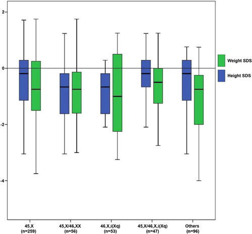
Birth length of 227 girls with TS was available. The results showed that the girls with TS were shorter than healthy neonates with a mean height of 48.1 ± 2.3 cm versus 49.4 ± 2.1 cm (P = 0.004). The mean length SDS of TS newborns was −0.6 (−4.5–1.7). The mean birth weight of 511 term TS neonates was lower than that of the normal population with 2.94 ± 0.56 kg versus 3.30 ± 0.40 kg, respectively (P = 0.001) (5). The weight SDS of newborns with TS was −0.88 (−5.75–3.00) (Fig. 1). Birth length and birth weight of term patients with TS were not different regarding karyotype distribution (P = 0.220, P = 0.420). In addition, we did not find any difference when we compared patients with 45,X karyotype against non-45,X patients (P = 0.143, P = 0.368).

Birth weight data of 48 preterm and 483 term neonates were available. The frequency of SGA birth was 4.2% (2/48) in preterm and 36% (174/483) in term neonates (P < 0.001). The rate of SGA in preterm were 2/40 in late preterm and 0/8 in very preterm respectively (P = 0.001) (Table II). SGA frequency in TS did not change with karyotype (P = 0.129).
| SGA birth present | SGA birth absent | Total | P* | |
|---|---|---|---|---|
| Term | 174 | 309 | 483 | <0.001 |
| Late preterm | 2 | 38 | 40 | |
| Extremely preterm | 0 | 8 | 8 | |
| P < 0.001**; P = 1.00*** | ||||
- * Overall comparison.
- ** Comparison between preterm and term neonates (late and extremely preterm neonates together) (χ2 test).
- *** Comparison between late preterms and extremely preterms (Fischer's exact test).
Characteristics at Presentation
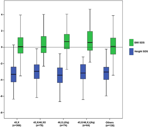
The mean age of presentation was 10.1 ± 4.4 years (range, 0–18 years) with no difference according to karyotype distribution. At presentation, the mean height SDS in 720 patients was −3.1 ± 1.7, body weight SDS and BMI SDS were −1.4 ± 1.5 and 0.4 ± 1.7, respectively. These parameters were analyzed according to karyotype (Fig. 2). There was no difference among karyotypes regarding height (P = 0.180) and weight (P = 0.309) at presentation. However BMI in the isochromosome Xq group was higher than in the non-isochromosome Xq group (P = 0.007).


DISCUSSION
The distribution of karyotype in our cohort revealed the most common karyotype as 45,X followed by mosaicism and/or isochromosome Xq. This was compatible with previous reports on karyotype diversity [Elsheikh et al., 2002].
The frequency of preterm birth in the healthy population has been recently reported about 4.8–10% over the previous 25 years and about 10% for Turkey [Onat et al., 2014]. The rate of preterm birth was 8.8% in our TS cohort. Data about preterm birth risk in TS are controversial. Although some studies have indicated increased risk of preterm birth in TS, no reason for this increase was proposed [Karlberg et al., 1991; Bernasconi et al., 1994; Hagman et al., 2010]. The rate of preterm birth in children with TS has been reported as 9.6–10.3% in some studies, slightly higher than we found in the present study [Wisniewski et al., 2007; Hagman et al., 2010]. Similar to previous reports [Bernasconi et al., 1994], we did not find any relation between karyotype and rate of preterm birth in our large TS cohort.
The main feature of girls with TS is short stature, which is thought to be due to three principal factors: the slow growth rate of the infancy component, which probably begins from the intrauterine period; a slow growth rate at the onset of the childhood component; and delayed onset of the childhood component. Newborns with TS have been reported about 500 g lighter and 3 cm shorter than their peers [Wisniewski et al., 2007]. Birth length of our patients (48.1 ± 2.3 cm) was about 1.3 cm shorter and birth weight of term newborns with TS in our cohort was 370 g lighter than that of healthy Turkish newborns. The birth weight and length SDS of our patients are consistent with those reported in the literature [Hagman et al., 2010]. Similar to our results, there was no difference among karyotypes and birth anthropometry in the published reports [Park et al., 1983; Ranke et al., 1988].
In developed countries, about 3–5% of healthy neonates are born SGA [Lee et al., 2003]. A Turkish national study found that 10.1% of neonates were born SGA [Kurtoğlu et al., 2012]. Girls with TS are more frequently born SGA compared with newborns in the non-TS population (17.8% vs. 3.5%) [Park et al., 1983; Wisniewski et al., 2007]. In some of these reports, a birth weight below −2 SDS (not below 10p) is defined as SGA. The frequency of SGA (birth weight below 10p) in our TS cohort was very high (33%). Low SGA frequency in preterm patients with TS compared with term patients suggested that fetal growth deceleration begins after the 37th gestation week. Factors that cause a reduction in fetal growth during the late pregnancy period in patients with TS should be investigated. Consistent with other studies, there was no association between karyotype and frequency of SGA in TS in our cohort [Park et al., 1983]. Girls with normal birth weight tend to be taller than girls with low birth weight during the whole growth period [Bernasconi et al., 1994]. However, we found no positive correlation between birth weight and childhood height SDS at presentation in children with TS (P = 0.065).
Short stature becomes more remarkable in TS during childhood and at peripubertal ages due to growth deceleration after birth and lack of pubertal growth spurt. The height of girls with TS drops below −2 SDS when they are aged about 2–5 years and the gap widens progressively. In a study conducted on 110 Turkish girls with TS [Bereket et al., 2008] it was reported that a final height of girls with TS without GH treatment was 142.8 cm (−3.1 SDS). In our cohort, the mean age at presentation and height SDS at presentation were 10.5 years and −3.1, respectively. Various studies report controversial results regarding height and 45,X karyotype. However, most of the studies, like our cohort, showed no effect of 45,X karyotype on growth compared with other karyotypes [Lenko et al., 1979; Pelz et al., 1991; Rochiccioli et al., 1994; Bereket et al., 2008; Isojima et al., 2010].
It is proposed that certain genes on the short arms of the X and Y chromosomes are responsible for normal stature. Some reports indicate that 45,X/46,XY or 46, X,del(Xq) TS patients are taller than others. Short arm (p) of X includes genes related to growth; long arm (q) has genes related to gonadal functions. Therefore, patients with 46, X,del (Xq) can be expected to be taller [Ferguson-Smith, 1993]. However, we found no difference in height between patients with deletions of p and q. Additionally, TS with SRY had similar height SDS compared with other karyotypes. In our cohort, similar to a previous study about Turkish girls with TS [Bereket et al., 2008] and some other studies [de Araújo et al., 2010], there was no relationship between karyotype and height SDS.
Growth deficit in TS girls is more severe for height than for weight. The frequency of overweight and obesity increased with age as reported in previous studies [Low et al., 1997]. Obesity is observed in 2–5% in healthy Turkish children [Bereket and Atay, 2012]; we detected a rate of 14% in our cohort of girls with TS. Obesity is more common with isochromosome Xq as previously reported. Higher rates of obesity in isochromosome Xq may be associated with autoimmune thyroiditis as this genotype has been shown to be prone to autoimmune diseases. However, in our cohort there was no difference in the rate of hypothyroidism between girls with TS and isochromosome Xq, and non-isochromosome Xq (P = 0.538) [Yeşilkaya et al., 2015].
Several reports indicate an association between parental height and the height of girls with TS at different ages. One study reported a final height of 146.3 ± 6.0 cm in a group of girls with taller parents (MPH 169.5 ± 3.4 cm), whereas the final height was 139.9 ± 6.5 cm in the group with shorter parents (MPH 159.4 ± 3.7 cm). The correlation between MPH and patient height is strong. Among girls with TS, MPH exerts the same role on statural growth as that observed in normal girls [Bernasconi et al., 1994]. We found a positive correlation between height SDS at presentation and MPH. It may be suggested that for the most part, the genes responsible for the influence of parental height on the height of their child are located on the autosomes.
Age at presentation was negatively correlated with height SDS at presentation (the younger the patient at presentation, the less short she was), which is in accordance with progressive height deficit in children with TS. Although some reports indicate that TS associated morbidities or spontaneous onset of puberty does not affect final height, some studies suggest that these abnormalities could impair growth parameters [de Araújo et al., 2010].
We did not exclude patients who had problems such as heart and kidney abnormalities, hypothyroidism, and celiac disease during data analyses. Additionally, we had no information on the number of patients with spontaneous puberty. Finally, it would be better to compare final height and karyotype. Another limitation of our study was that we cannot exclude ascertainment bias. Short stature was the main presenting symptom for most of our subjects. This may have led to having greater linear growth deficiency. Also, we had no data on the parental origin of the intact X chromosome, which has been shown to have an effect on phenotype in some studies [Hamelin et al., 2006]; however this has not been replicated in other studies [Devernay et al., 2012].
In conclusion we found that preterm birth rate was not increased in TS in our large cohort. One of three TS newborns was born SGA and this rate was significantly higher than in the normal population. Interestingly, SGA frequency was higher in term babies with Turner syndrome than preterm babies, which suggests that fetal growth deceleration happens later during pregnancy. However, the numbers are low and it is not possible to draw a definite conclusion regarding this significant difference. As expected, length at birth and height at presentation were statistically lower than that of the healthy population. However, there was no difference in birth length and birth weight according to karyotype distribution. With the exception of isochromosome Xq, which was found to have higher BMI SDS than other karyotypes, height and BMI SDS at presentation were similar according to karyotype. Physicians should pay special attention to patients with 46,X,i(Xq) and 45,X/46,X,i(Xq) for obesity. Parental height plays a major role in linear growth at all ages in TS. MPH predicts the height of children with TS, so girls with TS who have shorter parents are at increased risk for short stature.
ACKNOWLEDGMENTS
The following physicians contributed to this study by entering data of one or two patients to database: Atilla Büyükgebiz, Heves Kırmızıbekmez, Gül Yeşiltepe Mutlu, İhsan Esen. For technical support, we would like to thank the FAVOR Web-based registry system and its staff, and also the Turkish Society of Endocrinology and Diabetes.


