Volume 95, Issue 11 pp. 1430-1431
DIAGNOSTIC IMAGING IN HEMATOLOGY
Free Access

Primary plasma cell leukemia mimicking a Pancoast tumor

Yorick Sandberg

Corresponding Author

Yorick Sandberg

Department of Internal Medicine, Maasstad Hospital, Rotterdam, The Netherlands

Correspondence

Yorick Sandberg, Department of Internal Medicine, Maasstad Hospital, Maasstadweg 21, 3079 DZ Rotterdam, The Netherlands.

Email: [email protected]

Search for more papers by this author
Gert T. Verhoeven

Gert T. Verhoeven

Department of Pulmonary Medicine, Maasstad Hospital, Rotterdam, The Netherlands

Search for more papers by this author
Floor Weerkamp

Floor Weerkamp

Department of Clinical Chemistry, Maasstad Hospital, Rotterdam, The Netherlands

Search for more papers by this author
Annemiek Broyl

Annemiek Broyl

Department of Hematology, Erasmus Medical Center, Rotterdam, The Netherlands

Search for more papers by this author
Jasper Emmering

Jasper Emmering

Department of Radiology, Maasstad Hospital, Rotterdam, The Netherlands

Search for more papers by this author
Leo M. Budel

Leo M. Budel

Department of Pathology, Maasstad Hospital, Rotterdam, The Netherlands

Search for more papers by this author
First published: 24 August 2020

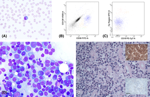
A 56-year-old non-smoking man was referred to the orthopedic surgeon because of a 5-month history of worsening pain and swelling of the left shoulder. Physical examination revealed a mass around the left shoulder. Magnetic resonance imaging and computed tomography (CT) demonstrated a large Pancoast tumor with rib destruction and invasion of the chest wall and soft tissue (Figure 1A,B), showing hypermetabolic activity on FDG positron emission tomography (PET)-CT (Figure 1C). Peripheral blood (PB) examination showed hypercalcemia, anemia, thrombocytopenia, and a normal leukocyte count (8.3 × 109/L). More thorough examination of PB smears illustrated aberrant plasma cells with nucleoli (Figure 2A; May-Grünwald-Giemsa (MGG) stain, original magnification ×1000). Flow cytometry detected a clonal plasma cell population of 23%, positive for CD56 and negative for CD19 (Figure 2B,C). The bone marrow aspirate smear identified nearly complete replacement of normal hematopoietic cells by clonal plasma cells (Figure 2D; MGG stain, original magnification ×1000). Serum free light chain kappa level was 732.5 mg/L. Further cytogenetic exploration showed t(4;14)(p16;q32). All findings were consistent with primary plasma cell leukemia (pPCL).

Details are in the caption following the image
Left-sided Pancoast tumor with destruction of the thoracic wall (including ribs 2-4). A, T2-weighted magnetic resonance imaging (MRI) with fat suppression; B, computed tomography (CT); C, fluorodeoxyglucose (FDG) positron emission tomography (PET)-CT [Color figure can be viewed at wileyonlinelibrary.com]
Details are in the caption following the image
A, Peripheral blood smear with May-Grünwald Giemsa staining shows an atypical plasma cell with two large nucleoli. B, Flowcytometry analysis of the peripheral blood demonstrates the CD38+/CD138+ plasma cells in blue. C, with the majority of plasma cells (weakly) positive for the kappa light chain and aberrant absence of CD19. D, Bone marrow aspirate smears show extensive infiltration of plasma cells with prominent nucleoli. E, Left apical lung tumor biopsy showing the morphology of the tumor with markable therapy-induced apoptosis and scattered plasma blasts (hematoxylin and eosin stain (H&E stain), 400x). E, insets, Immunohistochemical staining revealed cytoplasmic kappa light chain positivity and absence of lambda light chain expression [Color figure can be viewed at wileyonlinelibrary.com]

Left apical lung tumor biopsy demonstrated malignant cells that stained positive for CD138, CD38, CD56, and kappa light chains (Figure 2E; hematoxylin and eosin stain, original magnification ×200). Cells were negative for cytokeratin AE1/AE3, CD3, CD20, CD30, ALK1, EBV, PAX5, and CyclinD1. The patient started treatment with carfilzomib, lenalidomide and dexamethasone (KRd) which induced a complete response after four courses with regression of the pulmonary mass. Thus, KRd is a major treatment regimen in multiple myeloma1 and currently evaluated as induction therapy in a phase two study in pPCL.

Apical lung tumors are usually non-small-cell lung cancers, also known as Pancoast tumors.2 Other malignancies than primary lung cancer should always be considered in the differential diagnosis, and a histopathological diagnosis is essential for management of the patient.

CONFLICT OF INTEREST

None.

DATA AVAILABILITY STATEMENT

Data sharing not applicable to this article as no datasets were generated or analysed during the current study

    The full text of this article hosted at iucr.org is unavailable due to technical difficulties.