Quantitative magnetic resonance neurography in chronic inflammatory demyelinating polyradiculoneuropathy: A longitudinal study over 6 years
Abstract
Objective
To evaluate magnetic resonance neurography (MRN) for the longitudinal assessment of patients with chronic inflammatory demyelinating polyradiculoneuropathy (CIDP).
Methods
Prospective examination of twelve CIDP patients by neurological assessment, MRN, and nerve conduction studies in 2016 and 6 years later in 2022. Imaging parameters were compared with matched healthy controls and correlated with clinical and electrophysiological markers. The MRN protocol included T2-weighted imaging, diffusion tensor imaging (DTI), T2 relaxometry, and magnetization transfer imaging (MTI).
Results
Nerve cross-sectional area (CSA) was increased in CIDP patients compared to controls (plexus: p = 0.003; sciatic nerve: p < 0.001). Over 6 years, nerve CSA decreased in CIDP patients, most pronounced at the lumbosacral plexus (p = 0.015). Longitudinally, changes in CSA correlated with changes in the inflammatory neuropathy cause and treatment validated overall disability sum score (INCAT/ODSS) (p = 0.006). High initial nerve CSA was inversely correlated with changes in the INCAT/ODSS over 6 years (p < 0.05). The DTI parameter fractional anisotropy (FA) showed robust correlations with electrodiagnostic testing both cross-sectionally and longitudinally (p < 0.05). MTI as a newly added imaging technique revealed a significantly reduced magnetization transfer ratio (MTR) in CIDP patients (p < 0.01), suggesting underlying changes in macromolecular tissue composition, and correlated significantly with electrophysiological parameters of demyelination (p < 0.05).
Interpretation
This study provides evidence that changes in nerve CSA and FA reflect the clinical and electrophysiological course of CIDP patients. Initial nerve hypertrophy might predict a rather benign course or better therapy response.
Introduction
Diagnosis of chronic inflammatory demyelinating polyradiculoneuropathy (CIDP) typically relies on clinical and electrophysiological testing.1, 2 Magnetic resonance imaging (MRI) plays a supportive role in the diagnostic work-up of CIDP according to the recently published update of the EAN/PNS consensus guidelines.2 Specifically in cases of possible CIDP, it may help to confirm the diagnosis by showing nerve hypertrophy and increased T2-weighted signal of the plexus,3-5 while a routine clinical use is currently not recommended due to limited evidence from systematic studies.6
Besides early and specific diagnosis, the monitoring of disease progression and therapy response of CIDP to date remains an important challenge.7-9 So far, a broad range of tools have been implemented into clinical routine that provide guidance for identifying treatment effects and clinical decision-making depends on these outcome variables.10 The most widely implemented clinical tools are disability scales, such as the inflammatory neuropathy cause and treatment (INCAT) validated overall disability sum score (ODSS) scale (INCAT/ODSS)11, 12 and quality-of-life scales13 since these outcome measurements are easy to assess in clinical routine. While electrodiagnostic measurements (EDX) with proof of demyelination have indisputable diagnostic value in the early phase, the role of serial EDX for patients at later stages with non-excitable nerves is rather challenging.2
So far, the extent of which serial imaging may serve as a complementary tool in monitoring CIDP is also still under debate. There is significant evidence that high-resolution ultrasound (HRUS) may provide valuable information through quantification of nerve caliber and identification of echo texture, which may also aid to predict response to immune treatment.14-21 Most studies assessing the value of MRI in CIDP are cross-sectional studies that focused on the presence of nerve hypertrophy and increased T2-weighted signal intensity using conventional MR techniques (e.g., T2 short tau inversion recovery, STIR).4, 22, 23 Also, high-resolution imaging that is specifically developed for optimized imaging of peripheral nerves, so-called MR neurography (MRN), has been tested in CIDP patients and revealed changes even beyond the plexus structures.5 The great potential of MRN has been well documented through numerous prospective studies on various inflammatory, hereditary, and traumatic neuropathies.24-28 In addition to parameters of nerve morphology, MRN also offers quantitative parameters resulting from diffusion tensor imaging (DTI), T2 relaxometry, and magnetic transfer imaging (MTI). These techniques have been shown to offer a better insight into tissue pathology while increasing diagnostic accuracy.29-31 Several of these MR biomarkers have already been applied to CIDP patients in cross-sectional studies and mostly revealed changes consistent with demyelination.32-34
So far, the diagnostic and prognostic value of these biomarkers in the long-term follow-up of CIDP patients has not been investigated, and their adaption to therapy response and/or disease progression is still unclear. Therefore, we aimed to explore the potential of these MRN biomarkers with respect to the clinical course and treatment response through a long-term follow-up study in a cohort of CIDP patients covering a timespan of 6 years.
Materials and Methods
Subjects
This study has been approved by the local ethics committee of the Heidelberg University (No. S-146/2021) and the ethics committee of the Ruhr University Bochum (No. 18-6534-BR). Written informed consent was obtained from all participants.
In this prospective study, we enrolled 12 patients who have been diagnosed with CIDP using the 2010 EFNS/PNS diagnostic criteria (6 women, 6 men; mean age: 64.6 ± 5.9 years; range: 53–72 years) and a control group consisting of 12 healthy volunteers matched to sex and age (6 women, 6 men; mean age: 60.9 ± 5.9 years; range: 52–71 years). All CIDP patients had participated in an initial imaging study in 2016,5, 32 which comprised a clinical, electrophysiological, and initial MRN examination. For the current study, they were reexamined in 2022 by an extensive second MRN examination in addition to a second clinical and electrophysiological assessment. Six patients from the initial study, which comprised of 18 patients, were not able or did not consent to participate in the current study, resulting in 12 patients with longitudinal data.
Electrodiagnostic measurements and clinical examination
In all CIDP patients, a board-certified neurologist (K.P.) with over a decade of neurophysiology experience conducted EDX measurements. Nerve conduction studies (NCS) were performed with the Dantec™ Keypoint® Focus EMG device (Natus Medical GmbH, Planegg, Germany) using the standard techniques of percutaneous supramaximal stimulation and surface electrode in standardized conditions (skin temperatures: palm ≥33°C, external malleolus ≥30°C), as described before.35 Bilateral EDX covered the median (motor and sensory), ulnar (motor and sensory), radial (sensory), tibial (motor), and sural nerves (sensory). For correlation analyses, measurements of the median and tibial nerve were used. Motor nerve conduction velocity (NCV) was assessed in the wrist-to-elbow segments for the median nerve, and ankle-to-popliteal fossa for the tibial nerve, following reference values by Stöhr et al.36
The inflammatory neuropathy cause and treatment (INCAT) validated overall disability sum score (ODSS) scale (INCAT/ODSS)11 was used to clinically assess disease severity (ranging from 0 to 12 with higher scores indicating increased disability). Also, the Medical Research Council sum score (MRCSS) (ranging from 0 to 60 with higher scores indicating improved motor function), was assessed.37
Magnetic resonance neurography
All participants underwent MRN using a 3.0 Tesla MR-scanner (Magnetom Prismafit, Siemens Healthineers, Germany). Imaging details are listed in Table S1.
General image postprocessing and longitudinal assessment
Image analysis was performed by one reader (F.P.) with over 7 years of MRN expertise, blinded to clinical data, using a freehand region-of-interest (ROI) tool within the DICOM-viewer OsirixMD (Pixmeo Sarl, Switzerland). To track longitudinal MRN parameter changes, the initial 2016 study data were reanalyzed by the same reader using the current segmentation algorithm at corresponding anatomical sites. Changes over the 6-year timespan are indicated as Δ(parameter).
Dorsal root ganglion volume, nerve caliber, and normalized T2w signal
The plexus sequence and individual multiplanar reformations were used to calculate bilateral L5 and S1 dorsal root ganglion (DRG) volumes and the axial T2-weighted turbo spin echo (TSE) sequences were used to determine the cross-sectional area (CSA) for all peripheral nerves.
DRG volumes were calculated using an axis-corrected volume estimation approach (V = π/6 x A x B x C; A, axial plane diameter; B, axial plane diameter perpendicular to A; C, length in the long axis), as this results in reliable DRG volume estimation.38
For the lumbosacral plexus, anterior divisions of spinal nerves L5 and S1 were segmented bilaterally on two consecutive slices at presacral (L5) and intrasacral (S1) levels, using a 1 mm axial reconstruction. Then, CSA values for L5 and S1 were averaged and are further referred to as CSAPlexus. For the remaining peripheral nerves, mean CSA was calculated by averaging over three consecutive slices. If general or multifocal nerve enlargements were present, the segment with the peak enlargement was chosen for analysis. CSA of the sciatic nerve was calculated by adding the CSA values for the tibial and peroneal portion within the corresponding slice, to avoid inclusion of paraneurial fat. Additional ROIs were placed into adjacent non-denervated muscles to calculate normalized T2 nerve signal (nT2).39
Quantitative MR neurography parameters
Segmentation information for the remaining quantitative MRN parameters resulted from the high-resolution T2-weighted TSE sequence. For this purpose, ten (sciatic nerve and lower leg tibial nerve, respectively) or five (median, ulnar, and radial nerve) consecutive slices were evaluated by manually drawing ROIs along the target nerves. For the upper limbs, a lower number of slices was chosen to avoid any artifacts resulting from B1-field inhomogeneities or partial volume effects in case of oblique or rather superficial segments of upper extremity nerves in the volume. Quantitative analysis of the sciatic nerve was restricted to the tibial portion to prevent inclusion of individual paraneurial fat, as done in previous studies, and to be in accordance with EDX measurements.32
For DTI analysis, ROIs from upper and lower extremity nerves were transferred from the T2-weighted TSE sequence onto the corresponding b0-image and corrected manually for spatial misalignment, if necessary. Then, maps for the derived DTI scalars FA, radial diffusivity (RD), mean diffusivity (MD), and axial diffusivity (AD) were generated using the OsirixMD plugin DTI map with a noise threshold of 10 arbitrary units (regarding the signal intensity in the b0-image), as in previous studies on peripheral neuropathies and healthy volunteers.40-42
In this equation, S(TE) describes the signal intensity at a given TE, PSD is a dimensionless value and proportional to proton density per voxel and T2 is the transverse relaxation time.
All quantitative readout parameters were averaged over the analyzed slices upon further analysis. The respective mean values for the median, ulnar, and radial nerve were averaged and referred to as UAmean (UA, upper arm).
Statistical analysis
Statistical analyses were performed using GraphPad Prism (Version 9.5, La Jolla, CA). To test for Gaussian normal distribution, the Shapiro–Wilk test was applied. In the case of Gaussian normal distribution, two-sided t-tests were used for group comparisons between CIDP patients and controls from the current cohort (unpaired) as well as for comparisons between CIDP patients from 2022 and 2016 (paired) and Pearson correlation analyses were applied for correlation studies. In the case of non-Gaussian distribution or measurements with ordinal scale values, group analyses were performed using the Mann–Whitney test and nonparametric Spearman correlation was used for correlation studies. Regarding instances of F wave disappearance attributed to severe demyelination, specific ranks were assigned, similar to the previous study (a maximal rank if F waves were present in 2016 but not in 2022, a minimal rank if F waves were absent in 2016 but present in 2022; absent F waves in both 2016 and 2022 resulted in exclusion from F wave correlation analyses).32 Furthermore, simple linear regression analyses were performed to describe the relationship between changes in nerve caliber and changes in clinical scores. p values ≤0.05 were regarded as statistically significant. Values are displayed as mean ± standard deviation.
Results
Patients and clinical characteristics
A total of 12 CIDP patients underwent an extensive MRN examination as well as clinical and electrophysiological testing both in 2016 and 2022. In 2022, mean disease duration after initial diagnosis was 10.7 ± 5.3 years. Mean INCAT/ODSS in the current cohort was 3.83 ± 1.34 (range, 1–7; respective cohort in 2016: 3.0 ± 1.41) and individual Δ INCAT/ODSS ranged from −2 to 3. Mean MRCSS was 54 ± 5.3 (range, 46–60; respective cohort in 2016: 55.8 ± 5.1). Five of twelve patients were not physically able to undergo imaging of the upper extremity nerves due to the demanding patient positioning. Nine of 12 CIDP patients were under immune treatment (Table 1).
| Pseudonym | Sex | Age | Years since diagnosis | INCAT/ODSS (2022/2016) | MRCSS (2022/2016) | Therapy | ||
|---|---|---|---|---|---|---|---|---|
| P1 | female | 53 | 8 | 1 | 2 | 59.5 | 60 | None since 2017 |
| P2 | male | 71 | 8 | 3 | 4 | 60 | 60 | None |
| P3 | male | 66 | 26 | 4 | 3 | 52 | 55 | None since 2017 |
| P4 | female | 66 | 19 | 4 | 4 | 52 | 56 | IVIG (38 g every 4 weeks) |
| P5 | male | 65 | 13 | 3 | 5 | 53 | 48 | IVIG (90 g every 4 weeks) |
| P6 | male | 72 | 10 | 7 | 5 | 47 | 52 | IVIG (110 g every 4 weeks) |
| P7 | male | 67 | 7 | 4 | 2 | 60 | 60 | IVIG (90 g every 4 weeks) |
| P8 | male | 68 | 8 | 4 | 4 | 46 | 45 | IVIG (160 g every 4 weeks) |
| P9 | female | 54 | 8 | 4 | 1 | 47 | 60 | IVIG (100 g every 4 weeks) |
| P10 | female | 66 | 10 | 4 | 1 | 58.5 | 58 | IVIG (20 g every 4 weeks) |
| P11 | female | 66 | 10 | 4 | 2 | 56.5 | 60 | IVIG (53 g every 4 weeks) |
| P12 | female | 61 | 11 | 4 | 3 | 56 | 56 | IVIG (72 g every 4 weeks) |
- INCAT, inflammatory neuropathy cause and treatment; IVIG, intravenous immunoglobulin; MRCSS, Medical Research Council sum score (an increase is indicating clinical improvement); ODSS, overall disability sum score (a decrease is indicating clinical improvement over the observed timespan).
Magnetic resonance neurography
Descriptive statistics for all MRN parameters in CIDP patients (2022 and 2016) and healthy controls are provided in Table S2.
Diffusion tensor imaging
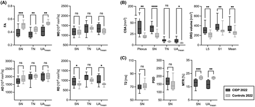
In 2022, FA was significantly decreased for all analyzed nerves when comparing CIDP patients with healthy controls (p < 0.01) (Fig. 1A). Furthermore, RD of CIDP patients was significantly increased for the sciatic nerve (p = 0.043) and the upper arm nerves UAmean (p = 0.033), compared to healthy controls, whereas no significant changes were detected for AD and MD. These results were relatively similar to the initial MRN of 2016.32

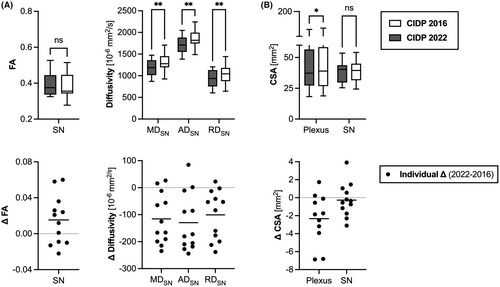
Regarding CIDP patients over the six-year timespan (Fig. 2A), mean FA values did not change significantly on a group comparison level (p value range: 0.077–0.22), whereas RD (p = 0.003) and AD (p = 0.002) decreased for the sciatic nerve and MD decreased for all observed nerves (p value range: 0.001–0.04).

Nerve cross-sectional area and DRG volume
In 2022, nerve hypertrophy was observed in 9 of 12 CIDP patients (75%) compared to healthy controls in at least one location, which we defined as an increase greater than mean + 2 standard deviations compared with the control group. Overall CSA values were significantly increased for the lumbosacral plexus (p < 0.001), the sciatic nerve (p < 0.001), and the upper arm nerves UAmean (p = 0.02) in CIDP patients compared with healthy controls, which was similar to 20165 (Figs. 1B and 3). Furthermore, DRG volumes were significantly increased in CIDP patients both for L5 (p = 0.002) and S1 (p = 0.005) as well as overall (mean L5 + S1) (p = 0.003) when compared with healthy controls.

Regarding the longitudinal course of CSA values in CIDP patients from 2016 to 2022 (Fig. 2B), mean CSA values significantly decreased for the lumbosacral plexus (p = 0.015), the tibial nerve at lower leg level (p = 0.027) and the upper arm nerves UAmean (p = 0.032).
Nerve T2-weighted signal, T2 relaxometry, and magnetization transfer imaging
In 2022, mean normalized T2-weighted signal (nT2) was significantly increased in CIDP patients both for the lumbosacral plexus (p = 0.035) and the sciatic nerve (p = 0.008). Similar to 2016, this was due to an increase in PSD, although currently the increase was not statistically significant (p = 0.158) (Fig. 1C). Over time, sciatic nerve PSD significantly decreased (p = 0.002) together with nT2 (p = 0.034). The T2 relaxation time for the sciatic nerve was not significantly different between CIDP patients and healthy controls (Fig. 1C) and did not show any significant changes in the observed course.
MTI, which had been added as a new contrast in 2022, revealed a significant decrease in mean MTR for the sciatic nerve (p < 0.001) and the upper arm nerves UAmean (p = 0.002) in CIDP patients compared with healthy controls (Fig. 1C).
Correlation of current MRN parameters with demographical, clinical, and EDX data
We found no significant correlations between 2022 MRN parameters and 2022 clinical scores (INCAT/ODSS, MRCSS), disease duration, or patient age. Nerve MTR inversely correlated with BMI (rs = −0.619, p = 0.043).
In contrast, strong linear correlations were observed between DTI of the sciatic nerve and EDX results (Table 2, Table S3): decreased FA was associated with decreased tibial NCV (rs = 0.803, p = 0.003), decreased compound muscle action potential (CMAP) (rp = 0.646, p = 0.023), and with increased F wave latency (rs = −0.711, p = 0.013). RD correlated inversely with tibial NCV (rs = −0.606, p = 0.041). Furthermore, decreasing MTR values were associated with decreasing tibial NCV (rs = 0.679, p = 0.027) and increasing tibial F wave latency (rs = −0.62, p = 0.048). In the upper arm, mean FA of all three nerves positively correlated with median nerve NCV (rp = 0.802, p = 0.03). Increased RD values were associated with decreased median nerve NCV (rp = −0.859, p = 0.013) and increased F wave latency (rp = 0.812, p = 0.026) (Table S4).
| CSA [mm2] Lumbosacral plexus | CSA [mm2] Sciatic nerve | FA Sciatic nerve | MTR [%] Sciatic nerve | |||||
|---|---|---|---|---|---|---|---|---|
| r | p | r | p | r | p | r | p | |
| Age at MRI [years] | −0.231s | 0.467 | −0.117p | 0.717 | 0.088 p | 0.785 | −0.21p | 0.536 |
| BMI [kg/m2] | 0.266s | 0.4 | −0.332 p | 0.291 | −0.223p | 0.486 | −0.619p | 0.043 |
| Years since diagnosis | 0.191s | 0.55 | −0.046s | 0.89 | 0.003s | 0.996 | −0.345s | 0.298 |
| Tibial NCV [m/s] | −0.718 s | 0.011 | −0.648 s | 0.027 | 0.803 s | 0.003 | 0.679s | 0.027 |
| Tibial CMAP [mV] | −0.617s | 0.037 | −0.608 p | 0.036 | 0.646 p | 0.023 | 0.469p | 0.145 |
| Tibial F wave latency [ms] | 0.551 s | 0.068 | 0.384 s | 0.218 | −0.711 s | 0.013 | −0.62s | 0.048 |
| INCAT/ODSS | −0.179s | 0.582 | −0.163s | 0.62 | 0.004s | 0.995 | −0.179s | 0.602 |
| MRCSS | −0.271s | 0.392 | 0.074s | 0.821 | 0.334s | 0.287 | 0.457s | 0.159 |
| Δ CSA [mm2] Lumbosacral plexus | Δ CSA [mm2] Sciatic nerve | Δ FA Sciatic nerve | Δ MTR [%] Sciatic nerve | |||||
|---|---|---|---|---|---|---|---|---|
| Δ Tibial NCV [m/s] | 0.118s | 0.735 | −0.105s | 0.749 | 0.594s | 0.046 | n.a. | n.a. |
| Δ Tibial CMAP [mV] | −0.064s | 0.854 | −0.018s | 0.961 | 0.669s | 0.02 | n.a. | n.a. |
| Δ Tibial F wave latency [ms] | −0.095s | 0.84 | 0.234s | 0.54 | −0.803 s | 0.012 | n.a. | n.a. |
| Δ INCAT/ODSS | 0.785s | 0.006 | 0.759s | 0.006 | 0.337s | 0.282 | n.a. | n.a. |
| Δ MRCSS | −0.33s | 0.323 | −0.416s | 0.182 | −0.416s | 0.182 | n.a. | n.a. |
- Bold text indicates statistically significant results (p < 0.05).
- CMAP, compound motor action potential; CSA, cross-sectional area; FA, fractional anisotropy; INCAT, inflammatory neuropathy cause and treatment; MRCSS, Medical Research Council sum score; MTR, magnetization transfer ratio; NCV, nerve conduction velocity; ODSS, Overall Disability Sum Score; p, Pearson correlation coefficient; s, Spearman correlation coefficient.
Furthermore, an increase in nerve caliber was associated with decreasing tibial NCV and CMAP, both for the lumbosacral plexus (NCV: rs = −0.718, p = 0.011; CMAP: rs = −0.617, p = 0.037) and the sciatic nerve (NCV: rs = −0.648, p = 0.027; CMAP: rs = −0.608, p = 0.036) (Table 2). In line with this, increased mean DRG volume inversely correlated with tibial NCV (rs = −0.669, p = 0.021), CMAP (rs = −0.727, p = 0.01) and positively correlated with tibial F wave latency (rs = 0.638, p = 0.03) (Table S3).
Longitudinal correlation of MRN parameters with clinical and EDX data
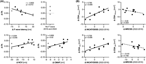
Over the 6-year timespan, changes in EDX data showed significant linear correlations with Δ FA of the sciatic nerve (Table 2, Fig. 4A). In this regard, an increase in FA was associated with an increase in tibial NCV (rs = 0.594, p = 0.046) and CMAP (rs = 0.669, p = 0.02) and a decrease in tibial F wave latency (rs = −0.803, p = 0.012).

Furthermore, Δ INCAT/ODSS showed a positive correlation with Δ CSA both for the lumbosacral plexus (rs = 0.785, p = 0.006) and the sciatic nerve (rs = 0.759, p = 0.006), suggesting that a decrease in nerve caliber was associated with clinical improvement (Table 2, Fig. 4B). Weaker negative correlations were observed for Δ MRCSS and Δ CSA, indicating a similar clinical trend.
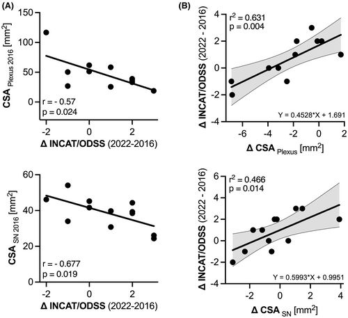
In addition, we observed that nerve hypertrophy in the initial study (CSAPlexus/SN2016) was associated with a rather decreasing INCAT/ODSS over time, suggesting that patients with initially enlarged nerves may potentially respond to therapy or experience a comparably benign course (Plexus: rs = −0.57, p = 0.024; SN: rs = −0.677, p = 0.019) (Fig. 5A). Additional simple linear regression analysis demonstrated that the relationship between Δ CSA and Δ INCAT/ODSS accounts for approximately 47–63% of the variation of the data (sciatic nerve: r2 = 0.466; plexus: r2 = 0.631) (Fig. 5B).

Changes in the remaining scalar DTI parameters and T2 relaxometry-derived parameters of the sciatic nerve neither showed any significant correlations with changes in EDX data nor with changes in clinical scores (Table S3).
In the upper extremity, correlations between changes in mean FA and changes EDX data were less pronounced and just missed significance (p value range: 0.06–0.669) (Table S4).
Discussion
In this longitudinal imaging study on CIDP, we investigated the utility of various MRN biomarkers with regard to clinical and electrophysiological parameters over a time course of 6 years. Our major findings were that changes of the DTI parameter FA over this timespan notably correlated with changes in electrophysiological parameters depicting demyelination (NCV) and axonal damage (CMAP), while alterations in nerve caliber significantly correlated with clinical changes as reflected by the INCAT/ODSS. Moreover, initial nerve hypertrophy was associated with a comparably milder or beneficial disease course over 6 years.
Previous cross-sectional MRN studies revealed proximal nerve enlargement and increased nerve T2 signal in CIDP.5 Simultaneously, DTI has emerged as a promising tool to deliver biomarkers of tissue integrity in the peripheral nervous system (PNS) by exploiting water diffusion characteristics along nerve fiber tracts.44, 45 Specifically, the main DTI parameter FA, which is a dimensionless parameter between 0 (isotropic diffusion, e.g., fluids) and 1 (anisotropic diffusion, e.g., fiber bundles), has been shown to be reduced in studies on various neuropathies, including CIDP.32, 46, 47 Cross-sectionally, both CSA and FA are known to correlate well with EDX data, and the observed changes are consistent with demyelination.5, 32 However, little is known about the development of these MR biomarkers over time and/or under therapy, as longitudinal MRN studies are overall scarce. So far, only one study performed a 6-month imaging follow-up in a cohort of 11 CIDP patients and found an unchanged decrease in sciatic nerve FA.33
In this study, we did not observe a correlation between nerve CSA and clinical scores when regarding a single timepoint, aligning with prior HRUS studies.19, 20, 48 Yet, longitudinal changes in nerve CSA significantly correlated with longitudinal changes in the ODSS, revealing a decrease in nerve caliber with clinical improvement. This MRN finding is in line with previous ultrasound studies, which reported a decrease in nerve caliber with clinical improvement or vice versa.14, 49-51
In our study, INCAT/ODSS improvement was more pronounced in patients who initially presented with enlarged nerves, as indicated by a significant negative correlation between baseline nerve CSA in 2016 and Δ INCAT/ODSS (Fig. 5A). This aligns with Härtig et al., who observed a better therapeutic response in patients with hypoechoic nerve enlargements detected by HRUS over 12 months.19 Furthermore, Fisse et al. found that CIDP patients with hypoechoic nerves tended to remain clinically stable or improve over time, which possibly presents a similar phenotype due to nerve edema, although echogenicity alone was not regarded a suitable follow-up tool.21 While the diagnostic and possibly prognostic value of HRUS in CIDP has been well demonstrated,14-18 this raises the question to what extent multiparametric MRN can add additional value to monitor disease activity.
Comparative studies have highlighted the complementary roles of HRUS and MRN in assessing peripheral neuropathies, with recent studies indicating higher sensitivity for MRN.52, 53 However, HRUS is a more cost-effective and readily available option, thus making it an excellent screening tool with high spatial resolution.54 A recent study on the diagnostic performance of HRUS and MRN in immune-mediated neuropathies pointed out, that brachial plexus sonography could complement MRI,55 while a recent study identified HRUS as the most valuable imaging tool to distinguish immune-mediated neuropathies from mimics through HRUS-CSA measurements.56 Although HRUS allows for a reliable CSA estimation of peripheral nerves in the upper and lower extremities, a study comparing HRUS and MRN revealed a bias for larger nerves, while MRN more frequently yielded higher CSA values compared to HRUS.57 This suggests caution when directly comparing CSA measurements between these modalities, although fair correlations were observed. Moreover, the lumbosacral plexus is only accessible for imaging via MRN.
Although the overall changes in nerve caliber were relatively small, they were more pronounced in the lumbosacral plexus (range Δ CSA: −6.9 to 1.73 mm2) than in the sciatic nerve (range Δ CSA: −3.11 to 3.9 mm2), suggesting that the caliber changes may be more pronounced proximally. Based on a linear regression analysis, a CSA decrease of 1 mm2 was corresponding to a decrease in INCAT/ODSS (indicating clinical improvement) of −0.45 (lumbosacral plexus) or −0.6 (sciatic nerve) in our study, while an unchanged nerve caliber was associated with an increase in INCAT/ODSS (indicating clinical progression) (+1.6 for the lumbosacral plexus, +1 for the sciatic nerve) (Fig. 5B). The correlations between initial nerve hypertrophy and clinical improvement (Fig. 5A) as well as between nerve caliber change and clinical improvement (Fig. 5B) might imply that patients with initial nerve hypertrophy could be more responsive to treatment, given their capability to exhibit a reduction in nerve CSA under treatment.
Another major strength of MRN is that it offers a combination of morphological and functional nerve information. Of all previously assessed quantitative MRN parameters, FA seems to be the most reliable MR biomarker in the long-term follow-up of CIDP. The persisting robust correlations of sciatic nerve FA (tibial portion) and tibial EDX data not only underline our previous findings,32 but also indicate that FA may serve as a useful longitudinal biomarker of nerve integrity. Notably, not only the absolute values of these parameters, but also their respective changes showed significant correlations. Interestingly, individual changes in FA values were smaller than expected and ranged between −0.022 and 0.06, which does not substantially exceed the range of a possible measurement error, as described in a previous reliability study.40 While FA appears to accurately depict chronic nerve injury, we noticed a tendency toward normalization in nerve PSD values. In our initial investigation, PSD values were significantly higher in CIDP patients.5 Given that an elevation in PSD is typically associated with increased water content, we hypothesize that PSD is better suited to reflect rather acute changes during inflammation, such as nerve edema.
Besides DTI, we introduced MTI and its deriving parameter MTR as a possible new biomarker in CIDP. This technique derives its information from two different pools of protons and their interaction: restricted protons bound to macromolecules (e.g., myelin) and protons in free water.58 Originally applied in the CNS, MTR has been described as a sensitive marker for demyelination in multiple sclerosis (MS),59 which has been confirmed in a MRI-pathologic correlation study.60 More recently, MTI has been applied to the PNS in patients with relapsing–remitting MS and revealed decreased MTR values in the sciatic nerve compared to healthy controls.61 Our results add to this by revealing significantly decreased MTR values in upper and lower extremity nerves of CIDP patients, with significant correlations between sciatic nerve MTR and EDX markers of demyelination (F wave latency, NCV). In concordance, decreased MTR values were more pronounced in patients with proximal nerve hypertrophy, and thus severe demyelination (rs = −0.7; Figure S1). A previously observed negative correlation between sciatic nerve MTR and BMI was confirmed in our study.62 Notably, BMI did not differ between CIDP patients and controls (p > 0.05).
Lastly, we also demonstrated that CIDP is associated with an increase in DRG volume on top of proximal nerve enlargement. Several studies on inherited and metabolic neuropathies have identified changes in DRG morphology and perfusion and hence these parameters are regarded as promising biomarkers.63-65 To our knowledge, increased DRG volume has not yet been described in CIDP but seems plausible considering that incoming and outgoing nerve fibers are part of the DRGs' nerve fiber rich area.
Our study has limitations, most importantly due to the small cohort of CIDP patients analyzed. This was a result of the nature of the study, as one-third of the initial patient group did not consent to or was not able to re-undergo an extensive second MRI examination. This circumstance unavoidably leads to potentially low statistical power in some of our analyses. Notably, this limitation becomes more pronounced when considering the upper limb data, as imaging of the upper extremity nerves turned out to be challenging for some patients, resulting in a further reduction in this subcohort size. Therefore, our conclusions primarily focus on the lumbosacral plexus and sciatic nerve data. Nevertheless, we believe that longitudinal data derived from a small cohort of 12 patients still possesses significant value, as discussions about MR biomarkers often prioritize understanding of individual patient perspectives. Furthermore, our data largely relates to treated CIDP patients, which might rather lead to an underestimation of the true effect when comparing MRN results to treatment-naive patients. Further longitudinal studies including treatment-naive patients are therefore desirable. Also, our observations were made under treatment with intravenous immunoglobulin (IVIg) and may not directly apply to patients treated with corticosteroids or plasma exchange. Given the explorative nature of our study, we did not adjust for multiple comparisons. While such adjustments help control Type I and family-wise error rates, in this exploratory phase, they might overly constrain the exploration of potential relationships of patterns within the data.
In sum, our study identified nerve CSA and FA as the most valuable longitudinal MR biomarkers in CIDP due to robust correlations with clinical and electrophysiological outcome measures. This could be particularly advantageous in cases where severe demyelination results in non-excitable nerve segments and for the evaluation of nerve segments not accessible with nerve ultrasound. Physicians conducting MRN follow-up examinations of CIDP patients in a clinical setting should be aware that nerve hypertrophy and increased T2-weighted signal can persist over extended periods and detecting subtle changes in these features require precise measurements.
Initially increased nerve CSA might predict a rather benign course or better therapy response and should be further investigated as a dedicated prognostic parameter in future longitudinal studies. Moreover, the addition of a DTI sequence into the clinical imaging protocol, and especially the assessment of nerve FA, can be recommended while the remaining DTI scalars and parameters derived from T2 relaxometry analysis appear to offer limited additional information in a longitudinal clinical setting.
Acknowledgments
This study was supported by a research grant from the GBS|CIDP Foundation International (FP and MK). FP was supported by a Physician-Scientist Fellowship of the Medical Faculty of the University of Heidelberg. SH (SFB 1118), DS (SFB 1118), and MB (SFB 1158) were supported by the German Research Foundation. For the publication fee we acknowledge financial support by the German Research Foundation within the funding programme “Open Access Publikationskosten” as well as by Heidelberg University. Open Access funding enabled and organized by Projekt DEAL.
Author Contributions
FP, KP and MK designed and coordinated the study. FP collected and analyzed the MR data. KP conducted the electrophysiological and clinical examinations. All authors participated in analysis and interpretation of data. FP and MK wrote the manuscript with relevant input from all co-authors.
Conflict of Interest
The authors declare that they have no relevant financial or nonfinancial interests to disclose.


