CAR T cells targeting αvβ3 integrin are effective against advanced cancer in preclinical models
Abstract
Integrins are heterodimeric receptors that convey cell-to-cell and cell-to-matrix interactions. Integrin αvβ3 is expressed in several tumor entities including melanoma, glioblastoma, breast, pancreatic, and prostate cancer, where it promotes tumor cell survival and metastasis. Here, we generated αvβ3-specific chimeric antigen receptor (CAR) T cells and analyzed their antitumor function in preclinical models in vitro and in vivo. αvβ3-CARs comprising a super-humanized hLM609 targeting domain with either high or low affinity (hLM609v7, Kd = 3 nM vs hLM609v11, Kd = 160 nM) and equipped with either a long or a short IgG4-Fc extracellular spacer (229 vs 12 amino acids) were expressed in CD8+ and CD4+ T cells through lentiviral transduction. αvβ3-CAR T cells eliminated αvβ3-positive tumor cells rapidly and specifically, produced IFN-γ and IL-2 (CD4+ > CD8+) and exhibited productive proliferation. In vitro, we observed the strongest reactivity with the higher affinity hLM609v7 αvβ3-CAR in the short spacer configuration, consistent with the tumor membrane-distal localization of the hLM609 epitope. In a murine xenograft model of metastatic A-375 melanoma, the strongest antitumor effect was mediated by the lower affinity hLM609v11 αvβ3-CAR. Notably, a single administration of hLM609v11 αvβ3-CAR T cells was able to induce complete elimination of melanoma lesions, leading to long-term tumor-free survival. These data establish αvβ3 integrin as a novel target for CAR T-cell immunotherapy and affirm our previous notion that binding domain affinity and spacer length can be calibrated to augment CAR reactivity. αvβ3-CAR T cells have therapeutic potential in several prevalent solid tumors, including melanoma and triple-negative breast cancer.
1 INTRODUCTION
Integrins are heterodimeric cell surface receptors expressed by all nucleated cells and involved in adhesion and signaling processes between cells and their microenvironment. They are formed by noncovalent association of one α- and one β-subunit.1 Upon binding of their respective ligands, integrins recruit large signaling complexes to their cytoplasmic domain and transmit information about the cell's functional state to the microenvironment by modulating the binding affinity to their ligands through conformational changes.2 Because integrins influence tumor cell migration, invasion, proliferation, and survival, they are being pursued as molecular and immunologic targets in cancer therapy.3 Integrin αvβ3, also known as vitronectin receptor, is one of the best-studied integrins in cancer research. It primarily interacts with ligand proteins containing an RGD-motif and can increase tumor cell survival.4, 5 Several mechanisms have been identified that link expression of integrin αvβ3 on tumor cells to increased metastatic spread.6, 7 First, the activated form of integrin αvβ3 enables binding to platelets, which protects tumor cells from shear stress and enhances their adhesion to blood vessels.8, 9 Second, recruitment and activation of the tyrosine kinase c-Src by the cytoplasmic tail of the β3 subunit of integrin αvβ3 promotes anchorage-independent cell survival.10 Furthermore, integrin αvβ3 promotes the epithelial-mesenchymal transition of tumor cells by direct interaction with the TGF-β receptor II and binding of TGF-β. Exposure to TGF-β also leads to increased integrin αvβ3 expression.11, 12 Expression of αvβ3 integrin has been demonstrated in several tumor entities such as melanoma and glioblastoma, as well as breast, pancreatic, and prostate cancer.7, 13-16 In melanoma, transition from benign radial growth to malignant vertical growth correlates with de novo expression of αvβ3 integrin.17 In addition to tumor cells, integrin αvβ3 is also expressed on cells that are essential components of the tumor environment including cancer-associated fibroblasts (CAFs), tumor-associated macrophages, and angiogenic endothelial cells.18-20
There have been prior attempts of exploiting αvβ3 integrin as a therapeutic target. This includes immunotherapy with monoclonal antibodies (mABs) that inhibit ligand binding to αvβ3 integrin, which has been reported to be safe but only of limited efficacy.21 Here, we report on the development of αvβ3-specific chimeric antigen receptor (CAR) T cells and their antitumor function in preclinical models. CARs are synthetic receptors that most commonly employ the variable heavy (VH) and variable light (VL) chains of a mAB for antigen targeting. To construct αvβ3 integrin-specific CARs, we utilized the VH and VL chains of a super-humanized mAB LM609 (hLM609), which we developed in previous work.22 We have recently demonstrated that binding domain affinity and extracellular spacer domain design affect tumor cell recognition and CAR T-cell function,23, 24 and are modulating both variables to derive an αvβ3-CAR with optimal antitumor reactivity. Through these iterations, we have obtained an αvβ3-CAR that confers potent reactivity against αvβ3-expressing hematologic and nonhematologic tumor cells in vitro and eliminates metastatic melanoma in a murine xenograft model in vivo.
2 MATERIALS AND METHODS
2.1 Construction of αvβ3-CAR encoding lentiviral vectors
Two codon optimized single-chain variable fragments (scFvs) were synthesized (GeneArt, ThermoFisher Scientific, Regensburg, Germany) based on mABs hLM609v7 and hLM609v1122 with a (G4S)3 linker to fuse VH and VL. The scFvs were cloned into epHIV7 lentiviral vectors to generate constructs with either a long (IgG4Fc Hinge-CH2-CH3, 229 amino acids) or short (IgG4Fc Hinge only, 12 amino acids) extracellular spacer domain, linked to the CD28 transmembrane domain, and the cytoplasmic signaling domains of CD28 and CD3ζ.24 A truncated epidermal growth factor receptor was expressed in cis with the CAR transgene, separated by a T2A element (EGFRt).25
2.2 Generation of αvβ3-CAR T-cell lines
CD8+ and CD4+ T cells were isolated by magnetic cell separation (Miltenyi, Bergisch Gladbach, Germany) from PBMCs of healthy donors and activated with anti-CD3/CD28 microbeads (ThermoFisher). T cells were lentivirally transduced 1 day after activation as described previously.23 The anti-CD3/CD28 microbeads were removed 4 to 7 days after activation. At 10 to 14 days after activation, EGFRt+ cells were enriched by magnetic cell separation using the in-house biotinylated (ThermoFisher) mAB Cetuximab (Bristol-Myers Squibb, New York, NY) and antibiotin microbeads (Miltenyi) and expanded using a rapid expansion protocol.26
2.3 In vivo experiments in xenograft mouse model
Six- to 8-week old female NOD.Cg-Prkdcscid Il2rgtm1WjI/SzJ (NSG) mice were obtained from Charles River (Sulzfeld, Germany). On day 0, 1 × 10⁶ A-375/ffluc_GFP cells were injected into the tail vein. On day 7, engraftment of the tumor cells was verified by bioluminescence imaging (BLI). Mice were then randomly assigned to treatment cohorts. Groups of mice (n = 3-6) were treated with 2.5 × 10⁶ CD8+ and 2.5 × 10⁶ CD4+ T cells, either unmodified or expressing the hLM609v7/short or hLM609v11/short αvβ3-CAR, by tail vein injection or remained untreated (n = 2). For weekly bioluminescence imaging, (IVIS Lumina; PerkinElmer, Waltham, MA) the mice received 0.3 mg/g body weight D-luciferin i.p. (Biosynth, Staad, Switzerland). Living Image Software (PerkinElmer) was used for data analysis. The experimental endpoint was reached, when mice presented with either >20% weight loss or when radiance obtained by BLI exceeded 3 × 10⁷ p/s/cm²/sr. Cytokine concentrations in mouse serum were measured using the MAGPIX System (ThermoFisher) and a Human Cytokine Magnetic 10-Plex kit (ThermoFisher). All mouse experiments were approved by the Institutional Animal Care and Use Committee of the University of Würzburg.
3 RESULTS
3.1 αvβ3 integrin is expressed on hematologic and nonhematologic tumor cell lines
We analyzed expression of αvβ3 integrin by flow cytometry on a panel of hematologic and nonhematologic tumor cell lines using a mAb directed against the αvβ3 integrin complex. We detected high-level αvβ3 integrin surface expression on the melanoma cell lines A-375, M14, Malme-3M, UACC-62 and UACC-257, and the triple-negative breast cancer cell line MDA-MB-231 (xMFI = MFI mAb/MFI isotype: 7.3-19.5); and intermediate-level expression on the T-cell acute lymphoblastic leukemia cell line Jurkat (xMFI: 3.6) (Figure 1A). The chronic myelogenous leukemia cell line K562 and the mantle cell lymphoma cell line JeKo-1, as well as the lung carcinoma cell line A-549 and the melanoma cell line LOX-IMVI showed only a minor increase in MFI compared to isotype control (xMFI: <2). To determine if this was due to low-level expression of αvβ3 integrin complex or rather unspecific binding, we analyzed expression of the αv and β3 subunits on these cell lines individually (Figure 1B, S1). We detected high-level expression of αv on LOX-IMVI, A-549, and K562, but not on the surface of JeKo-1 cells. The β3 subunit was not expressed on LOX-IMVI and A-549 cells and only detectable at extremely low levels on K562 and JeKo-1 cells. For comparison, on K562 cells that we transduced with a lentiviral vector encoding the β3 integrin subunit (K562_β3), we detected uniform, high-level expression of αvβ3 (xMFI: 13.3) (Figure 1A).
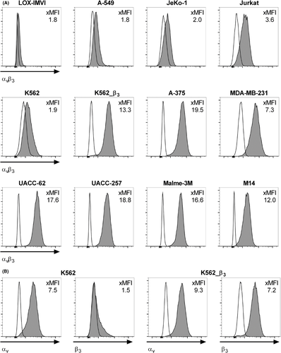
We also analyzed normal T cells, B cells, and monocytes from peripheral blood, and mobilized hematopoietic stem cells (HSCs) and did not detect relevant expression of αvβ3 integrin complex (xMFI: <2) (Figure S2, S3A). Taken together, these data affirm the prior notion, that integrin αvβ3 is expressed on malignant cells in hematologic and nonhematologic cancers, including prevalent high-level expression in melanoma cell lines. We selected the K562 and A-549 (negative); A-375, UACC-62, UACC-257 and MDA-MB-231 (positive), and K562_β3 cell lines (positive control) as target cells to analyze the specificity and antitumor potency of T cells that we gene-engineered to express an αvβ3-specific CAR.
3.2 Expression of αvβ3-CARs in human T cells
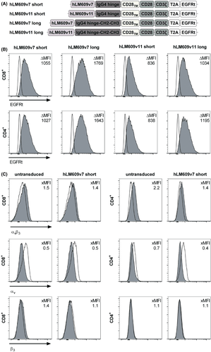
We designed a set of four αvβ3-CAR constructs comprising: i. a scFv targeting domain derived from a super-humanized variant of anti-αvβ3 integrin-specific mAb LM609 with either high (hLM609v7; Kd = 3 nmol/L) or low (hLM609v11; Kd = 160 nmol/L) monovalent affinity22; ii. an IgG4-Fc derived spacer of either Hinge only (short spacer, 12 amino acids) or of Hinge-CH2-CH3 (long spacer, 229 amino acids).23 Each of the constructs contained a CD28_CD3zζ signaling domain and was coexpressed with a truncated epidermal growth factor receptor (EGFRt) as transduction marker (Figure 2A).25 We transduced each of the constructs into CD8+ and CD4+ T cells and enriched CAR+ T cells using the EGFRt marker (Figure 2B). At the end of the manufacturing process and prior to functional testing, each of the CD8+ and CD4+ αvβ3-CAR T-cell lines was negative for αvβ3 integrin–expression by flow cytometry (Figure 2C).
3.3 αvβ3-CAR T cells elicit potent antitumor functions in vitro
We sought to determine the antitumor function of T cells expressing our αvβ3-CAR constructs. First, we analyzed the cytolytic activity of CD8+ αvβ3-CAR T cells and found that each of the four CAR constructs conferred specific recognition and elimination of A-375, UACC-62, UACC-257, MDA-MB-231, and K562_β3 target cells (Figure 3A). Overall, the four αvβ3-CAR constructs with high versus low affinity and short versus long spacer were similarly potent and not significantly different in their ability to induce cytolysis of target cells. K562_β3 cells were more susceptible to cytolysis compared to the melanoma cell lines and MDA-MB-231, especially at low effector-to-target cell ratios. We detected low-level recognition of native K562 cells, potentially due to very low-level expression of αvβ3 integrin complex, as suggested in our expression analysis. Accordingly, we did not detect cytolysis of A-549 cells (Figure 3A). We also evaluated recognition of normal HSCs, and consistent with their αvβ3-negative phenotype, viability was not affected by coculture with αvβ3-CAR T cells (Figure S3B).
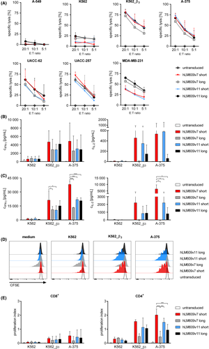
We confirmed that CD8+ and CD4+ αvβ3-CAR T cells produced IFN-ɣ and IL-2 after stimulation with αvβ3-positive tumor cells (Figure 3B,C). The αvβ3-CAR with high affinity and short spacer domain (hLM609v7/short) induced the strongest IFN-ɣ production, particularly in CD4+ T cells (Figure 3C). The αvβ3-CARs with short spacer domain induced more IL-2 production compared to the constructs with long spacer. In CD4+ T cells, the hLM609v7/short αvβ3-CAR induced the strongest production of IL-2 (Figure 3B,C). Overall, we observed stronger cytokine production by CD4+ compared to CD8+ CAR T cells. We also evaluated the proliferation of αvβ3-CAR T cells after stimulation with αvβ3-positive tumor cells (Figure 3D,E). Again, there was stronger proliferation of CD4+ compared to CD8+ CAR T cells, and the hLM609v7/short αvβ3-CAR was superior in inducing proliferation compared to the other receptor designs. Notably, there was some background proliferation of T cells equipped with the hLM609v7/long αvβ3-CAR, potentially due to tonic signaling.
In summary, the data show that αvβ3-CAR T cells confer specific and potent antitumor functions against αvβ3-expressing target cells in vitro. The data also show that the αvβ3-CAR construct with higher affinity and short spacer domain conferred the strongest effector functions, particularly cytokine secretion and proliferation, in line with our prior observation that these parameters affects the antitumor function of CAR T cells.23 On the basis of our analysis, we selected the αvβ3-CAR constructs with short spacer domain for evaluation in an in vivo model.
3.4 αvβ3-CAR T cells are effective against metastatic melanoma in vivo
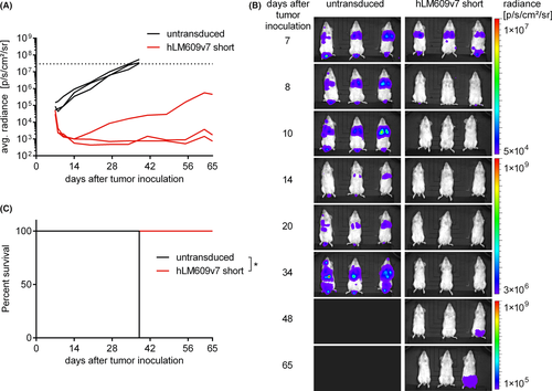
We established xenografts of metastatic melanoma in immunodeficient NSG mice using the aggressive A-375 cell line. We injected NSG mice with 1 × 10⁶ firefly luciferase-labeled A-375 cells i.v. on day 0 and treated them with a single dose of 5 × 10⁶ T cells i.v. after 7 days (CD8:CD4 ratio = 1:1). Mice were either treated with αvβ3-CAR T cells (hLM609v7/short) or untransduced control T cells derived from the same donor. We performed serial bioluminescence imaging and observed rapid tumor regression in all mice treated with αvβ3-CAR T cells (n = 3) and rapid tumor progression in all control mice (n = 3) (Figure 4A,B). All CAR T-cell-treated mice were alive until the end of the observation period on day 65 (Figure 4C); only one mouse had a slowly progressing, singular tumor lesion in the lower back (Figure 4A,B).
In the next set of experiments, we included a group of NSG/A-375 mice that received T cells expressing the hLM609v11/short αvβ3-CAR to compare the higher and lower affinity αvβ3-CAR constructs. On day 3 and day 7 after adoptive T-cell transfer, we analyzed peripheral blood samples and confirmed T-cell engraftment in all treatment groups (Figure 5A). Between day 3 and day 7, the percentage of αvβ3-CAR T cells increased, in particular in the group of mice that had received the lower affinity hLM609v11/short αvβ3-CAR. In serum samples, we measured elevated concentrations of IFN-ɣ and GM-CSF in mice that received αvβ3-CAR T cells, but not in mice that had received control T cells (Figure 5B). Interestingly, also the cytokine concentrations were higher in mice that had been treated with the lower affinity hLM609v11/short compared to the higher affinity hLM609v7/short αvβ3-CAR. Treatment with αvβ3-CAR T cells conferred a potent antitumor effect. On day 3 after T-cell transfer (ie, day 10 after tumor inoculation), we already detected lower levels of bioluminescence signal in all of the mice that had received CAR T cells, whereas there was increasing bioluminescence signal in all mice in the control groups that had either received untransduced T cells or no treatment (Figure 5C). By day 28, the bioluminescence signal had reached background levels in 4 of 6 mice in the hLM609v7/short, and 5 of 6 mice in the hLM609v11/short αvβ3-CAR T-cell groups, respectively, indicating that all tumor lesions had been eradicated (Figure 5C,D). Out of the 12 mice treated with αvβ3-CAR T cells, 3 did not reach the background bioluminescence signal and had a recurrent tumor at the same site as one of the first lesions. Three of the six mice in the hLM609v7/short treatment group were lost to follow-up for reasons unrelated to tumor progression. Kaplan-Meier analysis showed that by day 44, all of the mice in the control groups had to be sacrificed due to tumor progression (Figure 5E). In contrast, all of the mice that completed follow-up were alive in the hLM609v7 αvβ3-CAR (3 out of 3), and all of the mice were alive in the hLM609v11 αvβ3-CAR cohort (6 out of 6). Bone marrow analysis at the end of the experiment confirmed that CAR T cells and untransduced T cells had persisted for the entire duration of the experiment (Figure 5F).
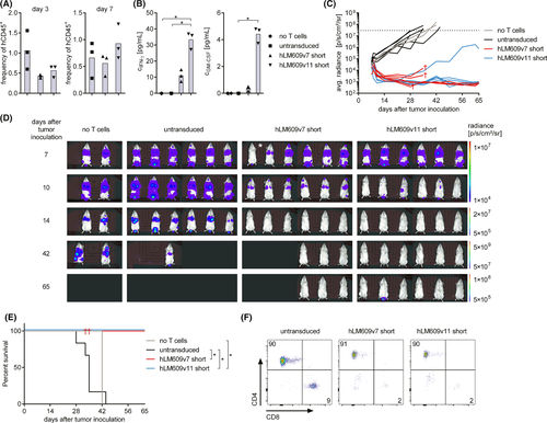
Taken together, these data show that αvβ3-CAR T cells confer potent antitumor efficacy in vivo. Several lines of evidence, including stronger CAR T-cell expansion and cytokine secretion, and a higher rate of complete responses indicate that αvβ3-CAR T cells with the lower affinity binding domain hLM609v11 conferred stronger antitumor reactivity than αvβ3-CAR T cells with the higher affinity hLM609v7 binding domain in vivo, which reverses the hierarchy established in our in vitro analysis.
4 CONCLUSIONS
Adoptive immunotherapy with gene-engineered CAR T cells has curative potential against advanced hematologic malignancies.27 At present, significant efforts are being invested to extend the clinical success that has been obtained with CD19-specific CAR T cells in B-cell leukemia and lymphoma, to prevalent nonhematologic tumors, which requires the identification and validation of novel target antigens. Here, we demonstrate that CAR T cells specific for αvβ3 integrin exhibit potent antitumor reactivity, including effective tumor cell lysis, as well as cytokine production and proliferation after stimulation with αvβ3-expressing cancer cells in vitro. Furthermore, we demonstrate that αvβ3-CAR T cells are capable of completely eradicating established A-375 melanoma in a xenograft model in mice.
Our data show that αvβ3-CAR constructs with hLM609-derived binding domains confer superior antitumor function when equipped with a short IgG-Fc Hinge rather than a long IgG-Fc Hinge-CH2-CH3 spacer domain. This is consistent with our previous observation with CD19- and ROR1-specific CARs, showing that the length of the extracellular spacer domain affects tumor cell recognition and CAR T-cell function, likely because of the spatial interaction of CAR and tumor antigen, and the localization of the targeted epitope.23, 24 The paradigm that emerged from our previous studies is that the “long” spacer domain is optimal for targeting epitopes that are proximal to the tumor cell membrane, whereas a “short” spacer domain is optimal for targeting membrane-distal epitopes. The mAB LM609 recognizes a membrane-distal epitope of integrin αvβ328 and accordingly, our data that αvβ3-CAR constructs with “short” spacer domain confer the strongest antitumor functions, are in line with this paradigm.
We show that αvβ3-CAR T cells are able to eradicate metastatic A-375 melanoma in an NSG mouse xenograft model. The A-375 melanoma model is commonly used to evaluate novel anticancer agents and notably, the efficacy that we observed with αvβ3-CAR T cells in our study is superior to the efficacy that has recently been reported for several alternative investigational treatments, including a combination treatment of oncolytic adenovirus and dacarbazine chemotherapy, and itraconazole, that both achieved tumor reduction but not tumor clearance like in our study.29, 30
Interestingly, we observed stronger CAR T-cell expansion and cytokine secretion in mice that had been treated with the lower affinity hLM609v11 αvβ3-CAR, indicating the lower affinity receptor had induced stronger T-cell activation in vivo. In contrast, in our in vitro analysis, the higher affinity hLM609v7 αvβ3-CAR had induced stronger cytokine secretion and proliferation. Other investigators have reported similar findings with CAR T cells targeting ErbB2.31 A potential explanation is that the lower affinity hLM609v11 binding domain has a three-times faster off-rate than the higher affinity variant hLM609v7 (16 × 10−4 s−1 vs 5.4 × 10⁻⁴ s−1 measured for the corresponding monovalent Fab) and therefore, hLM609v11 CAR T cells are more likely to sequentially interact with tumor cells and thus, may receive a higher net activation signal. Experiments with additional affinity variants of hLM609 are warranted in order to define the “sweet spot” of hLM609 affinity that permits maximum antitumor function of the αvβ3-CAR.
In this study, we used super-humanized LM609 VH and VL variants to derive αvβ3-CAR targeting domains with minimal immunogenicity. Indeed, we and others have demonstrated in previous work that immune responses directed against immunogenic epitopes located in murine VH/VL CAR targeting domain can lead to rejection of CAR T cells, which may limit or abrogate the antitumor effect.32, 33 Notably, the super-humanized hLM609v7 and hLM609v11 variants employed in our αvβ3-CARs retained only the third complementarity-determining region (CDR) of the parental mouse mAB LM609 VH and VL chains. All other CDRs and framework regions were replaced by human amino acid sequences.22
A previous study has reported on the construction of an αvβ3-CAR that instead of a mAB-derived scFv, utilized a modified echistatin peptide, a disintegrin as targeting moiety, which binds to both human and mouse αvβ3 integrin.34 These αvβ3-specific CAR T cells were tested in a syngeneic mouse model against B16 melanoma and inhibited tumor growth, but did not achieve tumor eradication. Furthermore, this study showed that echistatin-based αvβ3-CAR T cells damage tumor vasculature, due to αvβ3-expression on tumor endothelium, which was evident from hematomas in the tumor tissue. Encouragingly, the adoptive transfer of echistatin-based αvβ3-CAR T cells did not cause any evident toxicity to normal tissues.34 Expression of αvβ3 integrin has been reported on several healthy tissues including angiogenic endothelial cells, osteoclasts, vascular smooth muscle cells, platelets, macrophages, and HSCs.4, 35-37 The expression density and conformation of αvβ3 may be modulated, for example, under inflammatory conditions.1-3
Notably, clinical studies with the two humanized LM609 mABs vitaxin and abegrin did not report any severe adverse events and did not report toxicity to these normal tissues and cell subsets.38-41 While encouraging for the clinical translation of adoptive therapy with αvβ3-CAR T cells, the safety data obtained with mABs have to be considered with caution because of the substantially higher potency of CAR-T cells. In a first-in-human study with αvβ3-CAR T cells, the lower affinity hLM609v11 scFv may be a preferable targeting domain because of data obtained with other CAR constructs indicating that higher affinity may come at the expense of reduced selectivity, and that higher affinity scFvs are more likely to confer recognition of normal tissues that express the targeted antigen with low density.31, 42, 43 In a clinical study, it is advisable to equip CAR T cells with a depletion marker or suicide gene to provide a safety switch in case of toxicity.44 The αvβ3-CAR T cells presented in our study are equipped with an EGFRt marker that we have shown can be used to deplete CAR T cells by administering the anti-EGFR mAB cetuximab.25
Our data suggest that adoptive therapy with αvβ3-CAR T cells has the potential to confer greater therapeutic efficacy compared to immunotherapy with anti-αvβ3 mABs. The anti-αvβ3 mABs that were employed in previous clinical studies exerted their mechanism of action by sterically hindering the binding of RGD-ligands to αvβ3 integrin to inhibit tumor vascularization.28 We demonstrate that αvβ3-CAR T cells exert a distinct set of effector mechanisms including direct cytolytic activity against αvβ3-positive tumor cells. In addition, we anticipate that αvβ3-CAR T cells will also alter the tumor microenvironment and limit tumor angiogenesis by destruction of αvβ3-expressing cancer-associated fibroblasts and endothelial cells in tumor-associated blood vessels. Furthermore, the expression of αvβ3 on tumor vasculature should facilitate the migration of αvβ3-CAR T cells to and invasion into primary and metastatic tumor lesions. Indeed, in prior work, other investigators have equipped primary lymphocytes with a membrane-bound fusion-molecule that targets integrin αvβ3 through the disintegrin kistrin and observed increased lymphocyte infiltration of tumor sites.45
A recent observation is the strong resistance of αvβ3-expressing cancer cells to cisplatin and doxorubicin, the standard-of-care chemotherapy agents for several solid tumor malignancies.46 This observation suggests that adoptive immunotherapy with αvβ3-CAR T cells may be used either concomitantly or sequentially to chemotherapy in order to delete chemotherapy-resistant tumor cells and improve therapeutic outcomes.
Collectively, our data introduce αvβ3 integrin as a novel target for CAR T cells with potential utility in several prevalent solid tumors. Our data affirm our previous notion that binding domain affinity and spacer length can be calibrated to augment the antitumor reactivity of CARs. Because αvβ3 integrin is expressed on primary and metastatic tumor cells, endothelial cells of tumor vasculature and tumor stroma, αvβ3-CAR T cells bear significant therapeutic potential in a clinical setting.
ACKNOWLEDGMENTS
M.H. was supported by the Young Scholar Program of the Bavarian Academy of Sciences (Junges Kolleg, Bayerische Akademie der Wissenschaften), and is an extraordinary member of the Bavarian Academy of Sciences. This work was supported by grants from the German Cancer Aid (Deutsche Krebshilfe e.V., Max Eder Program 110313 to M.H.) and the IZKF Würzburg (Interdisziplinäres Zentrum für Klinische Forschung, Universitätsklinikum Würzburg, Projekt Z-4/109, D-244, B-354 to M.H.). The authors thank Professor Bastian Schilling (Universitätsklinikum Würzburg) for providing the Malme-3M, M14, UACC-62, UACC-257, and LOX-IMVI cell lines, and Professor Halvard Bönig (Goethe-Universität Frankfurt am Main) for providing normal hematopoietic stem cells.
CONFLICT OF INTEREST
M.H. is coinventor on a patent application (PCT/US2013/055862) related to CAR spacer design that has been filed by the Fred Hutchinson Cancer Research Center (Seattle, WA) and licensed by JUNO Therapeutics, Inc. C.R. is coinventor of U.S. Patent 7,087,409 that claims the humanized LM609 variants and is owned by The Scripps Research Institute (La Jolla, CA).
ETHICS STATEMENT
The authors confirm that the ethical policies of the journal, as noted on the journal's author guidelines page, have been adhered to and the appropriate ethical review committee approval has been received.
CLINICAL IMPLICATIONS
Adoptive immunotherapy with CAR T cells has been shown to be effective in advanced hematologic malignancies. Here, we demonstrate that αvβ3-expressing tumors can be targeted with αvβ3-specific CAR-T cells. Integrin αvβ3 is expressed on several prevalent epithelial, mesenchymal, and neuroectodermal cancers, including melanoma, glioblastoma, breast, pancreatic, and prostate cancer. CAR T cells exert a unique mode of action that can overcome resistance to conventional chemoradiotherapy and antibody-mediated immunotherapy. Intriguingly, αvβ3 integrin is also present on tumor vasculature and tumor stroma cells, for example, cancer-associated fibroblasts suggesting, that αvβ3-CAR T cells would be able to effectively migrate to and invade a solid tumor mass. Our data encourage the initiation of clinical studies to assess the safety and efficacy of αvβ3-CAR T-cell therapy.


