Surgical Treatment of Hemangioma on the Dorsum of the Penis
Supported by the project funded by the Development of Innovative Research Team in the First Affiliated Hospital of Nanjing Medical University and Key Medical Discipline of Jiangsu Province.
Abstract
Abstract: Therapies for hemangiomas of the penis include surgical excision, electrofulguration, cryotherapy, sclerotherapy, and neodymium:yttrium aluminum garnet laser treatment. Urologists often face difficulties in deciding whether to use surgery in penile hemangioma cases. In this study, we investigated the safety and feasibility of surgical treatment of hemangioma in the penis. The study included 6 patients, 19 to 42 years of age (median age 23 years), with hemangiomas on the dorsal aspect of the penis. All patients were treated with surgery. All operations were successful, and no complications were observed. Patients were followed up for 2 to 5 years (median 2.5 years) after discharge from the hospital. Five patients were assessed for sexual function. Lesions healed completely, and all patients were satisfied with the aesthetic results. All patients had returned to normal sexual activity within 3 months of the operation. Sexual function and sexual satisfaction were well maintained after the operation. A therapeutic reference standard for the treatment of penile hemangioma is still lacking because of the rarity of the disease. The results of our experience confirm that surgical treatment for penile hemangioma represents a safe radical curative procedure. Surgical treatment offers an alternative to the more conservative therapy previously advocated.
Hemangiomas are benign vascular malformations of enlarged dysplastic vascular channels with abnormal growth of the endothelial cells. They are classified into capillary, cavernous, arteriovenous, venous, and mixed subtypes. Cavernous and mixed varieties are the most common (Dinehart et al, 2001). Hemangiomas are most common in the musculoskeletal system, liver, and spleen (Lin et al, 2002). However, penile hemangiomas are very rare, and hemangioma localizations on the dorsum of the penis are even rarer (Jahn and Nissen, 1991). Generally, they are small in size and clinically irrelevant except for complaints about the aesthetic aspect and the possibility of bleeding during intercourse (Dinehart et al, 2001). The etiology of dorsum penile hemangioma is not completely understood. Some investigators maintain that it should be considered a congenital vascular anomaly or a benign vascular neoplasm (Lane, 1953; Dehner and Smith, 1970; Whitmore, 1970; Senoh et al, 1981); others believe that it could be produced by herniation of the cavernous body tissue (Mortensen and Murphy, 1950) or could grow up through the revascularization of a previous penile hematoma (Gibson, 1937). Treatment options are varied and include such techniques as surgical therapy, electrofulguration, cryotherapy, sclerotherapy, and neodymium: yttrium aluminum garnet (Nd:YAG) laser application (Goldwyn and Rosoff, 1969; Yamazaki et al, 1978; Angulo et al, 1993). More recently, laser fulguration and intralesional sclerotherapy have been proposed. However, all nonoperative therapies have their limitations, especially when attempting to resolve large lesions on the dorsum of the penis that are more than 1 cm in diameter (Senoh et al, 1981; Jimenez-Cruz and Osca, 1993; Lin et al, 2002). The objective of this study was to evaluate surgical treatment for patients with hemangiomas located on the dorsal aspect of the penis and report on operative techniques, clinical outcome, and implications for patients' cosmetic results and sexual function.
Materials and Methods
Patient Characteristics
Six patients with dorsum penile hemangioma, aged from 19 to 42 years (x̄ = 23 years), were recruited from January 2004 to May 2009. All patients went to the hospital because they had found masses on the dorsa of their penises (Figure 1). Preliminary diagnosis time ranged from 6 to 38 years (x̄ = 23 years). No patients complained of erectile dysfunction, bleeding, hemospermia, or macrohematuria. Preoperative evaluation included complete clinical history and assessment of comorbidities, such as diabetes, heart/vascular/coronary conditions, arterial hypertension, smoking, alcohol consumption, signs and symptoms of hypogonadism, and any regular medications that might affect erection. Upon physical examination, the lesions were soft, compressible, and not tender, with long diameters ranging from 0.6 to 2.8 cm (x̄ = 1.7 cm). No similar lesions were found on the rest of the body. The formation decreased considerably in size after slight digital compression. Four patients showed a slight penile lateral curve (less than 30°, 3 to the left and 1 to the right). Before the operation, cavernosography was performed to identify the depth, number, and location of the hemangiomas on the cavernous body of the penis (Figure 2). The 5 patients who had been sexually active were anxious about the psychosexual morbidity of surgery before the operation.
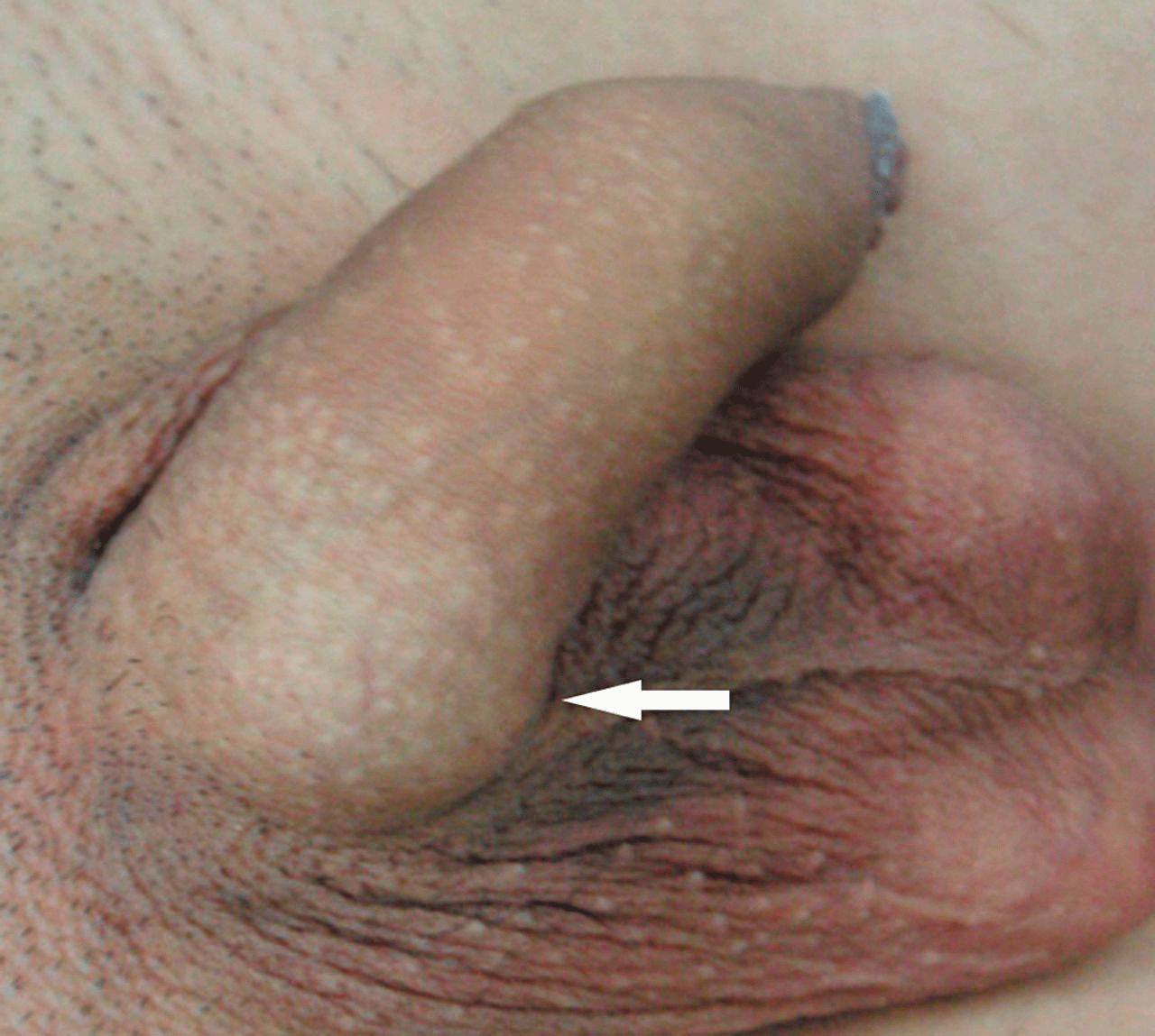
Appearance of the hemangioma in the dorsal aspect of the penis before surgery. Arrow indicates the hemangioma. Color figure available online at www.andrologyjournal.org.
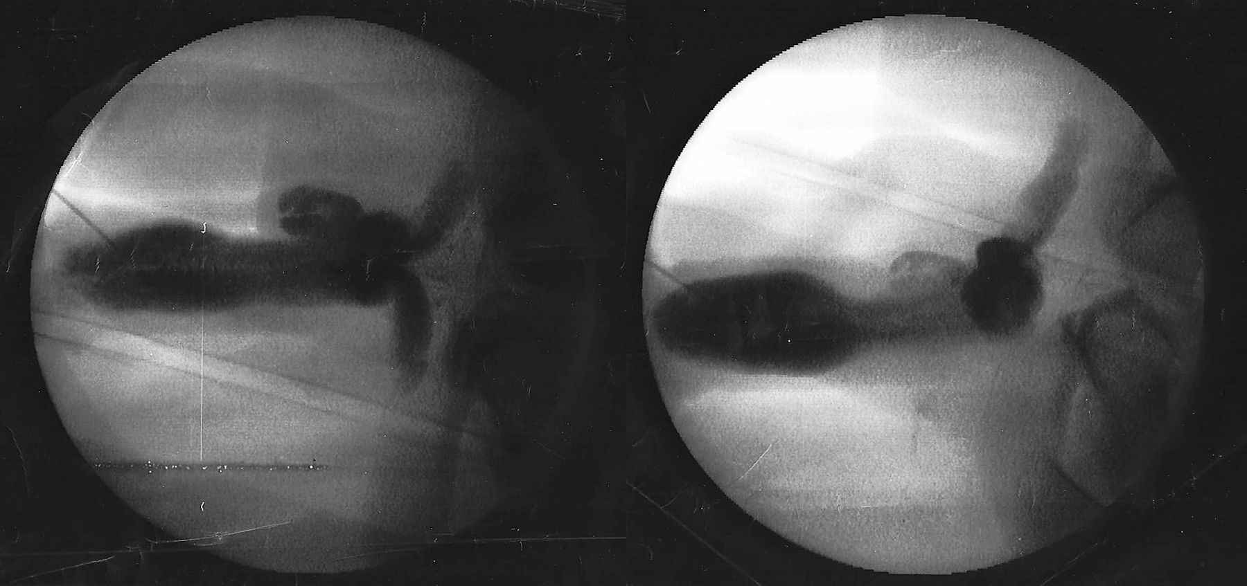
Cavernosography image showing hemangioma on the cavernous body of the penis.
All patients provided written informed consent and completed the entire trial, including treatment and follow-up examinations. The study protocol was approved by the ethical committee of First Affiliated Hospital of Nanjing Medical University, and agreements were signed by all patients.
Operative Technique
All operations were performed by the same highly experienced urologist. Patients were given epidural anesthesia and placed in a horizontal position. The skin of the dorsum penis was dissected above the hemangioma. Further isolation was performed to expose the hemangioma for integrated surgical ablation (Figure 3). Two cases of lesions were small in size (diameter < 1 cm) and had a narrow pedicel outside the corpus cavernosum of the penis. In these cases, the pedicel was ligated and the hemangioma excised (Figures 4 and 5). For other cases that had large defects of the tunica albuginea, surgical resection alone was not enough. We replaced the hemangioma with free autologous testis tunica vaginalis grafts for penile morphological reconstruction (Figures 6 and 7). After each hemangioma was exposed clearly, we measured its length and width, dissected it, and repaired the incisal margin regularly and smoothly, all while avoiding damage of the deep artery of the penis. Tunica vaginalis grafts were removed according to the size of tissue section removed from the albuginea penis after excision for hemangioma. If the hemangioma was near the urethra, we inserted a catheter and dissociated part of the urethra to remove the hemangioma and avoid damaging the urethra. The tunica vaginalis grafts were cropped to the required size and the mucosa side was placed facing inside, fixed in the former location of the defect, and sutured with absorbable stitches.
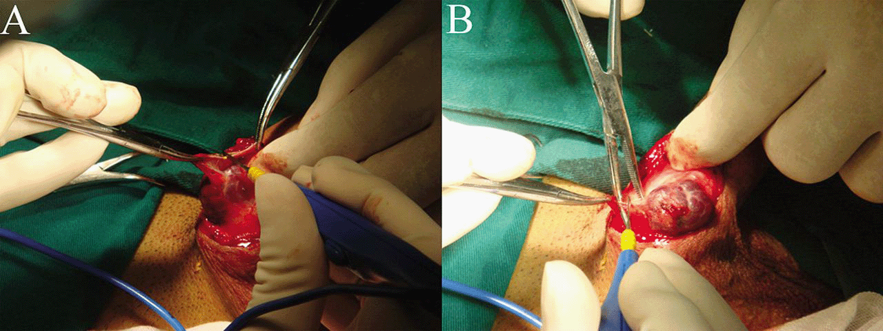
Dissociation of the hemangioma for further surgical resection (A, B). Color figure available online at www.andrologyjournal.org.
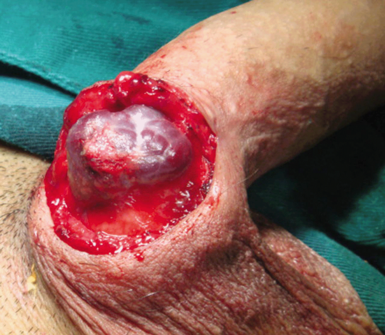
Appearance of the hemangioma on the dorsal aspect of the penis during surgery. Color figure available online at www.andrologyjournal.org.
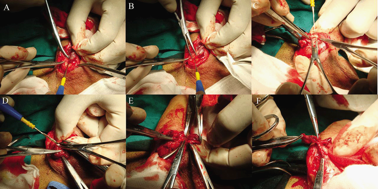
Excision of the hemangioma from the dorsum of the penis (A—F). Color figure available online at www.andrologyjournal.org.
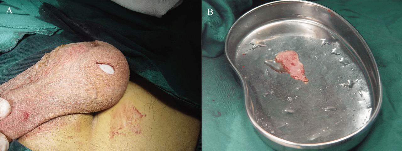
(A) Tunica vaginalis grafts removed according to the size of the (B) defect in the albuginea spongy body. Color figure available online at www.andrologyjournal.org.
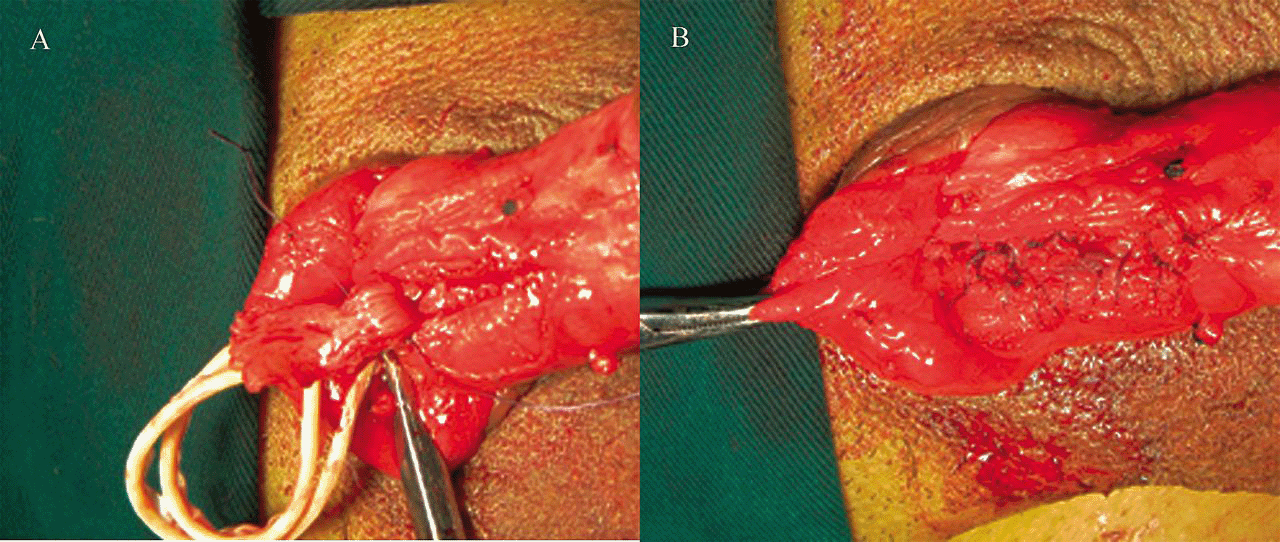
(A) The mucosa side was placed facing inward and fixed it in the location of defect by (B) suturing it with absorbable stitches. Color figure available online at www.andrologyjournal.org.
Follow-up and Evaluation
Clinical results were reviewed by examination of inpatient and outpatient hospital records. We paid attention to all patients and no one was lost to follow-up. The 5 patients were interviewed and filled out an (international index of erectile function, IIEF) questionnaire to determine erectile function, orgasmic function, sexual desire, intercourse satisfaction, and overall satisfaction with sexual life. All patients completed the IIEF questionnaire in less than 15 minutes and reported little or no difficulty in comprehending the items. Scores were computed for individual answers, and the final scores were compared before and after the operation. Cosmetic results were also assessed in all patients with a 5-graded scale ranging from very dissatisfied to very satisfied.
Statistical Analysis
The resulting numeric values for each domain of sexual function were compared statistically before and after surgery by paired t test. Statistical analysis was carried out with SPSS 16.0 for Windows (SPSS Inc, Chicago, Illinois). A value of P < .05 was considered statistically significant.
Results
Surgical Outcome and Follow-up
Mean blood loss was 11 mL, ranging from 6 to 15 mL, and the mean operation time was 30 minutes, ranging from 15 to 45 minutes. After surgery, the penises were dressed with elastic bandages and treated to prevent infection. The postoperative period was uneventful, and the patients were discharged after 3 to 5 days. No infection was observed. The wound healed completely after 7 days. All patients were followed for 2 to 5 years (x̄ = 3.5 years). No recurrences have been observed.
Cosmetic Results and Sexual Function After Surgery
The operated areas were flat and smooth and showed normal coloration. In some areas, the borders between the operated area and the healthy penis were not distinguishable. The degrees of curvature noted by the patients had been corrected. The cosmetic results were regarded as satisfying or very satisfying by 100% of the patients.
Five patients returned to normal sexual activity within 3 months after surgery without abnormal sensation. The IIEF scores from the IIEF questionnaire before and after the operation are listed in the Table. Former patients had the same erectile function (P < .05) or even better erectile function after surgery than before. Orgasmic function and sexual intercourse were resumed in all previously sexually active patients (P < .05). All of the 5 previously sexually active patients felt the grade of intercourse and overall satisfaction maintained (P < .05) when they had sexual stimulation or intercourse after surgery.
| Domain (n = 5) | Preoperation (median ± SD) | Postoperation (median ± SD) | P Valuea |
|---|---|---|---|
| Erectile function | 24.40 ± 1.12 | 24.80 ± 1. 30 | .178 |
| Orgasmic function | 8.00 ± 0.71 | 8.40 ± 0.55 | .178 |
| Sexual desire | 8.00 ± 0.71 | 7.80 ± 0.84 | .374 |
| Intercourse satisfaction | 13.20 ± 0.84 | 12.6 ± 0.55 | .070 |
| Overall satisfaction | 8.00 ± 0.71 | 8.80 ± 0.45 | .099 |
- Abbreviation: IIEF, international index of erectile function.
- a Paired t test.
Discussion
Hemangioma can be classified by depth within the skin, the number of lesions, and location on the body. Many hemangiomas consist entirely of cutaneous components and present clinically as bright red noncompressible papules, nodules, or plaques when fully developed. Hemangiomas of the penis are very rare, and only 7 cases have been reported (Senoh et al, 1981; Jimenez-Cruz and Osca, 1993; Lin et al, 2002). Whether penile hemangiomas are true tumors or come from herniation of the corpus spongiosum or from vascularization caused by a hematoma or thrombus remains controversial and unproven. We consider penile hemangiomas to be mulberry-sized, purple, elevated, compressible lesions that are associated with a defect in the tunica albuginea.
Neither ultrasonographic examination of the urinary system nor urine or blood analysis contributed significantly to the diagnosis (Kumar et al, 2008). Magnetic resonance imaging and computed tomography scans were not able to show whether there was a clear separation between the cavernous body and the angiomatous formation. In contrast, cavernosography clearly showed how a contrast medium injected into the corpus cavernosum was unable to reach the angiomatous formation and render it opaque. It was therefore assumed that the angiomatous formation was not related to the cavernous body.
From a review of published reports on penile hemangiomas, we found that most lesions occur in the glans, Several therapeutic options have been proposed. Among the many treatments for this disorder are surgical excision, laser therapy, sclerotherapy, electrofulguration, and cryotherapy (Goldwyn and Rosoff, 1969; Yamazaki et al, 1978; Angulo et al, 1993; Hemal et al, 1998). Laser treatment is a safe method for the treatment of vascular lesions in children. It has been performed on an outpatient basis with almost no blood loss or complications because it can be performed within a few seconds under local anesthesia. Reports of laser treatment for these lesions with the patient under local anesthesia have described good functional and cosmetic results (Amaro et al, 1997). Sclerotherapy is less expensive and more readily available than laser therapy. Hernal et al (1998) reported on 4 patients who were successfully cured after injecting 30% hypertonic saline into penile hemangiomas. This matches the results of a study that reported the results of repeated injections of 2% polidocanol (Savoca et al, 2000). In recent years, intralesional sclerotherapy and Nd:YAG laser therapy have seen significant, especially for superficial lesions. However, for cases with large albuginea penis defects, laser treatment and sclerotherapy have limited applicability. Repair and surgical treatment is necessary because neither of these therapies can deal with the defect. Sclerotherapy can cause Peyronie disease with such symptoms as persistent pain and severe curvature of the penile shaft. Surgery is the classical therapeutic approach. Some physicians considered surgical therapy to pose a high risk of bleeding, not only during the excision but also during the postoperative period, with sexual dysfunction and scar formation as additional complications. In each of our 6 patients, mean blood loss was only 11 mL, and after a mean follow-up period of 2.5 years, excellent cosmetic results had been achieved in every patient. The results were regarded as satisfying by 100% of the patients. Healthy, normal appearance after the operation helped patients regain their self-image and self-esteem, which might have led to their high levels of sexual satisfaction after the operation.
Surgical therapy is also an option, especially in peripheral hospitals, where it can be performed with minimal available infrastructure and produce acceptable results. Surgeons can operate with patients under local anesthesia through blockage of the dorsal nerve of the penis.
A therapeutic reference standard for treatment is still lacking because of the rarity of the disease. Although penile hemangiomas can continue to grow, most lesions require no treatment unless the patient is concerned with the cosmetic effects. The use of systemic steroids has been a common medical treatment methods for hemangioma, but it has failed in many clinical cases. We believe that surgical treatment is the most effective way to address penile hemangioma.
Conclusion
Cosmetic results can be well maintained after surgery, and patients tend to be satisfied with sexual function and sexual satisfaction. The outcome data suggest that this surgical technique is technically safe and functionally and cosmetically satisfying to the patient. Surgical management of hemangioma on the dorsum of the penis is safe and feasible in clinical practice. It is easily implemented and can be used with different kinds of penile hemangiomas.


