The Biphosphonate Aledronate (MK-217) Inhibits Bone Loss due to Ovariectomy in Rats
Abstract
Estrogen deficiency in mammals is known to increase bone turnover and result in reduced bone mass. The bisphosphonate, 4-amino-1-hydroxybutylidene-1,1-bisphophonic acid disodium salt, alendronate (MK-217), is a potent inhibitor of bone resorption and was evaluated in this study for its ability to inhibit bone loss following ovariectomy in rats. Alendronate was administered sc in doses of 0.0, 0.056, 0.28, 1.40, and 7.0 mg P/kg/month, divided into two, four, or eight monthly subcutaneous injections for each dose, to female Sprague-Dawley rats (250–280 g) that underwent bilateral ovariectomy. Rats were sacrificed 12 weeks postovariectomy, the femora ashed, and the tibiae prepared for static and dynamic histomorphometric analyses.
Femoral bone mass in vehicle-treated rats was reduced by 12% 12 weeks after ovariectomy compared to the nonovariectomized control group. In MK-217-treated rats femoral bone mass was significantly increased in a dose-dependent manner compared to either ovariectomized or nonovariectomized controls. Histomorphometric analysis showed significant increases in tibial trabecular bone volume with no decrease in osteoclast number. Doses delivered twice per month or eight times per month were equally effective in achieving the peak bone volume 12 weeks after ovariectomy.
In conclusion, alendronate (MK-217) was effective in inhibiting bone loss due to estrogen deficiency in rats, and the magnitude of its effect was related primarily to the total amount of compound administered rather than the frequency of its administration.
INTRODUCTION
Estrogen deficiency in mammals increases bone turnover as revealed by serum and urinary biochemical markers, including alkaline phosphatase, bone Gla protein, urinary calcium, and hydroxyproline,1, 2 and by bone histomorphomeric analysis.3-6 The result of the increased bone turnover and an imbalance between bone resorption and formation is a decrease in bone mass. In rats following ovariectomy bone mass is reduced by 8 weeks7-9; in humans bone loss is detected by 1 year.10, 11 Wronski et al. showed that rats may serve as appropriate models for estrogen-deficient osteopenia8 since skeletal events at the bone tissue level are similar to those in humans after surgical menopause.
Preventing bone loss following natural or surgically induced menopause is important for preventing later bone fractures. Estrogen replacement therapy has been proven efficacious12-15; however, in view of potential side effects and poor patient compliance, alternative therapies should be considered. Bisphosphonates are potent inhibitors of bone turnover and bone resorption16, 17 and may be therapeutic candidates for preventing bone loss in estrogen-deficient states. The bisphosphonate alendronate, 4-amino-1-hydroxybutylidene-1,1-bisphosphonic acid disodium salt, hereafter referred to as MK-217, has been proven effective in rats in preventing bone loss associated with immobilization by reducing bone resorption.18
Similarly, MK-217 was effective in lowering serum calcium in humoral hypercalcemia of malignancy in humans19, 20 and in the management of Paget's disease.21 In the present study MK-217 was evaluated in ovariectomized rats for its ability to inhibit bone loss associated with estrogen deficiency. Also evaluated in this study was the effect of using different dosing regimens to deliver the same skeletal load of drug.
MATERIALS AND METHODS
Female Sprague-Dawley rats weighing about 220 g were used for this study (Taconic Farms, NY). Animals were anesthetized intraperitoneally using ketamine hydrochloride (Ketaset, Aveco, 10 mg/kg) and acepromazine (PromAce, Aveco, 3 mg/kg), their abdomens shaved, and the skin cleaned using 70% ethanol followed by povidone-iodine (Betadine) solution. A longitudinal midline incision was made in the umbilical region exposing the rectus abdominus muscle and the abdominal cavity. The urinary bladder was retracted forward and the uterine horns identified. The ovarian arteries were ligated and bilateral ovariectomies were performed. The abdominal musculature was sutured and the skin closed with staples.
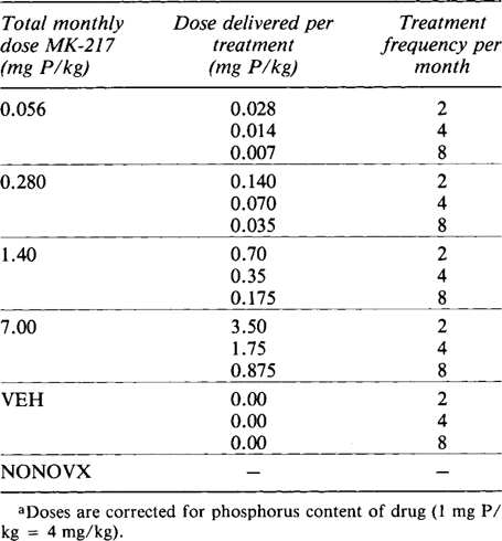
Ovariectomized animals (n = 6 per group) were assigned to one of three treatment regimens receiving subcutaneous (SC) injections twice, four times, or eight times per month. The doses of MK-217 used and expressed as the total MK-217 dose per month (28 days) were as follows: 0.0, 0.056, 0.28, 1.4, and 7.0 mg P/kg. Doses were corrected for phosphorus content of formula weight (alendronate = 24.9% P). Each total monthly dose of MK-217 was adjusted for weight gain and divided equally in either two, four, or eight injections as shown in Table 1. The drug was delivered using a saline vehicle, and treatment regimens were started 24 h postsurgery. A nonovariectomized untreated control group (NONOVX) was also maintained. Animals were treated intramuscularly (IM) with tetracycline HCl (Achromycin, Lederle, 20 mg/kg) 7 days before sacrifice and oxytetracycline (Terramycin, Pfizer, 20 mg/kg) 1 day before sacrifice. Following week 12 of treatment animals were weighed and then sacrificed using a CO2 inhalation chamber. A successful ovariectomy was confirmed at necropsy by examining the uterine horns for signs of atrophy. The hind limbs were removed, disarticulated, and denuded of muscle. Left bicondylar femoral length (anatomic length) was measured to the nearest 0.05 mm, and the left femora were ashed using a muffle furnace at 700°C for 24 h. The bone ash was weighed to the nearest 0.1 mg and ash weight/femoral length was calculated in mg/mm.
Proximal tibiae were fixed in cold 70% ethanol, dehydrated through graded concentrations of ethanol up to 100%, cleared in xylene, and embedded in methyl methacrylate.22 Coronal sections 6 and 10 μm thick were cut using a Reichert-Jung Polycut S microtome and mounted on gelatin-coated glass slides. Sections 6 μm were deplastified in warm xylene and stained with Masson's trichrome.
Histomorphometric analysis was performed in the secondary spongiosa of the proximal tibia in the nonovariectomized control group and in the groups treated two and eight times per month beginning 1.8 mm below the epiphyseal growth plate. The area of analysis extended distally for an additional 1.8 mm and spanned the corticoendosteal surfaces.
Measurements were recorded using the BONE software within the Joyce-Loebl Magiscan System. This is an interactive real-color video image analysis system in which the operator uses a light pen to indicate types and extent of bone surfaces and areas to be measured based on staining characteristics. The accuracy of color detection can be verified by a computer-generated overlay that identifies the surface for analysis, and further editing of the image is accomplished using the light pen. Accuracy of area measurement was confirmed by measuring objects of known dimension.23
The standardized histomorphometric parameters24 measured within the BONE software included cancellous bone volume (BV%), expressed as the percentage area occupied by trabecular bone; eroded surface (ES%), expressed as the bone surface containing evidence of bone resorption; and osteoid surface (OS%), expressed as bone surface containing osteoid and active osteoblasts. Multinucleated cells on bone surfaces were counted and used to calculate osteoclast number per mm bone surface (N.Oc/mm) and osteoclast number per tissue area analyzed (N.Oc/mm2).
Slides containing the 10 μm thick unstained sections were coverslipped with Fluormount-G and examined under ultraviolet (UV) illumination. The mean interlabel distance was calculated from six double-label intercepts at the tibial midshaft. The diaphyseal mineral apposition rate (MAR) was calculated by dividing the mean interlabel distance by the 6 day label interval, yielding MAR in μm/day.25 Metaphyseal mineral apposition rate and surface labeling extent were not measured because of diffuse labeling of primary spongiosa retained in the proximal tibia as a result of MK-217 treatment. Means and standard deviations (SD) were computed and significant differences at p < 0.05 determined by Student's two-tailed t-test.
RESULTS
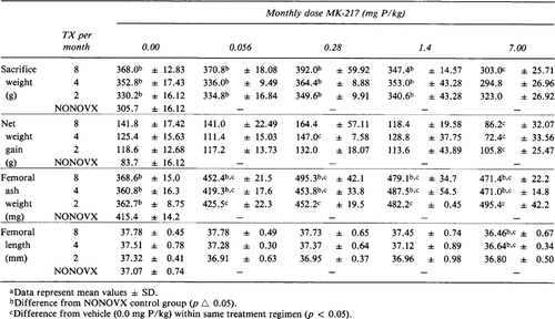
All rats in the study gained weight over the 12 week experimental period, as shown by the sacrifice weights in Table 2. Treatment with MK-217 generally did not produce changes in net weight gain except at the highest dose (7.0 mg P/kg).
Femoral ash weights in the ovariectomized vehicle-treated groups ranged from 361 to 369 mg and were 12% less (p < 0.05) than in the nonovariectomized group. The lowest MK-217 dose administered (0.056 mg P/kg) given in the two and four times per month regimens maintained femoral bone mass at 426 and 419 mg, respectively, and was not different from the femoral bone mass of 415 mg for the nonovariectomized group. Administration of 0.056 mg P/kg in eight treatments per month and all of the higher doses of MK-217 in all three treatment regimens produced significant (p < 0.05) increases in femoral bone mass compared to the nonovariectomized group. All MK-217 treatment regimens, regardless of dose, increased femoral ash weight significantly (p < 0.05) compared to ovariectomized vehicle-treated groups (Table 2).
With the exception of the highest MK-217 dose in the four and eight times per month treatment regimens, femoral length measurements did not show differences when compared to the nonovariectomized or vehicle-treated groups. The four and eight times regimens of 7.0 mg P/kg/month decreased (p < 0.05) femoral length by 2–3% (Table 2).
The femoral ash weight/length ratios in the nonovariectomized group and in all MK-217-treated groups was significantly greater (p < 0.05) than in the ovariectomized vehicle-treated groups (Fig. 1). The femoral ash weight/length ratio in animals treated with the low dose of MK-217 (0.056 mg P/kg), administered in the two and four times per month regimens, was not statistically different from that of the nonovariectomized group, whereas MK-217 doses of 0.28, 1.4, and 7.0 mg P/kg in the two, four, or eight times per month treatment regimens produced significantly greater femoral ash weight/length ratios compared to the nonovariectomized control group.
Histomorphometry
The tibial cancellous bone volume (BV) in the ovariectomized vehicle-treated group was 9.92% for the two times and 10.26% for the eight times per month regimens compared to 25.93% in the nonovariectomized group. These differences represent a loss of about 60% of cancellous bone volume (p < 0.05) following 12 weeks of estrogen deficiency produced by bilateral ovariectomy (Fig. 2).

Effect of MK-217 on femoral ash weight/length ratio in ovariectomized rats. Data presented as mean values ± SEM. *Indicates significant difference (p < 0.05) from NONOVX control. **Indicates significant difference (p < 0.05) from vehicle-treated (0.0 mg P/kg) group.

Effect of MK-217 on trabecular bone volume in ovariectomized rats. Data presented as mean values ± SEM. *Indicates significant difference (p < 0.05) from NONOVX control. **Indicates significant difference (p < 0.05) from vehicle-treated (0.0 mg P/kg) group.
Treatment with MK-217 at 0.056, 0.28, 1.4, and 7.0 mg P/kg/month significantly increased tibial BV compared to either vehicle-treated or the nonovariectomized group. Tibial BV for rats treated with MK-217 two times per month ranged from 36% for the low dose to 80% for the highest dose, and the BV for rats treated eight times per month ranged from 52% for the low dose to 80% for the highest dose. The tibial bone volume in both regimens increased dose dependently and began to plateau at about 75% at the two highest doses. This represents a threefold increase in BV over the nonovariectomized control and a sevenfold increase over ovariectomized vehicle-treated groups.
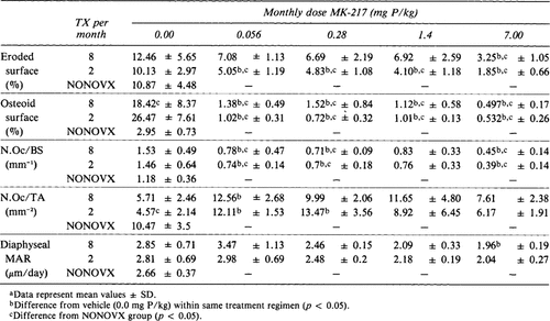
Active erosion surface per mm bone surface (ES) was not statistically different in the vehicle-treated and the nonovariectomized groups (Table 3). In the eight times per month regimen only the highest dose of MK-217 (7.0 mg P/kg/month) produced a significant decrease in ES compared to either vehicle or the nonovariectomized group. In contrast, in the two times per month treatment regimen all doses of MK-217 decrease ES compared to vehicle-treated or nonovariectomized control groups.
Osteoid surface per mm bone surface (OS) ranged from 18 to 26% in the ovariectomized vehicle-treated groups (Table 3) and was significantly higher (p < 0.05) than the nonovariectomized control OS of 2.9%. Treatment with MK-217 decreased the osteoid surface extent relative to the nonovariectomized and the vehicle-treated ovariectomized groups (p < 0.05).
The number of osteoclasts per mm bone surface (N.Oc/BS) in MK-217-treated groups was consistently lower than in the vehicle-treated and the nonovariectomized groups (p < 0.05; Table 3), ranging from about 0.7 for the lowest dose (0.56 mg P/kg/month) to 0.4 for the highest dose (7.0 mg P/kg/month). Although not significantly different, this compared to an osteoclast number per mm bone surface of 1.5 in the vehicle-treated groups and 1.2 in the nonovariectomized control group. The number of osteoclasts per mm2 of tissue area analyzed (N.Oc/TA) showed few differences between the MK-217-treated groups and the vehicle-treated ovariectomized groups and no differences compared to the nonovariectomized control group. Only osteoclast number per mm2 in the vehicle-treated nonovariectomized group in the two times per month treatment regiment was reduced (p < 0.05) compared to the nonovariectomized control group.
The mean diaphyseal mineral apposition rate of the vehicle-treated groups was 2.85 μm/day for the eight times per month treatment group and 2.81 μm/day for the two times per month treatment group. These were not significantly different from the 2.66 μm/day MAR in the non-ovariectomizsed control group (Table 3). Only the high dose of MK-217 in the eight times per month regimen was significantly reduced compared to the vehicle-treated ovariectomized control group. The diaphyseal MAR of all other groups did not differ significantly from the vehicle-treated or the nonovariectomized groups.
DISCUSSION
Bilateral ovariectomy in the rat decreases circulating serum estrogen levels, which results in increased bone turn-over. Wronski et al.26, 30 demonstrated by using histomorphometric methods that the tibial metaphysis of ovariectomized rats had reduced trabecular bone volume and increased erosion and formation surfaces. Within 4–6 weeks ovariectomized rats show osteopenic changes in both the axial and appendicular skeleton.31
Aminobisphosphonates are potent inhibitors of osteoclast-mediated bone resorption that suppress bone turnover through their ability to prevent the initiation of new erosion sites and to reduce ongoing excavation.32, 33 Immobilization of the rat hind limb by sciatic neurectomy produces a rapid increase in osteoclast number resulting in increased bone resorption and turnover.23, 24, 34, 35 Thompson et al. demonstrated that in the rat immobilization model treatment with the bisphosphonate MK-217 inhibited bone resorption and reduced bone turnover.35
The present study demonstrated that the bisphosphonate MK-217 effectively prevented bone loss in the rat resulting from estrogen deficiency. The vehicle-treated groups lost 12% femoral bone mass and the MK-217-treated groups had significantly increased femoral bone mass compared to either the nonovariectomized or ovariectomized control groups 12 weeks following ovariectomy. This increase in bone mass was also reflected in a greater femoral ash weight/length ratio. Since there were no significant changes in femoral length among groups, differences were due to an increase in bone mass produced by treatment with MK-217.
At the tissue level this increased bone mass corresponded to an increase in trabecular or cancellous bone volume of the tibial metaphysis. The metaphyseal bone volume was 60% lower in the ovariectomized vehicle-treated groups than the nonovariectomized group. However, groups dosed with MK-217 had metaphyseal bone volumes significantly greater than either the nonovariectomized or ovariectomized vehicle-treated groups. The increase in bone volume was dose dependent and peaked at about 80% in both the eight treatments per month and two treatments per month regimens. The amount of cancellous bone volume appeared to be dependent on the total monthly dose or load of drug received rather than the frequency of treatment administration, since both the eight and the two times per month treatment regimens attained the same peak bone volume. However, for doses that produced less than the maximal bone volume (0.28 and 0.056 mg P/kg) the eight times per month regimen appeared to be more efficacious than the two times per month regimen. Nevertheless, the total dose of drug the skeleton received was more indicative of the effect on bone volume than the frequency or number of treatments used to administer the total dose. Further studies with more infrequent dosing intervals are necessary to confirm this hypothesis.
MK-217 given at the dose of 0.056 mg P/kg in the twice per month regimen maintained or slightly improved the bone mass compared to the nonovariectomized rats. However, compared to the ovariectomized vehicle-treated groups this dose significantly decreased bone resorption and trabecular bone loss associated with estrogen deficiency in rats. Higher doses or more frequent treatment with MK-217 prevented resorption of the primary as well as the secondary spongiosa, and this contributed to the significantly increased bone volume measured in the metaphyses from these animals. The use of animals older than 6 months of age, after which little or no metaphyseal bone growth is observed, may alleviate the problem of retention of calcified cartilage due to treatment in future studies.
Examination of the tibial metaphyseal bone surfaces suggests that MK-217 reduced bone turnover. The percentage of eroded surface in the ovariectomized vehicle-treated groups was not different from that in the nonovariectomized control group, but the percentage of osteoid surface was significantly increased. In conjunction with the reduced bone volume in the vehicle-treated groups, this suggested that a significant amount of erosion occurred postsurgery and that these surfaces were still in a state of active bone turnover.
The MK-217-treated groups showed a slightly reduced percentage of eroded surface compared with the nonovariectomized control and the ovariectomized vehicle-treated groups, but no differences were observed between doses or treatment modalities. The number of osteoclasts per mm bone surface (N.Oc/BS) and the number of osteoclasts per mm2 tissue area (N.Oc/TA) followed similar patterns and the percentage of osteoid surface was decreased in all MK-217-treated groups as predicted for a state of reduced bone turnover.
The mechanism of action of MK-217 in preventing bone resorption is not known. Although bone surfaces contained osteoclasts, histomorphometrically no method is currently available to determine their functional state of resorption or the duration of time that osteoclasts occupy the bone surface. However, it is known from in vitro studies that MK-217 treatment impairs the ability of osteoclasts to resorb even though they are apposed to bone surfaces.36 Lowik et al. has shown that other bisphosphonates can interfere with the maturation of functional osteoclasts at the bone surface by inhibiting the development of tartrate-resistant acid phosphatase activity.37
Mineralization defects have been reported in ovariectomized rats treated with some bisphosphonates.38 MK-217, however, did not produce increased osteoid seam thickness and the diaphyseal mineral apposition rate was not altered. The effect of MK-217 treatment on metaphyseal microarchitecture made it difficult to reliably measure apposition rates in this region since the preservation of primary spongiosa due to inhibition of resorption of calcified cartilage caused labeling to appear diffuse and ill-defined.
In conclusion, the current study demonstrates that the bisphosphonate alendronate (MK-217) effectively inhibited bone loss associated with estrogen deficiency in rats. MK-217 treatment produced dose-dependent increases in cancellous bone volume as determined by the total monthly dose rather than treatment frequency. This increased bone mass was most likely the result of inhibition of osteoclast-mediated resorption and was not due to lack of osteoclast recruitment.
Acknowledgements
The authors wish to thank Dr. Gideon Rodan for advice on experimental design and critical reading of this manuscript, Ms. Pamela J. Wampler for her expert histologic assistance, and Ms. Dianne McDonald for manuscript preparation.
Appendix: JBMR Anniversary Classic: The Bisphosphonate Alendronate (MK-217) Inhibits Bone Loss due to Ovariectomy in Rats
JG Seedor, HA Quartuccio, DD Thompson
Originally published in Volume 6, Number 4 pp 339–346 (1991)
This and another study demonstrated the effectiveness of alendronate in blocking bone loss in two critical animal models, immobilization and ovariectomy.


