Bone Mass and Hip Axis Length in Healthy Asian, Black, Hispanic, and White American Youths†
Portions of this work have been presented in Wang M, Kendall CG, Marcus R, Bachrach LK. Ethnic differences in bone mass in children, adolescents and young adults [abstract]. Am J Epidemiol 143(11):S38, 1996.
Abstract
The primary objective of this study was to examine the associations of ethnicity, diet (calcium, protein, energy), and weight-bearing activity with dual-energy X-ray absorptiometry (DXA)-measured bone mass and hip axis length (HAL) in 423 Asians, blacks, Hispanics, and non-Hispanic Caucasians, aged 9–25 years. Bone mass was expressed as bone mineral content (BMC), bone mineral density (BMD), and bone mineral apparent density (BMAD). The data were analyzed using multiple linear regression, after stratifying for gender and pubertal stage and adjusting for height and weight. With few exceptions, Asians and Hispanics had comparable bone mass to whites at all pubertal stages. Greater femoral neck BMAD in black than white females was observed at all pubertal stages. Black males displayed greater BMD and BMAD than white males at all sites in early puberty and at the femoral neck in maturity. Calcium was positively and protein negatively related to BMAD at the femoral neck in early pubertal females. Among males, calcium was negatively associated with whole body BMC and BMD and spine BMD and BMAD in midpuberty. Weight-bearing activity was not associated with bone mass in females; in males, it was positively related only to femoral neck BMC in early puberty. There was an absence of evidence for ethnic differences in HAL among females. In males, we observed shorter HAL in mature Asians and blacks than whites. Neither diet nor activity was associated with HAL.
INTRODUCTION
BONE MASS AND BONE GEOMETRY independently predict fracture risk.1-6 Low bone mass increases skeletal fragility and fracture risk at all sites,1-4 while longer hip axis length (HAL) specifically increases the risk of hip fracture.5, 6
Low bone mass is believed to result from suboptimal attainment of peak bone mass during adolescence or young adulthood4, 7 and accelerated bone loss later in life.2, 4 Documented racial and ethnic differences in bone mass8-11 reflect both genetic and environmental influences. Twin studies demonstrate that genetic factors explain 60–80% of the variance in peak bone mass,8, 12, 13 suggesting that environmental factors are also important. In the United States, bone mass has been observed to be higher in black than in white adults8, 14, 15 and children.8, 16-19 Diet and physical activity are two important environmental determinants of bone mass. In particular, calcium and weight-bearing activity have been observed to promote bone mass4, 7, 20-23 while high protein intake may increase urinary calcium excretion.20
HAL, like bone mass, appears to be largely determined by genetic factors.24 Environmental correlates of HAL have not yet been described, but racial and ethnic differences in HAL have been reported. White American women have a longer mean HAL than their black, Asian, and Hispanic counterparts.6
Little is known about bone mineral acquisition and bone geometry in healthy, ethnically diverse adolescents. To examine the tempo and correlates of skeletal maturation, we initiated a longitudinal study of bone mineral acquisition in Asian, black, Hispanic, and white females and males. Entry data for Asian and white youths have been reported.25 For this paper, we analyzed similar data from the complete cohort of Asians, blacks, Hispanics and whites to examine cross-sectionally: (1) racial/ethnic differences in bone mass and HAL, and (2) relationships of bone mass and HAL with height, weight, calcium and protein intakes, and weight-bearing activity.
MATERIALS AND METHODS
We recruited a convenience sample of 103 non-Hispanic whites, 115 non-Hispanic blacks, 102 Hispanics, and 103 Asians, aged 9–25 years. For ease of reading, subjects will be referred to as white, black, Hispanic, or Asian. Further, the term “ethnicity” will be used interchangeably with “race.” A detailed description of the recruitment methodology is given elsewhere.25
Weight and height were measured using standard protocol.25 Pubertal stage was assessed by self-rating, using Tanner classifications of breast (females) and genital (males) development.26 Pubic and axillary hair rating was not used due to concerns about ethnic differences in the timing and quantity of body hair development. Pubertal stages were combined as follows: early puberty (Tanner stages 1 and 2), midpuberty (Tanner stages 3 and 4), and maturity (Tanner stage 5). On the basis of breast/genital rating alone, some of the older subjects scored themselves as Tanner stage 3 or 4, placing them in the midpubertal category. Based upon reference data for normal children,26 and the presence of menses in females, we reassigned 17 females aged 17 years and older and 16 male subjects aged 18 years and older to the mature group.
Diet was assessed using the self-administered 97-item National Cancer Institute Food Questionnaire.27 Nutrient analysis was performed using the Dietary Analysis System (Dietsys) version 3.0, 1994 (National Cancer Institute, Bethesda, MD, U.S.A.). Questionnaires reporting energy intakes of less than 500 kcal or more than 6000 kcal (n = 19) were excluded from all analyses involving nutrient intake. Physical activity was assessed by self-report of time engaged in recreational activities during the past year. Activities were categorized as non–weight-bearing and weight-bearing (defined as any exercise that loads the body with at least body weight). Walking was excluded because subjects had difficulty estimating time spent on this activity.
Bone mass was measured for the lumbar spine (L2–L4), left hip (femoral neck), and whole body, using dual-energy X-ray absorptiometry (DXA; QDR-1000W, software version 6.10, Hologic Corp., Waltham, MA, U.S.A.) in the pencil beam mode. Due to changes in hip geometry with age and the potential for inconsistencies in longitudinal measurements, special attention was given to the development of a standard protocol for measuring bone mass at the femoral neck. A single investigator (S.K.) visually inspected the femoral neck scans of all subjects and selected an appropriate neck box width which was then maintained for all subsequent studies. In our laboratory, the in vivo coefficient of variation for replicate measurements of BMD is approximately 0.6% for the femoral neck, lumbar spine, and whole body in adolescents and young adults. Bone mass was expressed as bone mineral content (BMC, g) as well as areal bone mineral density (BMD, g/cm2). Because BMD may be influenced by bone size, we also used an estimate of volumetric bone density, bone mineral apparent density (BMAD, g/cm3), for the spine and femoral neck.28
HAL, defined as the distance along the femoral neck axis from the greater trochanter to the inner pelvic brim, was measured as described by Faulkner and colleagues.5 The measurement was made using a modified hip analysis software program supplied by Hologic.29 With this version of the original software, the region of interest (ROI) was increased to include the adjacent and upper sides of the head of the femur. Each hip scan was inspected to ensure that it included enough of the pelvis for measurement, with soft tissue surrounding all areas of the bone scan. Scans containing insufficient soft tissue or hip bone in the ROI were excluded from the study. The coefficient of variation of the HAL measurement was 0.7%. All hip scans were analyzed for HAL by the same investigator (M.A.).
This study was approved by the Administrative Panel on Human Subjects in Medical Research at Stanford University. Written informed consent was obtained from all participants and from the parents of subjects under 18 years of age.
Statistical analysis
Gender, pubertal stage, and ethnic differences in bone mass and HAL were determined using analysis of variance; comparisons between two groups were assessed using Bonferroni's multiple comparisons procedure at a significance level of 0.05. Ethnic comparisons of other relevant characteristics of the subjects were assessed using the nonparametric Wilcoxon test,30 since the distributions of several of these variables deviated from normality.
Multiple linear regression analysis was applied to consider simultaneously the effects of ethnicity, height, weight, calcium, protein, energy intake, and weight-bearing activity on each bone mass measurement and HAL. In addition, regressions involving BMC included bone area as an additional independent variable. Ethnicity was treated as a dummy variable—Asian, black, and Hispanic compared with white.31 Energy intake was included to take into account its potential dietary and skeletal effects. The analysis was stratified by gender and pubertal stage.
Subject recruitment was often possible only if siblings were allowed to participate. Since the inclusion of siblings theoretically defies the statistical assumption of independence, we randomly removed all but one of the same-gender siblings from each pubertal group and reanalyzed the data. Exclusion of same-gender siblings did not change our findings substantially. Therefore, we present results pertaining to the complete cohort.
RESULTS
Clinical characteristics
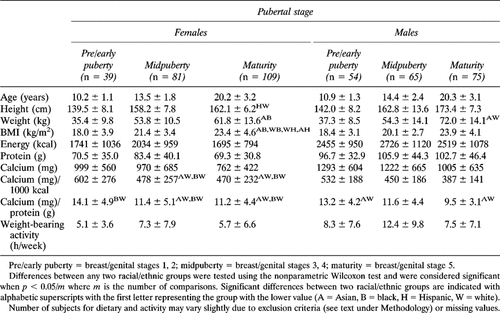
Table 1 provides summary data for the cohort by gender and pubertal stage. Mean age was comparable across all ethnic groups within each gender/pubertal stratum. Ethnic differences in height, weight, and body mass index (BMI) were not detected in early and midpuberty. At maturity, Hispanic females tended to be shorter than white females; Asians weighed less than blacks; and black and Hispanic females had greater BMI than Asian and white females. Mature Asian males weighed less than their white counterparts; however, racial differences in BMI were not significant. Early and midpubertal blacks consumed more calories than other racial groups, but differences did not always reach statistical significance. Calcium/protein and calcium/energy ratios of blacks and Asians tended to be lower than those of Hispanics and whites. Asians reported less weight-bearing activity than whites but the differences were not statistically significant.
Bone mass
Figures FIG. 1A., FIG. 1B. display mean bone mass at the spine and femoral neck for each ethnic group by pubertal stage for females and males, respectively. Data for whole body bone mass are not presented in the figures but will be discussed narratively when relevant.
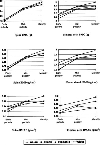
Bone mass at the lumbar spine and femoral neck by puberty and ethnicity—females. Significant pair-wise differences are as follows: Spine BMC, no differences at any pubertal stage; Spine BMD, no differences in early puberty and mid-puberty, maturity: Blacks > Asians; Spine BMAD, no differences in early puberty and mid-puberty, maturity: Blacks > Asians and Whites; Femoral neck BMC, early puberty: Blacks, Whites > Asians, mid-puberty: no differences, maturity: Blacks > all others; Femoral neck BMD, no differences in early puberty and mid-puberty, maturity: Blacks > all others; Femoral neck BMAD, early puberty: no differences, mid-puberty: Blacks > Whites, maturity: Blacks > Asians, Whites.
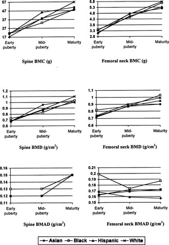
Bone mass at the lumbar spine and femoral neck by puberty and ethnicity—males. Significant pair-wise differences are as follows: Spine BMC, early puberty: Whites > Hispanics, mid-puberty: Asians > Blacks, maturity: no differences; Spine BMD, early puberty: Blacks > Hispanics, no differences in mid-puberty and maturity; Spine BMAD, early puberty: Blacks > Hispanics, Whites, no differences in mid-puberty and maturity; Femoral neck BMC, no differences at any pubertal stage; Femoral neck BMD, early puberty: Blacks > Asians, no differences in mid-puberty and maturity; Femoral neck BMAD, early puberty: Blacks > all others, mid-puberty: no differences, maturity: Blacks > Asians. Differences of means between any two ethnic groups were tested using ANOVA and Bonferroni's inequality (α = 0.05).
Pubertal patterns:
For most females, BMC and BMD at all sites tended to increase more rapidly from early to midpuberty than from midpuberty to maturity. Spine BMAD also increased more during the earlier than later stages of puberty. Black females showed an exception: they tended to experience greater increases in bone mass, particularly at the spine between mid- and late puberty than the other ethnic groups. Mean femoral neck BMAD did not change significantly from one pubertal stage to the next.
In contrast, males, especially blacks and whites, tended to experience late pubertal increases in BMC and BMD at all sites, as well as in spine BMAD, that were as great as or greater than gains during early puberty. As in females, femoral neck BMAD did not change significantly throughout puberty. Asian and Hispanic males demonstrated greater increases in BMC and BMD during the earlier than later stages of puberty.
Gender differences:
For completeness, we briefly describe gender differences in the following paragraphs (data not shown). No consistent pattern of gender differences emerged across all ethnic groups, but compared with females, males tended to display higher BMC and BMD for the whole body and femoral neck, but lower BMD and BMAD at the spine. Femoral neck BMC was higher in males than females among early pubertal Asians, Hispanics, and whites, and among mature subjects in all ethnic groups. For femoral neck BMD, males also had higher mean values than females among early pubertal Asians, Hispanics, and whites, and mature Asians and whites. In contrast, femoral neck BMAD was greater in females than males among midpubertal Asians and mature Hispanics. At the spine, females were observed to have greater BMD (in midpubertal and mature blacks), and BMAD (in early pubertal Hispanics, all midpubertal subjects, and mature blacks and Hispanics) than males.
Ethnic differences:
In early puberty and midpuberty, ethnic differences among females were observed only at the femoral neck. In particular, early pubertal black and white girls showed greater femoral neck BMC than Asian girls, while midpubertal black girls had greater mean femoral neck BMAD than their white counterparts. At maturity, mean values for BMD and BMAD at all measured sites were higher in blacks than at least one other ethnic group. Mature black females also had higher whole body and femoral neck BMC than all other females.
In males, ethnic differences were observed mostly in early puberty. Early pubertal whites had higher spine BMC than Hispanics, while blacks had higher spine BMD than Hispanics and higher femoral neck BMD than Asians. Higher BMAD at the spine and femoral neck was also noted in early pubertal blacks than other males. At midpuberty, only Asians showed higher spine BMC than blacks. At maturity, femoral neck BMAD was significantly greater in black than Asian males; ethnic differences at other skeletal sites were not observed.
Hip axis length
Pubertal patterns:
As with bone mass, we noticed that increases in HAL appeared to occur mostly during the earlier stages of puberty in females, whereas in males, HAL increased throughout puberty (Fig. 2). Interestingly, mean HAL adjusted for height did not increase from one pubertal stage to the next (Fig. 2).

Mean hip axis length by pubertal stage and gender in an ethnically diverse cohort of 9- to 25-year-old Americans.
Gender differences:
Mean HAL was comparable between early pubertal boys and girls in all ethnic groups and also between midpubertal males and females in nearly all ethnic groups; only among midpubertal Asians was HAL noted to be longer in males than females. At maturity, males in all ethnic groups exhibited longer HAL than females.
Ethnic differences:
Mean HAL was longer in white than Asian and Hispanic girls at all stages of puberty. However, these comparisons did not always achieve statistical significance. Among males, the only significant ethnic difference observed was that of shorter HAL in mature Asian compared with white males (data not shown).
Multiple linear regression analysis
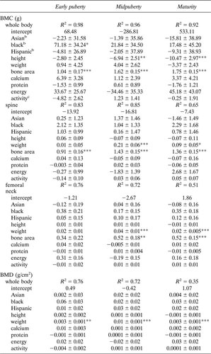
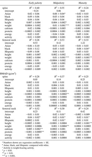
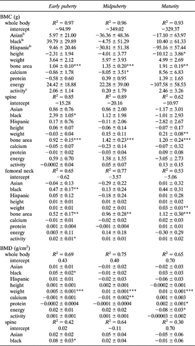
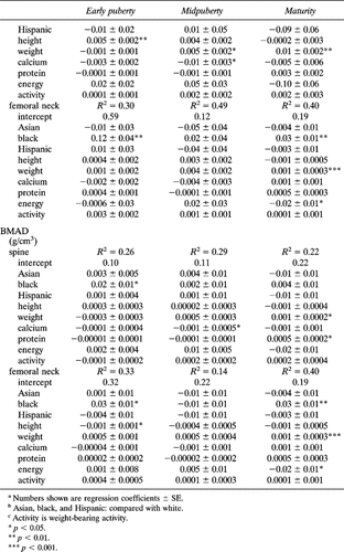
Multivariate associations of bone mass measurements with ethnicity, height, weight, calcium, protein, energy intake, and weight-bearing activity for females and males are shown in Appendices A and B as regression coefficients ± SE.
Bone mass in females:
In early puberty, black girls showed higher whole body BMC (p < 0.05) and femoral neck BMAD (p < 0.05) than white girls after taking into account the effects of relevant variables such as height, weight, diet, and activity. The bone mass of early pubertal Asian and Hispanic girls was comparable to that of whites at all sites. Height was not associated with any measure of bone mass while weight was related only to whole body BMD (p < 0.01). Diet was associated only with femoral neck BMAD, with calcium bearing a positive (p < 0.01) and protein a negative (p < 0.01) association. Weight-bearing activity was not related to bone mass in early pubertal girls.
In midpuberty, there were few ethnic associations with BMD; only at the spine was an ethnic difference observed between Asian and white females (p < 0.05). In contrast, spine BMAD was not associated with ethnicity, but femoral neck BMAD was lower in white than all other females (p < 0.05). Height was related to BMD (p < 0.05) but not to BMAD at the spine, while weight was positively associated with nearly all bone mass measures (p < 0.01). Neither diet nor activity was associated with any bone mass measure in midpubertal females.
At maturity, black-white differences in femoral neck BMD and BMAD were observed (p < 0.05). Asian- and Hispanic-white differences in bone mass were not detected in mature females. Height correlated negatively with BMAD at the femoral neck (p < 0.01). Weight was associated with BMD but not BMAD. Associations of diet and weight-bearing activity with bone mass were not significant.
Bone mass in males:
Early pubertal black boys were observed to have higher bone mass at all sites than white boys. Height was positively associated with lumbar spine BMD (p < 0.01) but negatively associated with femoral neck BMAD (p < 0.05). Weight was related only to whole body BMD (p < 0.001). Neither diet nor activity was found to be associated with bone mass in early pubertal boys.
Unlike in females, there was no evidence of ethnic differences in bone mass among midpubertal males. Further, weight, a strong determinant of all bone mass measures in midpubertal females, was positively related only to whole body and spine BMD. Height was not associated with any bone mass measure. Whole body BMC and BMD and spine BMD and BMAD were negatively related to calcium intake (p < 0.05). Weight-bearing activity was not associated with any measure of bone mass.
In mature males, black-white differences in bone mass were observed only at the femoral neck. As in midpuberty, height was not related to any bone mass measure in maturity. Weight was consistently positively related to all bone mass measures. Relationships with diet were inconsistent. Protein was positively associated with whole body BMD and spine BMAD while energy was negatively related to whole body BMD and femoral neck BMD and BMAD (p < 0.05). Weight-bearing activity was not associated with bone mass in mature males.
Hip axis length:
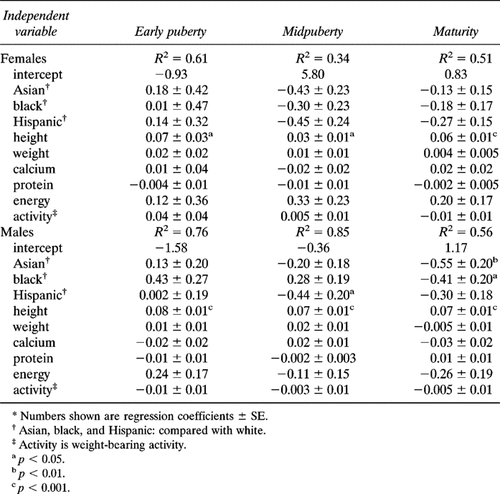
In females, height was the only variable consistently correlated with HAL at all pubertal stages. After adjusting for height, weight, diet, and activity, midpubertal Asian and Hispanic females and mature Hispanic females had shorter HAL than whites, but the differences failed to reach statistical significance. Neither diet nor weight-bearing activity was associated with HAL at any stage of puberty (Table 2).
In males, height was also strongly associated with HAL throughout puberty. Compared with whites, HAL was shorter in Hispanics (p < 0.05) in midpuberty, and in Asians (p < 0.01) and blacks (p < 0.05) at maturity. Calcium, protein, energy intake, and weight-bearing activity were not associated with HAL (Table 2).
DISCUSSION
Bone mass
We note that the tempo of bone mineral acquisition differed by gender and ethnicity. For all but black females, the gains in BMD tended to be greater between early and midpuberty than between midpuberty and maturity. Males and black females appeared to gain bone mass at closely similar rates throughout puberty. Within each pubertal/gender group, we observed few differences in bone mass among white, Hispanic, and Asian youths after controlling for diet, weight-bearing activity, height, and weight. Black-white differences in bone mass were clearly evident across puberty only for femoral neck BMAD.
Data on race/ethnic associations with bone mass among children are scanty. It is unclear whether reported black-white differences in adolescents and young adults16-19 reflect the tempo of bone acquisition, the magnitude of peak bone mass achieved, or both. Gilsanz et al.18 has observed that black-white differences in volumetric bone density at the spine emerged only in late puberty among females. Among mature females, we observed black-white differences in BMD and BMAD at the femoral neck but not at the spine or whole body, after controlling for potential confounders. However, we noted as Gilsanz did, that black females continue to gain bone mass at the spine during later puberty. Higher calcium absorption rates and lower urinary calcium excretion rates32, 33 in black compared with white girls may contribute to racial differences in bone gains.
Few reports examine ethnic differences in the bone mass of male children. In our study, all measures of bone mass except whole body BMC were greater in black than white boys in early puberty. However, by maturity, only BMD and BMAD at the femoral neck differed between black and white males.
There is only one published report on Hispanic American children,19 and no data other than those published by our group25 on Asian American children. McCormick et al. reported no differences in spine BMD between Hispanics (in Texas) and whites, aged 5–19 years.19 Our findings also indicate that BMD as well as BMAD were comparable between Hispanics and whites. In our earlier study comparing Asian and white children from the same cohort,24 only femoral neck BMAD was observed to be higher in Asian than white females; in this analysis, we show that this difference is observed only in midpuberty. The bone densities of adult Asians living in the U.S. appear to be comparable to those of whites after adjusting for height and weight.34-36
Gender differences in bone mass have been noted by others. In a recent study of Caucasian children and adolescents, Gilsanz et al. concluded that gender differences in vertebral bone mass reflect differences in vertebral size rather than volumetric BMD.37 We observed greater spine BMAD in females than males in all midpubertal subjects and in mature blacks and Hispanics. No gender difference in femoral neck BMAD was observed in most pubertal/ethnic groups, although midpubertal Asian and mature Hispanic females had greater mean values than their male counterparts. BMD was greater in females than males at the spine but lower in females than males at the femoral neck in some ethnic groups. These observations are in partial agreement with those of Kröger et al.38 who reported higher spine BMD but lower femoral neck BMD in females than males among older Finnish adolescents. However, while we observed no gender difference in femoral neck BMAD in most ethnic groups, Kröger et al. continued to observe lower femoral neck BMD, corrected for bone size, in females. In another study of Australian children and young adults, aged 5–27 years, volumetric BMD was higher in males than females at the femoral shaft but not different between genders at the femoral neck.39 In adults, there is evidence that females have greater spine BMD and femoral neck BMD after adjusting for BMI.40
We noted that height was positively associated with lumbar spine BMD in early pubertal males and midpubertal females, and weight was positively associated with all measures of bone mass in midpubertal females and mature males. Since areal BMD (as measured by DXA) may artifactually increase with bone size, these observations may partially reflect rapid pubertal gains in height, weight, and bone size, as well as independent effects of weight, lean body mass, or fat mass on bone density.41, 42 In a recent study of 500 individuals living in the Netherlands, aged 4–20 years, weight and height were associated with lumbar spine BMD, but only weight was related to spine BMAD.43 Although height was positively related to spine BMD in early and midpuberty, it was inversely associated with femoral neck BMAD in mature females. This observation may reflect Type II error or suggest that height is an independent risk factor for reduced volumetric BMD at the hip.
Associations between diet and bone mass were observed as expected among females only. Femoral neck but not spine BMAD was associated positively with calcium and negatively with protein in early pubertal females. These findings are in agreement with the biology of bone and calcium metabolism.20 Further, because there is proportionately more cortical than trabecular bone in the femoral neck than the spine,44 it is possible that dietary effects are more likely to affect skeletal sites with more cortical than trabecular bone. Among males, we found calcium to be negatively associated with whole body and spine bone mass in midpuberty. It is possible that before sexual maturity, gains in bone volume are more rapid than increases in bone mineral, resulting in transient decreases in BMD.45
Positive effects of childhood physical activity on bone density have been observed.46, 47 However, we did not find significant associations between weight-bearing activity and any measure of bone mass. We note that our activity assessment instrument did not take into account habitual walking, which may be an important determinant of bone density. In their recent study of children and adolescents in the Netherlands, Boot et al. also found no associations between physical activity and bone mass for the spine and total body.43
Hip axis length
We have made several observations about pubertal and gender differences in HAL. First, HAL increased mostly during the earlier stages of puberty in females, with little increase after midpuberty. This observation is in agreement with the findings of Flicker et al.24 and Goulding et al.48 who have reported that female adult values for HAL (or a variation of HAL) are reached by age 15 years. In males, we noted that HAL increased throughout puberty. Second, males had longer HAL than females, but only at maturity. Faulkner and colleagues49 have reported longer HAL in adolescent males than females. Third, as reported by others,5, 6 HAL was strongly correlated with height. In fact, since mean height-adjusted HAL did not change throughout puberty, it appears that increases in HAL in growing children and adolescents are proportional to linear skeletal growth. Fourth, HAL was not associated with weight. This contradicts a recent report on the association between weight and femur axis length (a variation of hip axis length) in growing females.48 However, since pubertal stage was not assessed in that study, it is possible that weight was a marker for puberty.
Perhaps the most important finding of this study is the absence of strong evidence for ethnic differences in HAL, especially among females. After adjustment for height and other potential confounders, we noted that only midpubertal Hispanic and mature Asian and black males had significantly shorter HAL than their white counterparts. To our knowledge, there are no published reports of ethnic comparisons of HAL in children. There are several possible explanations for our failure to find shorter HAL in Asian and black than white female subjects as previously observed in adults.6 We may not have had adequate statistical power for detecting small ethnic differences in HAL. Alternatively, environmental changes over time may have reduced ethnic differences in bone geometry. It has been suggested that 20–30% of the variance in HAL may be due to nongenetic variables.24 Hypothetically, improved nutrition may be associated with longer HAL. Better nutrition has been linked to longer leg length in Japanese American children compared with native Japanese children,50 and it is conceivable that leg length may be a determinant of HAL. Secular increases in HAL have been reported in New Zealander and British women,51, 52 and these may be partially attributed to improved nutrition during puberty.51 We did not find associations between calcium, protein, or energy intake and HAL. If our findings are confirmed in a larger study, the disappearance of racial differences in HAL among American youth may have important clinical implications since shorter HAL has been associated with reduced hip fracture risk.
The conclusions drawn here are limited by the cross-sectional design. The longitudinal data from this cohort will allow us to better define the correlates of bone mineral acquisition and bone geometry in healthy individuals. Although the sample size of 423 makes this the largest multiethnic study of bone mineral acquisition reported to date, the number of females and males at each pubertal stage for each ethnicity was small. Further multiethnic studies will provide opportunities to confirm our observations and better understand the role of genetic-environmental interactions in bone mass and geometry.
Acknowledgements
This study was supported by National Institutes of Health grant DK45226 (L.K.B.), NIH National Research Service Award DK09003 (M.C.W.), the Berry Child Health Fellowship (M.C.W.), and the Research Service of the Department of Veterans Affairs (R.M.). We also thank the subjects and their families for their interest and cooperation.


