Quantification of Biochemical Markers of Bone Turnover by Kinetic Measures of Bone Formation and Resorption in Young Healthy Females
Abstract
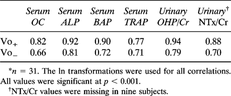
The quantification of biochemical markers of bone formation and resorption with kinetic measures of bone turnover is an essential step in their validation. Some biochemical markers have been validated by quantification against formation and resorption rates measured by calcium kinetics in adults with bone disease. However, none has been validated in healthy individuals who are undergoing skeletal growth and bone consolidation. Therefore, we have measured biochemical markers of bone formation (serum osteocalcin [OC], bone-specific alkaline phosphatase [BAP], and total alkaline phosphatase [ALP]) and resorption (serum tartrate resistant acid phosphatase [TRAP], urinary cross-linked N teleopeptides of type I collagen/creatinine [NTx/Cr], and hydroxyproline/creatinine [OHP/Cr]) in healthy females aged 11–32 years (n = 31) after an overnight fast to determine their relationship with bone formation (Vo+) and bone resorption (Vo−) as measured by calcium kinetics and balance. All biochemical markers were highly intercorrelated (r > 0.6, p < 0.001) as were Vo+ and Vo− (r = 0.91, p < 0.001). Highly significant correlations were present between bone formation measured by calcium kinetics (Vo+) and serum levels of bone biochemical markers (OC, r = 0.82, p = 0.001; ALP, r = 0.92, p = 0.001; and BAP, r = 0.90, p = 0.001) and between bone resorption measured by calcium kinetics (Vo−) and fasting serum levels and urine creatinine ratios of biochemical markers (TRAP, r = 0.77, p < 0.001; OHP/Cr, r = 0.79, p < 0.001; and NTx/Cr, r = 0.70, p < 0.001). Thus, biochemical markers of bone formation and resorption can be used to predict calcium kinetic rates during skeletal growth and the early years of formation of peak bone mass, ages at which strategies to build peak bone mass are important. Biochemical markers of formation and resorption are equally useful in predicting either the bone formation rate or the resorption rate.
INTRODUCTION
NET BONE ACCRETION during skeletal growth and consolidation and any subsequent age-related bone loss is the difference between the rate of bone formation and bone resorption. The gold standard for quantification of bone turnover rates is calcium kinetics in conjunction with calcium balance studies,1,2 although histomorphometric analysis of timed labels in bone biopsies1,3 can also be used. Both techniques are useful for research but are not suitable clinically for testing interventions designed to alter bone formation and/or bone resorption where inexpensive, easily repeatable, and noninvasive techniques are desirable.
Currently, several biochemical variables that mark for different aspects of bone formation and bone resorption are well characterized and relatively easily measured from blood and urine samples.4 Among the markers of bone formation, serum alkaline phosphatase (ALP) and serum osteocalcin (OC) have been most extensively studied. ALP, reflecting the mineralization activity of osteoblasts,5 can be measured enzymatically. However, specificity can be a problem, particularly in the presence of liver disease, because of the presence of isoenzymes.6 To overcome this, a double monoclonal antibody assay for the bone isoenzyme with less than 10% cross-reactivity for liver isoenzyme has been developed.7 Osteocalcin, a bone protein of unknown function that is uniquely produced by the osteoblast, is measured by immunoassay,8 although each assay tends to give different absolute values.9 Bone resorption can be assessed by serum tartrate-resistant acid phosphatase (TRAP),10 reflecting the enzyme activity of osteoclasts, or by the urinary excretion of bone collagen products released into the circulation by osteoclastic bone resorption. Hydroxyproline excretion has been widely used in the past. However, it suffers from lack of specificity because it reflects total collagen rather than bone collagen breakdown, and more specific assays based on molecules cross-linking the type I collagen chains in bone are available.11
A limitation to the use of biochemical markers is that they provide only a qualitative assessment of bone resorption and formation. Their level in serum and urine cannot be directly translated into rates of bone formation and resorption. To do so it is first necessary to establish the relationship between concentrations of the biochemical markers and more direct measures of bone turnover, such as those obtained by calcium kinetics combined with calcium balance1,2 or by histomorphometric analysis of double-labeled bone biopsies.1,3 Such studies have been undertaken in adults with various diseases that alter bone turnover rates, but the relationships have not been established in healthy young subjects from the period of maximal skeletal growth to the time of achieving peak bone mass.
Previously, we reported the rate of bone formation and resorption using calcium kinetics12,13 in a group of adolescent girls and young women studied in the same controlled living environment. The purpose of the present study was to establish the relationships between the level of biochemical markers of bone turnover in blood and urine and bone formation and resorption rates measured by kinetic analysis in these same subjects and for a subset restudied in late adolescence during pubertal and postpubertal growth and to test whether a measure of bone formation and a measure of bone resorption could provide a measurement of calcium retention during growth.
MATERIALS AND METHODS
Subjects
Twenty-five healthy, white female subjects aged 11–32 years were studied under a controlled living environment for 3 weeks as previously described.12,13 Six subjects initially studied at age 12–14 years were restudied at age 15–17 years, providing 31 sets of observations. Daily calcium balances were determined for 2 weeks following a 1-week adaptation. Exclusion criteria included daily dietary calcium intakes of <800 mg, disease affecting calcium metabolism, use of sex steroid contraceptives, smoking, and weight outside 85–120% of ideal body weight for age and height. The protocol was approved by Purdue University and Indiana University School of Medicine Use of Human Subject Research Committees, and informed consent was obtained from subjects and their guardians in the case of minors.
Calcium kinetics
Following a 1-week adaptation to a controlled diet of 1330 ± 101.5 mg/day, subjects were admitted to the General Clinical Research Center, and stable isotopes were administered orally (38 mg of44Ca as CaCO3), with a breakfast containing 250 mg of calcium, and intravenously (40 mg of42Ca as CaCl2) 1 h after the oral dose. Complete urine and fecal collections were obtained for 2 weeks, and 12 blood samples were collected during the first 12 h postdose and then daily for 2 weeks. Isotope ratios were determined by fast atom bombardment mass spectrometry.14 Data were fitted simultaneously to a three-compartmental model using the Simulation, Analysis, and Modeling program.15 Bone turnover parameters were determined as previously described13 including bone formation (Vo+), the calcium deposited in slowly exchanging compartments; bone resorption (Vo−), the calcium returned to the labile pool, calculated as the difference between Vo+ and calcium being excreted to keep the system in steady state; and bone balance, calculated as Vo+ − Vo−. Compartmental analysis was selected over noncompartmental analysis to gain additional information about the size of individual pools and transport between pools. Jung et al.16 compared compartmental and noncompartmental models and found that values for Vo+ did not vary systematically among models but did vary according to the length of the study.
Biochemical markers of bone turnover
After an 8-h overnight fast, blood was drawn and the second morning urine void was collected. Blood was left to clot for 30 minutes, serum was removed immediately and stored at −70°C, and urine was stored at −30°C until assayed.
Bone formation:
OC was measured by radioimmunoassay (RIA) using a rabbit polyclonal antiserum raised against bovine osteocalcin, a bovine osteocalcin as standard, and radioiodinated bovine osteocalcin as label.17 The interassay coefficient of variation (CV) is 9% at a serum concentration of 16 ng/ml. Total serum alkaline phosphatase (TAP) was determined enzymatically by standard techniques using paranitrophenol as substrate. The interassay CV is 5%. Serum bone alkaline phosphatase (BAP) was measured by a sandwich radiometric assay using two monoclonal antibodies and ALP from a human osteosarcoma cell line as standard.7,18 Reagents were supplied by Hybritech (San Diego, CA, U.S.A.). The CV is 3.2% at 45 U/l.
Bone resorption:
Serum TRAP was measured enzymatically using paranitrophenolphosphate as substrate. Serum was incubated for 1 h at 36°C before assay to destroy inhibitors.10 The interassay CV is 12% at a serum concentration of 10.8 U/l. Cross-linked N-teleopeptides of type I collagen (NTx) in urine were measured by an enzyme-linked immunoabsorbant assay using a monoclonal antibody to human NTx (Osteomark, Ostex International, Inc., Seattle, WA, U.S.A.). The interassay CV is 7.1% at a concentration of 406 nM bone collagen equivalents. After acid hydrolysis of urine, hydroxyproline (OHP) was derivatized with phenylisothiocyanate and quantitated by ultraviolet absorbance following high performance liquid chromatography (HPLC) separation using a Waters PICo Taq column eluted with a sodium acetate buffer.19 The interassay CV is 9.6% at a urinary concentration of 1.6 mg/ml. Urine creatinine (Cr) was measured by a Roche multichannel analyzer (Roche, Somerville, NJ, U.S.A.) by standard methods and expressed as the hydroxyproline/creatinine milligram ratio (OHP/Cr).
Statistics
Multiple regression analysis was used to determine the ability of bone biochemical markers to predict bone formation and resorption rates. Pearson's correlations were used to determine relationships between variables. Natural logarithm (ln) transformations were used to normalize the biochemical marker, Vo+, and Vo− data. Multiple linear regression models were used to model Vo+ and Vo−. To satisfy the independence assumptions needed for statistical inference, the repeat observations available for six individuals are not used in the calculation of correlations, p values, or prediction limits. These observations are included in the plots and regression equations, however, and clearly conform to the patterns of relationships seen for the other data. Statistics were performed using Statistical Analysis System (version 6.10, SAS Institute, Cary, NC, U.S.A.).
RESULTS
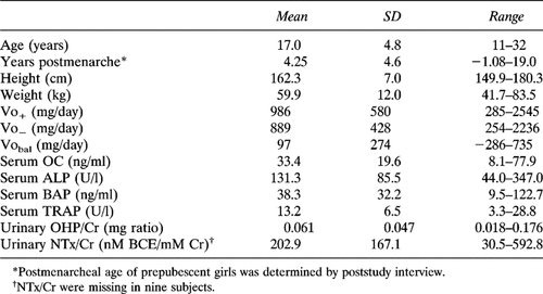
Means, standard deviations, and ranges for calcium kinetic parameters and biochemical markers of bone turnover for this healthy, young female population are given in Table 1. Menarcheal age for the seven girls who were prepubescent at the time of the study was determined through follow-up interviews.
Formation and resorption biochemical markers of bone turnover were highly intercorrelated. Between formation markers, the r values ranged from 0.85 to 0.97, and between resorption markers the r ranged from 0.63 to 0.91. Bone formation and resorption measured by kinetics, Vo+ and Vo−, were highly correlated (r = 0.91, p < 0.001).
Associations between biochemical markers of bone turnover and calcium kinetic rates were high (Table 2). Vo+ was strongly correlated with markers of bone formation, serum OC, serum ALP, and serum BAP (Fig. 1). However, the relationships between biochemical markers of resorption and Vo+ were also highly correlated (Table 2). Vo− was strongly correlated with markers of bone resorption, serum TRAP, urinary OHP/Cr, and NTx/Cr (Fig. 2), but also with biochemical markers for bone foration (Table 2). Use of natural logarithm transformations did not change the overall relationships. Serum ALP and uine OHP/Cr were the best predictors of Vo+ and Vo−.

Relationship between kinetic rates of bone formation and biochemical markers of bone formation in young females. (A) Vo+ and OC. (B) Vo+ and ALP. (C) Vo+ and BAP. Dashed lines represent 95% prediction intervals.

Relationship between kinetic rates of bone resorption and biochemical markers of bone resorption in young women. (A) Vo− and TRAP. (B) Vo− and OHP/Cr. (C) Vo− and NTx/Cr. Dashed lines represent 95% prediction intervals.
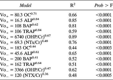
All the bone biochemical markers could be used in models using regression analysis to predict Vo+ and Vo− (Table 3). The range in R2 was from 0.44 to 0.89. Theoretically, a biomarker of bone formation could be used to predict Vo+, and a biomarker of bone formation could be used to predict Vo−, and the difference would estimate net calcium accretion. Correlations between estimated Vobal determined by the difference between predicted Vo+ and Vo− from biochemical markers using the models in Table 3 and actual Vobal determined by kinetics were significant for all pairs containing ALP to predict Vo+ and for all pairs containing TRAP to predict Vo−.
Two parameter regression models were also developed to predict bone formation and bone resorption. A model containing OHP/Cr and weight predicted Vo+ at an R2 of 0.94, whereas two biochemical markers (OHP/Cr and ALP) predicted Vo+ at R2 = 0.90. The best two parameter model for predicting Vo− contained weight and OHP/Cr at R2 = 0.75.
DISCUSSION
The primary goal of this study was to quantify the serum and urine levels of biochemical markers of bone formation and resorption by their relationship with bone formation and resorption rates determined by calcium kinetics in females during the period of skeletal growth and attainment of peak bone mass. The validity of using biochemical markers to estimate the rate of bone turnover has been reported in populations with various diseases characterized by abnormal bone turnover rates. In patients aged 26–73 years who had bone formation and resorption measured by45Ca kinetics and balance in a 7-day study, bone mineralization was significantly correlated with serum osteocalcin (r = 0.69, p < 0.001) and serum ALP (r = 0.45, p < 0.05), and bone resorption was highly correlated with urinary OHP (r = 0.78, p < 0.001).2 Nordin20 reported a similar relationship between bone resorption and OHP/Cr in 59 patients (r = 0.84). In 18 people with metabolic bone disease who had bone formation and mineral apposition rates measured by histomorphometry of iliac crest biopsies,3 serum carboxy-terminal propeptide of type I precollagen correlated positively with the mineral apposition rate (r = 0.53) and volume-referent bone formation rate (r = 0.61).
Similarly, urinary pyridinoline cross-links correlated significantly with iliac crest histomorphometric measurements of the bone formation rate (r = 0.69, 0.80, p < 0.001, and resorption (r = 0.35, p < 0.05) in 36 postmenopausal women with vertebral osteoporosis.21 In 15 patients with osteoporosis and 6 patients with Paget's disease, serum ALP was highly correlated with Vo+ (r = 0.97) and osteoblast layers in iliac bone biopsies (r = 0.85) and OHP was highly correlated with the sum of Vo+ and Vo− (r = 0.97).1 The very high correlations in this study probably resulted from the repeated measures on all of the kinetic and biochemical variables and the very wide range in variables by combining two disease states. One study of normal young (aged 30–41 years, n = 12) and older (aged 55–73 years, n = 11) women found no significant relationship between bone formation and either OC or BAP, but relationships were significant (p < 0.05) between OC and formation when age groups were analyzed separately.22
The present study is as far as we know the first to examine the relationship between biochemical markers of bone turnover to calcium kinetic rates measured by stable calcium kinetics in healthy subjects during growth. The wide ranges in body size, calcium balance, and kinetic rates and the level of bone markers reflect the large skeletal changes that occur with puberty and with subsequent achievement of peak bone mass during young adulthood. Traditional markers of bone formation (ALP) and bone resorption (OHP/Cr) correlated with Vo+ and Vo− to the same extent as studies of older populations. Charles2 reported a stronger relationship between biochemical markers and kinetic rates of bone turnover when the biochemical markers were adjusted for creatinine clearance. Renal function influences both the level of the bone biochemical markers in serum and bone turnover rates23 in an elderly, diseased population. However, in the healthy young subjects in the present study, renal function was normal. Adjusting for a number of variables relating to body size and glomerular filtration rate (lean body mass, 24-h urine creatinine and nitrogen output, total body bone calcium, and nitrogen balance) did not improve the relationships of bone biochemical markers to calcium kinetic parameters (data not shown).
Biochemical variables in serum and urine are being used clinically to assess the rate of bone formation and resorption. Biochemical markers of bone formation and resorption predicted Vo+ equally well and better than Vo−. The ability of biochemical markers to predict Vo+ better than Vo− is likely due to the fact that there is less variability in measuring Vo+ than in Vo− (9% vs. 19% precision, respectively).1 Single parameter models predicted Vo+ almost as well as two or more parameter models. Single parameter models can also predict net calcium retention; thus, serum osteocalcin predicted 63.4% of the variability in calcium retention in this population.24 The difference between bone formation and bone resorption estimated from biochemical markers generally significantly correlated with net calcium accretion (Vobal).
A study of biochemical markers of bone turnover in 91 healthy pubertal girls revealed that bone turnover was maximal in midpuberty and decreased in late puberty.25 The authors suggested that bone ALP, osteocalcin, and urinary deoxypyridinoline were more sensitive indicators of skeletal health because they showed a greater pubertal increase than other biochemical markers studied including type I procollagen carboxy-terminal propeptide, TRAP, carboxy-terminal pyridinoline cross-linked teleopeptide, immunoreactive urinary pyridinoline, and urinary galactosyl hydroxylysine. In the present study, correlations between all of the biochemical markers of bone turnover and age or postmenarcheal age were similar (r = −0.60 to −0.84, p < 0.001).
This study indicates that biochemical markers of bone turnover are as useful in predicting bone formation and resorption during growth as during the skeletal changes associated with metabolic disorders or aging. The best predictors of Vo+ were BAP, ALP, and OHP/Cr. While biochemical markers of bone formation could accurately predict bone formation, they were less able to predict Vo− (R2 < 0.65). We conclude that bone deposition can be determined quantitatively reasonably well from levels of biochemical markers in serum and urine.
Acknowledgements
The authors gratefully acknowledge the technical assistance of Ron McClintock in the analysis of the biochemical markers. This work was supported in part by Public Health Service grants R01 AR40553 and MO1 RR00750.


