Quantitative Ultrasound Assessment in Children With Fractures†
The authors have no conflict of interest
Abstract
BMD of children with fractures was compared with healthy controls using QUS. We found significantly lower SOS values in children suffering from fractures. None of the studied environmental factors could explain the difference in BMD measurements.
Introduction: The aims of this study were to compare the results of quantitative ultrasound (QUS) in children with fractures with the respective values in children without fractures and to identify possible environmental factors influencing speed of sound (SOS) in our study cohort.
Materials and Methods: BMD was measured by QUS in 50 children who had sustained an acute fracture and in 154 healthy children as controls. SOS values were obtained from the proximal phalanges of the last four fingers of the dominant hand. Nutritional habits and activity level of the children were documented by a standardized questionnaire.
Results: Children with fractures had a significantly lower SOS compared with children without a history of fractures. This difference in SOS could not be explained by differences in diet, body mass index, or physical activity.
Conclusions: Previous studies have suggested that low BMD levels might contribute to an increased prevalence of fractures in patients with systemic diseases. Our study showed that, in an otherwise healthy pediatric population, the SOS values are lower in children with fractures compared with healthy controls. Despite statistical significance, the biological impact of the results remains unclear. The difference in SOS values could not be explained by any of the studied environmental factors.
INTRODUCTION
FRACTURES CONSTITUTE 10-25% of all pediatric trauma. The calculated risk for a boy to sustain a fracture before reaching skeletal maturity is between 42% and 60% and for girls is between 27% and 40%, respectively.1, 2 The peak of incidence of fractures in children occurs at the time of peak height velocity: 11.5 and 13.5 years of age in girls and boys, respectively.3 Higher prevalence of fractures in boys has been suggested to be caused by personality and behavioral factors. Boys also participate in sports more frequently than girls.4 On the other hand, BMD is reaching a plateau at the peak of fracture incidence and is lower in boys compared with girls.5 Therefore, structural characteristics of the bone such as reduced bone mass, lower BMC, or decreased BMD, as well as architectural changes, may contribute to increased bone fragility and predispose a child to sustain a fracture.6
BMD in children can be measured by DXA, QCT, or quantitative ultrasound (QUS). Of these, both DXA and QCT are based on the emission of ionizing radiation, whereas QUS measures energy changes in ultrasound waves, resulting in speed of sound (SOS). QUS can be performed by a portable scanner and is technically simpler and more economical compared with DXA and QCT.7 QUS has been shown to be as reliable a technique as DXA in measuring BMD.8
The purpose of this study was to investigate the hypothesis that phalangeal SOS values in healthy children with fractures are lower than respective values in a control group of healthy children without fractures.
MATERIALS AND METHODS
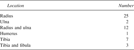
Fifty children (30 boys and 20 girls) with different acute fractures were treated between January and April 2003. The distribution of the fractures is presented in Table 1. All fractures were caused by low-energy trauma; high-impact trauma such as car accidents was excluded. Twenty-four fractures occurred during sporting activities (ball games, 16; skiing, 8) and 26 occurred at school or during leisure (falling on the ground, 17; falling from a height <2 m, 9). The mean age of the patients was 11 years (range, 9-12 years). A total of 154 healthy children (72 boys and 82 girls) without a history of a fracture was recruited from a school within the same period. The mean age of these children was 10.4 years (range, 8-12 years). Statistically significant differences between genders were not found concerning age, height, or weight.
Only healthy children were included in the study, with the exception of children with allergies. A clinical examination was performed on all children, including oral and visual status, which may reflect mineral uptake or possible impairment contributing to the mechanisms of accident, respectively. General medical history with pre- and postnatal details, nutritional habits, and supplementation (vitamin D, fluoride) were documented. Tegner activity score (TAS)9 was used to evaluate the level of physical activity. SOS was measured by QUS. Informed written consent was given by the parents of all participants.
Assessment of BMD
SOS was measured with a QUS device (DBM Sonic Bone Profiler BP01; IGEA, Carpi, Italy) consisting of two 1.25-MHz ultrasound transducers assembled on a high precision (±0.02 mm) caliper. The transducers were positioned at the distal end of the proximal phalanx on both sides of the condyles of the last four fingers of the dominant hand. The SOS through the phalanx in relation to the width of the finger was automatically calculated by the device (width of the finger/time of flight).10 The final result is presented as a mean value of these measurements (m/s). All measurements were performed by the second author to exclude interrater error. To determine the intrarater error, we measured the SOS of two children, five times each. The calculated mean percentage deviation results in the intrarater error, which was 1.3%.
Statistical analysis
All data were entered into a computerized database (Microsoft Excel) and analyzed. The results are reported as means ± SD, and for categorical data, frequencies (with percentages) are displayed. The Spearman correlation coefficient was calculated to assess the relationship between continuous variables, and cross-tabulations (two-tailed Fisher's exact test, χ2 test) were used to assess the relationship between categorical data. To determine the statistical significance of group differences in the case of continuous variables, a t-test was performed for normally distributed data; otherwise, the Mann-Whitney test was used. All computations were done using the statistical software package SPSS for Windows, version 11.0.1. A p value of <0.05 was considered significant.
RESULTS
Of all children, 46% had two or more inlays, 16% used prescription glasses, 11% were born preterm, and 6% used anti-allergic medication regularly. Daily milk consumption was reported by 62% of the participants, whereas 2% of the children refused to drink milk at all. Vitamin D supplementation during the first year of life had been carried out (84%) with few exceptions. Fluoride had been used by 61% of the children. Mean TAS score was 6.4 (range, 4-8) in children with fractures and 6.3 in the control group (range, 3-9). None of these aspects were significantly related to SOS values.
Mean SOS was 1914 ± 43 m/s (range, 1827-2040 m/s) in children with fractures and was significantly lower (p < 0.05) compared with the mean SOS of 1928 ± 39 m/s (range, 1816-2036 m/s) in the control group (Fig. 1).

Box-plot of SOS in fracture vs. nonfractured group.
In the control group, the mean SOS in boys was significantly lower than in girls (p < 0.001). SOS correlated positively with age in females (Fig. 2). In girls, the mean SOS measured 1940 ± 40 m/s (range, 1863-2036 m/s). In boys, the respective figures were 1914 ± 34 m/s (range, 1816-1998 m/s).

Age-related SOS.
DISCUSSION
BMD is defined as the average concentration of mineral per unit area of bone and is commonly measured by DXA.11 The measurement with DXA provides 2D data and is affected by the bone diameter in the considered bone section. QUS may provide BMD values independent from the bone size.12 Furthermore, it is believed that QUS values are highly correlated with the ultimate strength of the cortical bone, and therefore, they allow measurement of bone “quality.”13 In cortical bone, SOS may not only be correlated to BMD but also to bone structure and mechanical function.14, 15 In contrast, it has been shown that tibial SOS measurement may depend on BMD as well as on cortical thickness.16 Njeh et al.17 stated that the information about bone structure provided by QUS may be limited, but QUS is still a good predictor for bone strength. Njeh et al.17 concluded that QUS is useful for fracture risk prediction.
SOS values measured by QUS for 9- to 15-year-old children have been recently reported in a Polish population: SOS correlated positively with age, and a significant sex-related difference was found in children >12 years of age, with a mean SOS being higher in girls than in boys.18 Our results are consistent with the Polish study, but in our study, the sex-related difference in SOS was already evident at the age of 10 years. This might reflect a different pace of skeletal maturation in these two populations, the Austrians reaching more mature stages earlier than the Poles.
Goulding et al.19 reported that the mean BMD measured with DXA in girls with forearm fractures has been found lower than the respective values in control girls without a history of a fracture. According to Baroncelli et al.,10 the mean SOS values in children with bone and mineral disorders are lower in patients with fractures compared with patients without fractures. Our study confirmed these results in a group of healthy school-age children.
It is still controversial to what extent exogenous and endogenous factors influence bone quality.20-23 According to Cvijetic et al.,24 body weight and physical activity correlate positively with BMD. On the other hand, Halaba and Pluskiewicz18 found no correlation between dietary intake, physical activity, and bone mineralization values. Our results are in accordance with the results of Halaba and Pluskiewicz.18 We used the TAS score to register the level of physical activity. This scoring system was originally designed to determine the sports activity level after anterior cruciate ligament injuries. Hence TAS scoring has been used in evaluation of the activity level in patients with others injuries as well.25, 26 We realize that TAS scoring system is not an optimal method to register the actual amount and intensity of physical activity in children. Therefore, our results, where no relationship of TAS with SOS was found, should be interpreted with caution. For further prospective studies, a standardized activity testing with practical exercises could be more reliable.
In conclusion, in this study, children suffering from extremity fractures have lower SOS values compared with healthy controls. Despite statistical significance, the biological impact of the different SOS values remains unclear. Further investigations focusing on the factors influencing BMD in healthy children are necessary and may contribute to fracture prevention.


