Alopecia areata universalis in a dog
Sources of Funding: PAIDI (Plan Andaluz de Investigación, Desarrollo e Innovación).
Conflict of Interest: No conflicts of interest have been declared.
Part of this study was presented as a poster (PL-128) at the 31st Meeting of the European Society and College of Veterinary Pathologists. London, 2013.(http://www.esvp.eu/images/downloads/pdf/Proceedings2013.pdf).
Abstract
enBackground
Alopecia areata is a T-cell mediated autoimmune disease that occurs in humans and various other mammalian species. When the disease progresses to total alopecia it is defined as alopecia areata universalis (AAU), although this outcome has only been described in humans.
Hypothesis/objectives
To describe a case of canine alopecia areata universalis and its clinical outcome after 22 months of follow-up.
Animal
A 9-year-old intact male cross-breed hunting dog was presented with generalized and complete noninflammatory alopecia of 12–14 months duration.
Methods
Clinical examination; histopathological and immunohistochemical examination of skin biopsies.
Results
There was loss of all body hair including eyelashes and vibrissae. The histopathological and immunohistochemical findings supported a diagnosis of long-standing alopecia areata. Treatment with oral ciclosporin was associated with hair regrowth but muzzle hair, most eyelashes and whiskers were still lacking after 17 months of therapy.
Conclusions and clinical importance
To the best of the author's knowledge this is the first documented case of canine AAU. The clinical and histopathological features were consistent with a diagnosis of AAU as defined in humans. Treatment with oral ciclosporin resulted in near complete resolution of the alopecia, but after 5 months without treatment the alopecia did not relapse and spontaneous resolution cannot be ruled out.
Résumé
frContexte
L'alopecia aerata est une dermatose auto-immune médiée par les cellules T, décrite chez l'homme et des mammifères variés. Lorsque la maladie progresse en une alopécie généralisée on parle d'alopecia aerata universalis (AAU). Cette entité a été uniquement décrite chez l'homme.
Hypothèses/Objectifs
Décrire un cas d'alopecia universalis chez un chien et son évolution clinique après 22 mois de suivi.
Sujet
Un chien de chasse croisé mâle entier de 9 ans présenté avec une alopécie non-inflammatoire complète et généralisée évoluant depuis 12-14 mois.
Méthodes
Un examen clinique, un examen histopathologique et immunohistochimique des biopsies cutanées.
Résultats
Il y avait une perte de tout le pelage corporel y compris les cils et les vibrisses. Les données histopathologiques et immunohistochimiques supportaient le diagnostic d'alopecia aerata chronique. Le traitement à la ciclosporine orale a été associé à une repousse pilaire mais les poils du chanfrein, les cils et les vibrisses étaient toujours absents après 17 mois de traitement.
Conclusions et importance clinique
A la connaissance des auteurs, ceci est le premier cas décrit d’AAU canine. Les données cliniques et histopathologiques étaient compatibles avec un diagnostic d’AAU comme décrit chez l'homme. Le traitement à la ciclosporine orale a résulté en une presque complète résolution de l'alopécie mais après cinq mois sans traitement, l'alopécie n'a pas récidivé et une résolution spontanée ne peut être exclue.
Resumen
esIntroducción
la alopecia areata es una enfermedad autoinmune mediada por linfocitos T que se produce en humanos y otras especies animales. Cuando la enfermedad progresa alopecia total se define como alopecia areata universalis (AAU) aunque esta progresión sólo se ha descrito en humanos.
Hipótesis/Objetivos
describir un caso de alopecia areata universal y su progresión clínica tras 22 meses de seguimiento
Animales
un perro macho cruzado entero de nueve años de edad se presentó con alopecia generalizada y completa, no inflamada de 12 a 14 meses de duración
Métodos
examen clínico, examen histopatológico e inmunohistoquímico de las muestras de biopsias.
Resultados
hubo pérdida de todo el pelo del cuerpo, incluidas pestañas y vibrisas. Los hallazgos histopatológicos e inmunohistoquímicos indicaban un diagnóstico de alopecia areata de larga duración. El tratamiento con ciclosporina oral se asoció con crecimiento del pelo, pero el pelo del hocico, pestañas y vibrisas aún no estaba presente tras 17 meses de terapia.
Conclusiones e importancia clínica
es nuestro entender que este es el primer caso documentado de AAU en perros. Las características clínicas e histopatológicas apoyaron diagnóstico de AAU tal y como se define en humanos. Al tratamiento con ciclosporina oral resultó en casi completa resolución de la alopecia, pero tras cinco meses sin tratamiento la alopecia no se reprodujo así que una resolución espontánea no puede ser completamente descartada.
Zusammenfassung
deHintergrund
Die Alopecia areata ist eine T-Zell mediierte Autoimmunerkrankung, die beim Menschen und bei verschiedenen anderen Säugetierarten vorkommt. Wenn die Erkrankung zu einer totalen Alopezie fortschreitet, wird sie als Alopecia areata universalis (AAU) definiert, obwohl dieser Verlauf bisher nur beim Menschen beschrieben wurde.
Hypothese/Ziele
Beschreibung eines Hundes mit Alopecia areata universalis und der klinische Verlauf nach einem Follow-Up von 22 Monaten.
Tiere
Ein 9 Jahre alter intakter männlicher Mischlingsjagdhund wurde mit generalisierter und gänzlicher nicht-entzündlicher Alopezie von einer 12-14 monatigen Dauer vorgestellt.
Methoden
Klinische Untersuchung; histopathologische und immunhistochemische Untersuchung der Hautbiopsien.
Ergebnisse
Es bestand ein totaler Verlust des gesamten Haarkleids inklusive der Augenbrauen und der Wimpern. Die histopathologischen und immunhistochemischen Befunde unterstützten die Diagnose einer langandauernden Alopecia areata. Eine Behandlung mit oralem Ciclosporin ergab nachwachsendes Haar, aber Haar an der Schnauze, sowie die meisten Wimpern und Barthaare waren nach 17 monatiger Therapie nach wie vor nicht nachgewachsen.
Schlussfolgerungen und klinische Bedeutung
Nach bestem Wissen der Autoren handelt es sich hier um den ersten dokumentierten Fall einer AAU des Hundes. Die klinischen und histopathologischen Befunde waren kompatibel mit einer Diagnose von AAU, wie sie beim Menschen definiert ist. Die Behandlung mit oralem Ciclosporin ergab eine fast völlige Resolution der Alopezie , wobei nach fünf Monaten ohne Behandlung die Alopezie nicht wiedergekehrt war und eine spontane Resolution nicht ausgeschlossen werden kann.
Abstract
jaAbstract
zhIntroduction
Alopecia areata (AA) is a T-cell mediated autoimmune disease that occurs in humans and various other mammalian species.1-3. Clinically, human AA presents as patchy, isolated circular areas of complete hair loss without signs of inflammation. The disease can progress to the point where all of the hair is lost on the scalp (alopecia areata totalis) or even on the whole body including eyebrows and eyelashes (alopecia areata universalis).4, 5 In veterinary medicine one of the first reported cases of AA was categorized as alopecia areata universalis (AAU),6 but the use of this term was not appropriate because the alopecia was not complete.3, 7 In dogs, AA appears to be a rare condition.8 A previous retrospective study reports 25 cases of canine AA with initial focal or multifocal lesions mainly located on the face.1 Although in four cases hair loss had a generalized albeit incomplete distribution, complete hair loss including eyelashes and vibrissae resembling human AAU was not reported.1
The purpose of this case report is to describe the histopathological features of AA in a dog with a clinical presentation of complete hair loss consistent with AAU.
Case Report
A 9-year-old intact male cross-breed hunting dog was presented with noninflammatory alopecia. Hair loss began on the face and over 12–14 months progressively extended to the head, trunk and all limbs. Canine leishmaniosis was suspected and serology revealed a weakly positive ELISA titre (1–80; negative titre ≤1–40; IDDEX; Barcelona, Spain). There was no response to a 20-day course of treatment with meglumine antimoniate (Glucantime, Sanofi; Paris, France), at a dosage of 100 mg/kg once daily, followed by a 3 month maintenance treatment of 15 mg/kg once daily of oral allopurinol (Zyloric, Faes Farma; Leioa, Spain).
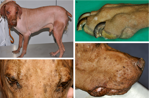
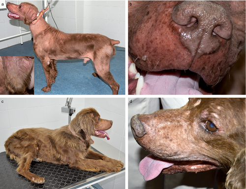
Upon presentation the general physical examination was unremarkable. The alopecia was complete except for some hair tufts on the caudal thighs (Figure 1a,b). The alopecia was not associated with any signs of inflammation and the eyelashes and whiskers were absent (Figure 1c,d).
Differential diagnoses included metabolic and endocrine diseases, and immune-mediated folliculitis. Blood and urine samples, as well as 8.0 mm skin biopsy samples from the face, trunk and limbs were taken for histopathological examination. Haemogram and routine clinical biochemistry were within normal laboratory reference ranges. An indirect immunofluorescence test (IIFT) (IDDEX) for leishmania antibodies was negative (titre 1–20). Serum thyroid hormones (total T4 and TSH) were also normal. Urine cortisol/creatinine ratio of 80.07 × 10⁻⁶ was considered higher than normal based on laboratory normal range values (normal 34 < 50 × 10−6; IDDEX). An ACTH stimulation test was performed 1 week later and both basal and post-ACTH serum cortisol (4.6 and 17.8 μg/dL, respectively) were within normal limits. Allopurinol treatment was stopped because clinical leishmaniosis was considered unlikely based on the negative IIFI titre, absence of characteristic clinical and histopathologic cutaneous lesions, normal clinicopathological results and lack of response to specific therapy.
Histopathology
Tissue samples were fixed in 10% neutral buffered formalin for 24 h, paraffin-embedded and sectioned at 4–5 μm thick for staining with haematoxylin and eosin (H&E), periodic acid Schiff (PAS) and Giemsa according to routine histological procedures.
Microscopic examination showed lesions common to all samples. Severe follicular atrophy was characterized by thin, telogenized and sometimes distorted follicles (Figure 2a). Discrete infundibular hyperkeratosis and occasional vacuolar degeneration of follicular keratinocytes was observed, as well as mild peribulbar and perifollicular fibrosis. Occasional lymphocytic infiltration involving the isthmus and/or the follicular anagen bulbs was seen (Figure 2b–d) in areas where patches of hairs remained, but was absent in more chronic alopecic skin. The epidermis was normal or moderately acanthotic and hyperpigmented. The sebaceous glands were normal. An immunohistochemical study, using formalin-fixed, paraffin-embedded tissue from serial sections from all biopsies, was performed by the avidin-biotin-peroxidase complex method (ABC)9 with the polyclonal CD3 and monoclonal CD79a antibodies (DAKO; Glostrup, Denmark). Briefly, endogenous peroxidases were blocked by incubating sections in methanol with 3% hydrogen peroxide for 30 min. Slides were incubated with primary antibodies overnight at 4°C; then incubated with secondary goat anti-rabbit (CD3ε) and anti-mouse IgG (CD79a) (DAKO) antibodies for 45 min at room temperature followed by incubation with streptavidin-conjugated horseradish peroxidase, 1:200 in Tris-buffered saline, for 45 min. Afterwards, immunoreaction was developed with diaminobenzidine tetrahydrochlorid. Slides were not counterstained with haematoxylin.
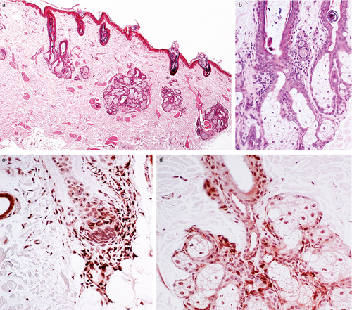
Immunohistochemical staining showed that the bulbar and lower follicular epithelium infiltrating lymphocytes were CD3+ (T lymphocytes) (Figure 2c,d). Morphological and immunohistochemical characteristics confirmed the diagnosis of severe follicular atrophy with T cell bulbitis and isthmus mural folliculitis consistent with late-stage AA.
Treatment and follow-up
The dog was treated with oral ciclosporin 5 mg/kg once daily (Atopica®, Novartis; Barcelona, Spain) and generic ketoconazole (10 mg/kg once daily). After 3 months new hair growth was observed on the digits and distal pinnae. The pattern of hair regrowth followed the pattern of hair loss in a reverse way, so that the last skin regions affected by the alopecia were the first to recover. From the distal limbs, hair growth progressed proximally. After 9 months of therapy the head, neck, most of the pinnae and large areas of the trunk were still alopecic (Figure 3a,b). After 14 months of therapy only the muzzle and periocular skin were still alopecic (Figure 3c) and eyelashes were also absent. Because of the marked hair regrowth on the body and the lack of growth on the muzzle after a further three more months of therapy, treatment was stopped. The dog was re-evaluated again 5 months post-treatment and there were no signs of relapse (Figure 3d). In view of the dog's positive clinical response further testing for leishmaniosis was considered unnecessary and its former diagnosis discarded.
Discussion
The histopathological pattern and immunohistochemical identification of T lymphocytes (CD3+, CD79a-) forming the unique inflammatory infiltrate involving hair bulb and follicular isthmus were consistent with a diagnosis of T-cell mediated AA.10 Contrary to other previously described cases, this dog lost its hair coat over the entire body including eyelashes and vibrissae, similar to human AAU. This progression of AA to AAU has not, to the best of the authors’ knowledge, been previously described in dogs.
In human AA, it has been hypothesized that AA develops in a previous healthy follicle because its constitutive immune privilege collapses. According to this theory, patients with a genetic predisposition are prone to abnormalities in the microenvironment of the follicle, allowing follicular autoantigens to be presented to pre-existing autoreactive CD8+ T cells. In our case, the dog had been formerly diagnosed and treated for leishmaniosis on the basis of a low positive ELISA titre but, although the alopecia had been present for many months, the dog never developed clinical signs of leishmaniosis. Moreover, progression of the alopecia continued in spite of the treatment of leishmaniosis and serum antibody titre was negative upon first presentation. When AAU was diagnosed no clinicopathological or histopathological changes compatible with leishmaniosis were present and the alopecia improved during ciclosporin therapy. Subclinical infection prior to referral cannot be completely ruled out, but any relationship with AAU would be unlikely in view of the dog's clinical evolution.
Dogs with AA may self-cure, respond to immunosuppressive therapy or be resistant to therapy.1, 8, 11 Spontaneous and complete hair regrowth was reported for 12 of 20 dogs (60%) where information on disease outcome was available.1 In very severe forms like human AAU, spontaneous remissions are infrequent.2 The general consensus is that no particular therapy is routinely successful; assessment of therapeutic benefit is difficult due to the possibility of spontaneous regrowth of hair.6
In one dog unresponsive to oral immunosuppressive doses of glucocorticoids, administration of low dose oral ciclosporin was followed by complete remission within 3 months,1 and several years later one more case of canine AA was successfully treated with oral ciclosporin.12 In our case, alopecia was extensive and long standing and it would be reasonable to attribute hair regrowth to ciclosporin administration; nevertheless, it has been reported that dogs can recover spontaneously from AA after a course of 6 months to 2 years.11 In the present case of AAU the owner dated the onset of alopecia around May–June 2011 and hair regrowth was first reported on 22 April 2013, 2 months after initiating ciclosporin treatment and within 2 years of the disease becoming apparent. In contrast with the common relapses reported in human cases treated with ciclosporin,13 after 5 months without treatment the dog remained stable with no signs of relapsing, a fact that increases the probability of spontaneous partial resolution.
In conclusion, we described a case of canine AAU. The clinical and histopathological features were consistent with a diagnosis of canine AA and the total hair loss, including eyelashes and whiskers, was consistent with classic human AAU. Treatment with oral ciclosporin was associated with clinical resolution but muzzle hair, most eyelashes and whiskers were still lacking after 17 months of therapy; once ciclosporin was discontinued there were no signs of relapse and spontaneous resolution cannot be ruled out.


