Uninterrupted growth in a non-polar hadrosaur explains the gigantism among duck-billed dinosaurs
Data archiving statement
Data for this study are available in the Dryad Digital Repository: https://doi.org/10.5061/dryad.d2547d7z3
Abstract
Duck-billed dinosaurs (Hadrosauridae) were the most common ornithopods of the Late Cretaceous. Second only to sauropods and in many cases exceeding the sizes of the largest land mammals (such as indricotheres or proboscideans), they are among the largest terrestrial herbivores to have walked the Earth. Despite their gigantic size, diversity and abundance, their growth strategies remain poorly understood. Here, we examine the bone microstructure of several Mongolian hadrosauroids of varied adult sizes. The small and middle-sized species have lines of arrested growth (LAGs). On the other hand, one of the largest duck-billed dinosaurs, Saurolophus angustirostris, shows uninterrupted growth, comparable with other big hadrosaurs for which the lack of cyclical growth arrests was interpreted as a result of living in the polar region. Since both of the studied taxa inhabited warmer, continental, monsoon-influenced environments of the Late Cretaceous Mongolia, we propose that the absence of LAGs is not a climatic-driven condition but rather connected with the animal's size (i.e. ontogeny). Our results show that, like sauropods, hadrosaurs changed their growth dynamics from cyclical to continuous during their evolution, which made it possible for them to achieve comparable body sizes.
Recently, it has been shown that growth strategy played an important role in achieving gigantism in sauropods (Cerda et al. 2017; Apaldetti et al. 2018). It seems that prolongation of fast and uninterrupted growth as a single process or in few stages was one of the important ways to achieve large body sizes, because this growth strategy is not only restricted to dinosaurs (Botha & Chinsamy 2001; Botha-Brink & Smith 2011; Sulej & Niedźwiedzki 2019) but has also been noted in large Therapsida and non-archosaurian Archosauriformes (Botha & Chinsamy 2001; Botha-Brink & Smith 2011; Sulej & Niedźwiedzki 2019). Similar adaptations have also been observed in the largest modern and fossil turtles: protostegids and dermochelyids (Rhodin 1985). Another dinosaur group, hadrosaurs, also tended to gain large body size (up to 16.5 m; Shantungosaurus giganteus Hu, 1973; Zhao et al. 2007) similar to some medium-sized sauropods (e.g. Cetiosaurus oxoniensis Phillips, 1871; Paul 2010). Unlike sauropods and despite their abundance in the fossil record, hadrosaurs are poorly known in terms of their bone microstructure (Sander 2000; Sander et al. 2004, 2011; Waskow & Sander 2014; Cerda et al. 2017). The main studies concerning hadrosaur histology thus far have been focused on growth dynamics of particular species (Horner et al. 2000; Vanderven et al. 2014; Woodward et al. 2015; Woodward 2019). Previous studies have shown that, for example, Maiasaura peeblesorum Horner & Makela, 1979 exhibits cyclical growth (Horner et al. 2000; Woodward et al. 2015) characterized by cyclical changes of bone vascularization (Chinsamy-Turan et al. 2012; Vanderven et al. 2014), in contrast to Edmontosaurus regalis Lambe, 1917 which had uninterrupted growth. However, the different growth strategy of the latter species was attributed to climate changes and migrations (Chinsamy-Turan et al. 2012). Similar assumptions had been made for the Alaskan Ugrunaaluk kuukpikensis Mori et al., 2015, which is closely related to Edmontosaurus. Lines of arrested growth (LAGs) were reported only in radii but not in other long bones (femora, tibiae, humeri) (Erickson et al. 2018). However, recently Woodward (2019) pointed out that the tibiae of M. peeblesorum exhibit both LAGs and zonal changes in vascular pattern (modulations sensu de Ricqlès (1983; Rimblot-Baly et al. 1995; Curry 1999), or localized vascular changes, LVCs (Woodward 2019)) which are present before the first LAG. The LVCs and the absence of LAGs are common in sauropods (Curry 1999; Sander 2000; Cerda et al. 2017). During the evolution of Sauropodomorpha, their growth dynamics changed from cyclical to uninterrupted (Cerda et al. 2017) and our research indicates that Hadrosauriformes show convergent growth strategy changes, which in both groups are connected with the achievement of large body sizes (Apaldetti et al. 2018).
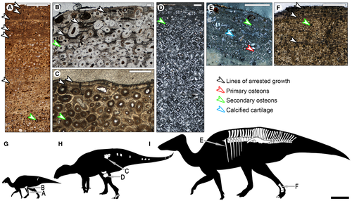
Thin sections of the Hadrosauroidea from the Baynshire Formation reveal that there were at least two forms, as previously noted (Norman & Kurzanov 1997; Tsogtbaatar et al. 2011). Recently, a bigger species has been described (Gobihadros mongoliensis Tsogtbaatar et al., 2019) and here we present remains of a smaller indeterminate Hadrosauroidea, the long bones of which already show marks of adulthood (Fig. 1B, C). Moreover, we also analysed the bone microstructure of Barsboldia sicinskii Maryańska & Osmólska, 1981 (Fig. 1E–F) and Saurolophus angustirostris Rozhdestvensky, 1952 (Fig. 2A–L), both from the Nemegt Formation. The latter is the most abundant Maastrichtian dinosaur of Asia, which allows us an insight into its ontogeny. We compare our results with data published for other Hadrosauriformes (Horner et al. 1999, 2000; Chinsamy-Turan et al. 2012; Vanderven et al. 2014; Fowler & Horner 2015; Prieto-Márquez et al. 2016a; Bertozzo et al. 2017; Stein et al. 2017; Erickson et al. 2018; Woodward 2019) and non-hadrosauriform members of Iguanodontia (Werning 2012). Finally, we examined the changes in growth strategy of duck-billed dinosaurs and checked whether the ‘typical’ sauropod growth strategy (Sander et al. 2004; Apaldetti et al. 2018) could also occur in hadrosaurs. The observed growth schemes seem to be universal across all amniotes and have implications for the interpretation of migratory behaviours of fossil animals.
Institutional abbreviations
PIN, Paleontological Institute, Russian Academy of Sciences, Moscow, Russia; ZPAL, Roman Kozłowski Institute of Paleobiology, Polish Academy of Sciences, Warsaw, Poland.
Geological setting
Locality
Specimens of the indeterminate Hadrosauroidea were found in 1963 during Polish–Mongolian Palaeontological Expeditions in Bayn Shire (also known as Bayan Shireh; ZPAL MgD-III/2, MgD III/7, MgD-III/17, and MgD-III/19) and Char Teeg (also known as Khar Teg; MgD-III/10), where the Baynshire Formation (also called the Bayanshiree Formation (Svita) or the Bayanshirenskaya Formation) is exposed. The fossils of Gobihadros mongoliensis analysed herein (ZPAL MgD-III/3) were found also in 1963, but in Khongil Tsav (also Changil Tsaw), in the same formation. Gobihadros mongoliensis has previously been reported from this locality (Tsogtbaatar et al. 2019).
Bayn Shire is the stratotype locality of the Baynshire Formation. From this locality mainly dinosaur bones and turtle remains are known (Kielan-Jaworowska & Dovchin 1968; Danilov et al. 2014; Tsogtbaatar et al. 2019).
At the Char Teeg locality sandy and clayey sediments crop out with numerous fragments of dinosaur skeletons, turtle shells and bivalves (Kielan-Jaworowska & Dovchin 1968). Khosatzky & Młynarski (1971) and Danilov et al. (2014) considered an indeterminate Trionychidae ZPAL MgCh/71 from Char Teeg, supposedly found in the Nemegt Formation. We identified other fossils found in Char Teeg during the Polish–Mongolian Palaeontological Expeditions in 1963, some of them as remains of Gobihadros mongoliensis (pes ungual ZPAL MgD-III/5; see Słowiak et al. 2019), which was described from the Baynshire Formation. Moreover, the lithology description given by Kielan-Jaworowska & Dovchin (1968) is also more consistent with the Baynshire rather than the Nemegt Formation.
All the fossils of Saurolophus angustirostris analysed here were found in the Nemegt Formation, but from different localities (N Nemegt, Altan Ula, Tsagan Khushu, Naran Bulak; see Słowiak et al. 2019). The remains of the Barsboldia sicinskii holotype were found in Northern Sayr, were the Nemegt Formation is also present.
Geology
The Upper Cretaceous Baynshire Formation is considered to be late Cenomanian to Santonian in age, lying between the older Sainshand Formation (Cenomanian) and younger Djadokhta Formation (middle Campanian) (Hicks et al. 1999; Jerzykiewicz 2000). The Baynshire Formation is considered to be similar in age to the Iren Dabasu Formation of Inner Mongolia, because of its similar fauna (Jerzykiewicz & Russell 1991). However, some studies do not agree with this age estimation (Van Itterbeeck et al. 2005), suggesting that the Iren Dabasu Formation is late Campanian to early Maastrichtian in age. The lithology of the Baynshire Formation is characterized by the presence of yellowish-brown medium-grained sandstones and grey mudstones (Watabe & Suzuki 2000). Watabe & Suzuki (2000) point out that the Baynshire Formation is composed of heterolithic interbedded units characteristic of meandering rivers (point bar deposits). On the other hand, Jerzykiewicz & Russell (1991) suggested that the Baynshire Formation can be divided into two units: the upper and lower beds. The latter are composed of conglomerates, indicating a high-energy fluvial environment. In turn, the upper beds have mudstones and claystones interbedded with sandstone suggesting the presence of rivers and a more lacustrine environment. All the fossils of the indeterminate Hadrosauroidea described herein have on their surfaces some adhering mudstone, suggesting therefore the upper part of the Bayn Shireh Formation. Based on turtle remains which are common along with dinosaur fossils in the Baynshire Formation, Danilov et al. (2014) suggested that the lower beds are Cenomanian to early Turonian, and the upper part is late Turonian to Santonian in age.
Material
The available material of Saurolophus angustirostris (PIN 551/8; PIN 552/2; ZPAL MgD-I/157; MgD-I/158; MgD-I/159), Barsboldia sicinskii (ZPAL MgD-I/110), Probactrosaurus gobiensis Rozhdestvensky, 1966 (PIN 2232/1; PIN 2232/10; PIN 2232/18-1) and a cast of Olorotitan arharensis Godefroit et al., 2003 were examined for comparative purposes. Comparisons with other taxa are based on their initial descriptions, unless otherwise specified.
Hadrosauroidea gen. et sp. indet
ZPAL MgD-III/2
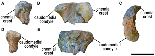
Proximal end of the right tibia (Fig. 3). The specimen is broken just below the proximal head. Found in Bayn Shire.
ZPAL MgD-III/7
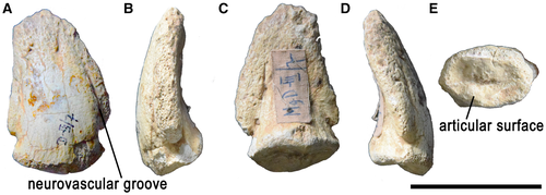
The ungual of the second digit of the left pes (Fig. 4). This specimen is mostly complete, showing only some minor damage around some of its edges. Found in Bayn Shire.
ZPAL MgD-III/10
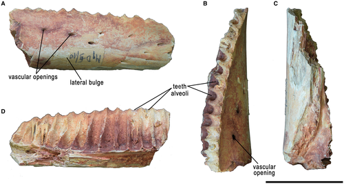
A small fragment of the right dentary representing the lateral (labial) part of the mandible, close to the symphysis (Fig. 5). Only remnants of teeth are preserved. Found in Char Teeg.
ZPAL MgD-III/17
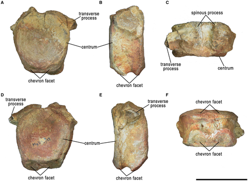
A proximal caudal vertebra (Fig. 6). The centrum is mostly complete but the processes and the neural arch are broken. Found in Bayn Shire.
ZPAL MgD-III/19
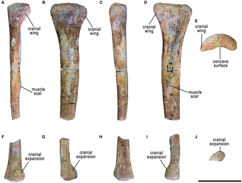
Most of the right fibula (Fig. 7). Approximately 15% of the bone is missing, separating the proximal and distal parts. Found in Bayn Shire.
Method
3D imaging
The 3D images of the specimens were prepared using the Shining 3D EinScan Pro 2X 3D scanner fixed on a tripod with EinScan Pro 2X Color Pack (texture scans), EinTurntable (alignment based on features), and EXScan Pro 3.2.0.2 software. The number of turntable steps was varied, chosen depending on the specimen. The models were meshed using the Watertight Model and High Detail presets.
Ontogenetic age criteria
In many cases it is difficult to determine accurate ontogenetic ages for fossil vertebrates. The most obvious basis for ontogenetic interpretations is the size of the specimens; on average, the larger the individual, the older it is. This is most useful for animals with indeterminate growth and it allows relatively safe interpretations of relative ages of individuals differing significantly in size. Another clue about the ontogenetic age of the individuals is the advancement of their osteological development; for example, the degree of ossification of the epiphyses, closure of sutures between the neural arches and vertebral centra or within the skull, ossification of carpal bones, development of sexually dimorphic features, etc. It must be kept in mind, however, that individuals of similar ontogenetic age may differ in size and developmental advancement due to intraspecific variability, sexual dimorphism, or environmental factors (Gilliland & Ankney 1992; Eaton & Link 2011; Hone et al. 2016; and references therein). Finally, the number of the lines of arrested growth (LAGs) is frequently used for ontogenetic age determination (Castanet et al. 2004; Horner & Padian 2004; Woodward et al. 2015). LAGs roughly correspond to annual growth cyclicity (Castanet et al. 2004; Woodward et al. 2015) and are independent of metabolic rate and climatic background (Huttenlocker et al. 2013) but at least in some animals may be influenced by photoperiodic cyclicity (Castanet et al. 2004). However, the record may be obscured by bone remodelling and by the fact that young animals do not arrest their growth but have some annular changes of vascularity pattern (Woodward 2019). Obviously, the animal must exhibit LAGs in its bone tissue to begin with and this is not the case for many taxa. Our interpretations of the relative ontogenetic ages of the studied individuals are based on all of the aforementioned clues, but mostly on the relative sizes. The differences in histological images obtained from the individuals interpreted as being of different ontogenetic ages and homogeneity of the images obtained within each of the inferred age groups suggest correctness of our interpretations, but some degree of caution must be exercised, as the recognition of the age groups is hypothetical.
We obtained most of our samples from incomplete bones of Saurolophus angustirostris; most of them had already been described by Maryańska & Osmólska (1984). To distinguish rough ontogenetic classes, we had to estimate the length of the individuals. We measured the preserved distal or proximal ends of long bones and compared them to the most complete skeleton of Saurolophus angustirostris (PIN 551/8, holotype) and the most complete skeleton of that animal housed ZPAL collection (ZPAL MgD-I/158). Therefore, we were able to obtain an approximate total length of each sampled fragmentary bone proportionately to a complete bone of PIN 551/8. The total length of the holotype is about 10 m, so the length of remaining specimens was proportionally approximated based on the length of each individual bone to the length of the appropriate bone of PIN 551/8 (Słowiak et al. 2019). The size of the specimen ZPAL MgD-I/168, from which we had taken the rib sample, was estimated based on its pelvis length. We classified the rib sample of ZPAL MgD-27 + 178 as taken from an adult individual because of its skull features. So far, the biggest known Saurolophus angustirostris individual is PIN 662/2. It was impossible to take histological samples from this individual, but we regarded it as the maximum or close to maximum size of S. angustirostris, which we estimate proportionally to the holotype (PIN 551/8) as 12.6 m (Słowiak et al. 2019). We are aware that dinosaurs grew allometrically, but the bones which we used for histological sampling were highly incomplete, so the approximations of the size or weight of the owners of the sampled bones will always be biased. The main purpose of our estimations was to obtain an approximation of the size of the animal.
The holotype of Barsboldia sicinskii (ZPAL MgD-I/110) was already considered to be an individual of advanced age (Brett-Surman 1989). The length of its tibia was estimated to be 140 cm (Prieto-Márquez 2011) indicating that it was similar in size to the biggest hadrosaurs, such as Shantungosaurus giganteus.
The holotype of Gobihadros mongoliensis (MPC-D100/763) was approximately 3 m long when it died (Tsogtbaatar et al. 2019). It was not a fully grown individual. In the supplementary materials attached to the article describing Go. mongoliensis, a specimen (MPC-D100/744) is mentioned with a femur measuring 72.8 cm, so the approximate length of the whole dinosaur was about 5.3 m. The individual ZPAL MgD-III/3 had a larger tibia and femur in comparison to specimen MPC-D100/744. The approximate length of the femur ZPAL MgD-III/3 was 104 cm, so the full length was about 7.5 m. The old age of ZPAL MgD-III/3 is also supported by the presence of many pathologies in the postcrania and the presence of an external fundamental system in the femur, described in detail below. We tentatively consider the individual ZPAL MgD-III/3 to be a representative of Go. mongoliensis because the remains were found in Khongil Tsav, in the Baynshire Formation, and the proximal end of tibia and femur, caudals, and pes fit into the morphology of Go. mongoliensis described by Tsogtbaatar et al. (2019). These elements are in many cases considered to be of little diagnostic value, but since there are no characters refuting this attribution, we consider it more parsimonious to refer the specimens to an already known species. The specimen ZPAL MgD-III/3 will be described in detail separately.
The tibia of the indeterminate hadrosauroid bears the external fundamental system in the outer cortex, and we therefore regard it as a fully-grown animal (Horner et al. 2000).
Bone histology and sections
Long bones (the humerus, ulna, radius, tibia, fibula), scapula, ribs and metacarpal of Saurolophus angustirostris, long bones (femur, tibia) of Gobihadros mongoliensis, tibia and fibula of the indeterminate hadrosauroid, and a rib and metatarsal of Barsboldia sicinskii were sectioned (Słowiak et al. 2019), in most cases as close as possible to the middle of the shaft. However, some sections close to the metaphysis are also included herein to document changes in histology along the bone length. The sections were obtained using standard procedures (Padian & Lamm 2013) in the Institute of Paleobiology, Polish Academy of Sciences and the Faculty of Geology, University of Warsaw. Large-scale photographs of thin sections were obtained using a Nikon Eclipse LV100 POL polarizing microscope with a DS-Fil camera in transmitted normal and polarized light, including a quartz wedge. The pictures were combined together in NIS-Elements microscope imaging software.
For the description of bone histology we used standard terminology and definitions following Chinsamy-Turan (2005) and Padian & Lamm (2013). The abbreviations used in the histological descriptions are:
LAGs, lines of arrested growth
A result of a pause in bone deposition visible in histological images by a line indicating a complete stoppage of growth with some resorption of bone tissue on the bone surface (Padian & Lamm 2013).
EFS, external fundamental system
Several closely spaced LAGs in the outermost cortex. This is understood to be an indicator of adulthood, because when the full size is approached by an animal, the rings become tightly packed (Horner et al. 2000).
RTB, relative bone wall thickness
A calculation of the ratio of the cross-sectional bone wall thickness to the cross-sectional bone diameter, expressed as a percentage (Ray et al. 2004). Because bones sectioned herein are mostly not round, for the cross-sectional bone diameter we calculated the mean of the diameter in two perpendicular axes.
Phylogenetic analysis
The phylogenetic analysis was performed using the matrix of Sues & Averianov (2009) with the updates by Tsogtbaatar et al. (2014), with 7 taxa (Eotrachodon orientalis Prieto-Márquez et al., 2016b, Gilmoreosaurus mongoliensis (Gilmore, 1933), Gobihadros mongoliensis, Probrachylophosaurus bergeri Fowler & Horner, 2015, Tenontosaurus tilletti Ostrom, 1970, Ugrunaaluk kuukpikensis, and Hadrosauroidea gen. et sp. indet.) and 14 characters added. In total, the matrix includes 49 taxa and 152 characters. The matrix was analysed in TNT (Goloboff et al. 2008a) with all characters unordered, Hypsilophodon foxii Huxley, 1870 as an outgroup, 10 000 replications, and 100 trees saved per replication. Initially, a traditional search (TBR) was used, resulting in 730 equally parsimonious trees (best score 450, CI = 0.384, RI = 0.805). Since the resolution of the obtained consensus tree was unsatisfactory with many taxa lumped into two large polytomies and only Probactrosaurus gobiensis, Protohadros byrdi Head, 1998, Lambeosaurinae and some clades of Saurolophinae recovered as separate branches, we then used the implied weighting option with K = 12, as suggested for morphological datasets by Goloboff et al. (2008b). This run gave 15 equally parsimonious trees (best score 18.42903, CI = 0.384, RI = 0.805). The jackknife values were calculated using default settings with 1000 replications.
Systematic palaeontology
DINOSAURIA Owen, 1842 ORNITHISCHIA Seeley, 1887 ORNITHOPODA Marsh, 1881 HADROSAURIFORMES Sereno, 1997 sensu Sereno 1998 HADROSAUROIDEA Cope, 1869 HADROSAUROIDEA gen. et sp. indet. Figures 3-7
Material
ZPAL MgD-III/2, pes ungual of the left 2nd digit; ZPAL MgD-III/2, right tibia; ZPAL MgD-III/10, right dentary; ZPAL MgD-III/19, right fibula; ZPAL MgD-III/17, proximal caudal vertebra (Figs 3-7).
Locality and horizon
Bayn Shire (type locality) and Char Teeg, south-eastern Mongolia. Upper part of the Baynshire Formation, late Turonian–Santonian in age (Jerzykiewicz 2000).
Osteological description
Dentary (ZPAL MgD-III/10)
In dorsal view, the tooth row is gently bowed. The lateral convex bulge is prominent but does not form a pronounced shelf. It is rounded, the curves are gentle, and its depth increases caudally in a straight line. The distance between the tooth row and the top of the bulge is proportionally smaller than in Saurolophus angustirostris (ZPAL MgD-I/159, PIN 551/8). Above it and (in the anterior part) on it, 11 neurovascular openings can be seen. The three most caudal of these openings are the largest, and all of the neurovascular canals are turned slightly anteriorly. Fourteen closely packed tooth alveoli are preserved (the anteriormost and posteriormost only partially), some of which still contain fragments of teeth (Fig. 5).
The walls separating the teeth are slender, as in Probactrosaurus gobiensis (PIN 2232/18-1). Vascular openings along the dorsolateral surface of the dentary are present in many hadrosauroids and their number appears variable; for example, in Gilmoreosaurus mongoliensis (minimum 7; see Prieto-Márquez & Norell 2010), Pr. gobiensis (minimum 12; PIN 2232/18-1), Eolambia caroljonesa Kirkland, 1998 (minimum 8; see McDonald et al. 2012) and Gobihadros mongoliensis (minimum 18; see Tsogtbaatar et al. 2019). In some species, such as Plesiohadros djadokhtaensis Tsogbaatar et al., 2014 or Saurolophus angustirostris (ZPAL MgD-I/159, PIN 551/8), the openings are missing. The lateral bulge is very close to the tooth row, in contrast to the situation seen in Eo. caroljonesa (see McDonald et al. 2012) and Bactrosaurus johhnosi Gilmore, 1933 (see Prieto-Márquez 2011). It is more similar, although still narrower and more pronounced laterally, to that of Gi. mongoliensis (see Prieto-Márquez & Norell 2010), Go. mongoliensis (see Tsogtbaatar et al. 2019), Pr. gobiensis (PIN 2232/18-1). In contrast to the co-occurring Go. mongoliensis, the indeterminate Hadrosauroidea has the dentary tooth row convex medially as in Bactrosaurus johnsoni (see Prieto-Márquez 2011) instead of being straight or concave medially (Tsogtbaatar et al. 2019). Moreover, the whole dentary of Go. mongoliensis bears 18 tooth positions, while the partial dentary of ZPAL MgD-III/10 already had 14 alveoli and they seem to be proportionally shallower and narrower than in Go. mongoliensis (see Tsogtbaatar et al. 2019). This brings it closer to Gilmoreosaurus mongoliensis (Iren Dabasu Formation, Santonian), having more than 30 tooth alveoli.
Tibia (ZPAL MgD-III/2)
Only the expanded craniocaudally proximal end of the right tibia is preserved (Fig. 3). The articular surface is comma-shaped in proximal view. The caudomedial condyle is massive, wide and rounded in the caudal view. In lateral view its most proximal part expands caudally. The lateral condyle is not preserved. The cnemial crest is short and strongly curved laterally (stronger than in Eolambia caroljonesa (see McDonald et al. 2012) and is therefore almost invisible in medial view, but in cranial view it forms a rounded expansion. The craniolateral margin of the cnemial crest is rugose, similar to the proximal articular surface, suggesting the presence of cartilage.
The caudomedial condyle is caudally expanded and the dorsal surface of tibia is also straight and flat, similar to Gilmoreosaurus mongoliensis (see Prieto-Márquez & Norell 2010). In dorsal view, the lateral margin of tibia is rounded in contrast to the straighter margin of Eolambia caroljonesa (see McDonald et al. 2012) or Probactrosaurus gobiensis (PIN 2232/1 and 2232/10). The cnemial crest is slender, and curved laterally, in similar manner to that of most Hadrosauroidea (e.g. Gi. mongoliensis (see Prieto-Márquez & Norell 2010), Saurolophus angustirostris (PIN 551/8), Edmontosaurus annectens (Marsh, 1892) (see Prieto-Márquez 2014). In contrast to Gobihadros mongoliensis (ZPAL MgD-III/3 and Tsogtbaatar et al. 2019), instead of being robust, the proximal end of tibia in the indeterminate Hadrosauroidea is slender, as in Pr. gobiensis (PIN 2232/1 and 2232/10). Moreover, the medial surface of the proximal end of tibia is gently arched and has a smooth surface, without any of the depressions that occur in Go. mongoliensis (ZPAL MgD-III/3 and Tsogtbaatar et al. 2019). Also, the cnemial crest of the indeterminate Hadrosauroidea is restricted to the proximal end and placed close to the base of the missing lateral condyle. This is similar to Pr. gobiensis (PIN 2232/1 and 2232/10) and Bactrosaurus johnsoni (see Prieto-Márquez 2011), but in Go. mongoliensis (ZPAL MgD-III/3 and Tsogtbaatar et al. 2019) the depression separating the dorsoventrally long cnemial crest from lateral condyle is wider and shallower. All those features differentiating the indeterminate Hadrosauroidea from Go. mongoliensis may appear ontogenetically dependent at first glance, but the thin sections taken from tibia ZPAL MgD-III/2 show a greatly expanded secondary remodelling throughout whole cortex (as in Go. mongoliensis ZPAL MgD-III/3) and the external fundamental system present in the outermost cortex indicates the adult state of the individual (see the description below).
Fibula (ZPAL MgD-III/19)
The proximal end of the right fibula is more expanded craniocaudally than the distal end (Fig. 7). The cranial expansion of the proximal end forms a triangular wing slightly curving medially. The lateral surface of the proximal end is convex, while the medial is concave, forming an articulation surface for the proximal condyle of the tibia. The generally straight shaft is flat mediolaterally in the proximal part and becomes triangular in the distal part. In the middle of the shaft, on the medial surface, a noticeable muscle scar is present, possible an attachment for the m. iliofibularis. The distal end is flat caudally, but expanded craniomedially and flattened craniolaterally, forming a tear-shaped, gently convex end in medial view.
The shape of the proximal fibula in lateral view is similar to that of Tenontosaurus tilletti (see Forster 1990), in which the proximal shaft is slightly bowed cranially. The proximal end is more expanded cranially than caudally forming a boot-shaped expansion, similar to that in most representatives of Hadrosauridae (e.g. Saurolophus angustirostris PIN 551/8), and Eolambia caroljonesa (see McDonald et al. 2012), Gobihadros mongoliensis (see Tsogtbaatar et al. 2019) or Tanius sinensis Wiman, 1929 (see Borinder 2015). In contrast, the proximal end of the fibula in Probactrosaurus gobiensis (PIN 2232/1 and 2232/10), Olorotitan arharensis (see Godefroit et al. 2011) or Probrachylophosaurus bergei, is moderately flared. The medial surface of the proximal part of the tibia is concave, as in most representatives of Hadrosauridae (e.g. Saurolophus angustirostris (PIN 551/8) or Edmontosaurus annectens (see Prieto-Márquez 2014)). In some basal representatives of Hadrosauridae, such as Eo. caroljonesa (see McDonald et al. 2012) or Pr. gobiensis (PIN 2232/1 and 2232/10), the fibula is flat medially. The distal end of the fibula in the indeterminate Hadrosauroidea is consistent with most of Hadrosauridae in being club-shaped in ventral view (e.g. Saurolophus angustirostris (PIN 551/8), Plesiohadros djadokhtaensis (see Tsogbaatar et al. 2014)). The large muscle attachment in the middle of the fibula shaft of the indeterminate Hadrosauroidea and its cranial bowing is not present in other Hadrosauroidea, such as Eo. caroljonesa (see McDonald et al. 2012), Gilmoreosaurus mongoliensis (see Prieto-Márquez & Norell 2010), Gobihadros mongoliensis (see Tsogtbaatar et al. 2019) and Pr. gobiensis (PIN 2232/1 and 2232/10).
Among Hadrosauriformes in general, the fibula has a very conservative morphology, so it is hard to distinguish different genera based on its macrostructure. The histological section of the fibula (see below) shows at least eight LAGs placed close to each other and the vascularization is very low in the outermost cortex. This suggests that the animal was almost fully grown. The longest fibula of the Gobihadros mongoliensis individuals described by Tsogtbaatar et al. (2019) was 65.8 cm. The proximal end of the tibia of the large Go. mongoliensis individual ZPAL MgD-III/3 indicates that its fibula could reach about 74.5 cm (based on the proportion between the tibia and fibula). This makes it over 20% larger than the fibula ZPAL MgD-III/19, the total length of which is estimated to be about 50 cm. Because the latter fibula is almost fully grown and it is much smaller than the fibulae of Go. mongoliensis, we assume it belongs to the smaller species of Hadrosauroidea co-occurring with Go. mongoliensis.
Pes ungual (ZPAL MgD-III/7)
The left ungual of second digit (Fig. 4) is dorsoventrally flattened, arrow shaped, bears neurovascular grooves on its dorsal surface and has an elliptical proximal surface, as in Gilmoreosaurus mongoliensis (see Prieto-Márquez & Norell 2010), Probactrosaurus gobiensis (PIN 2232/10, also see Norman 2002) and Lophorhothon atopus (see Langston 1960). In Gi. mongoliensis and Pr. Gobiensis the ungual of digit III has a short proximal region and it is symmetrical, unlike the unguals of digits II and IV, which have long proximal part and are asymmetrical (Norman 2002; Prieto-Márquez & Norell 2010; PIN 2232/10). The ungual of the indeterminate Hadrosauroidea is strongly asymmetrical in comparison to the unguals of digit II of Gi. mongoliensis (see Prieto-Márquez & Norell 2010) and Pr. gobiensis (PIN 2232/10, also see Norman 2002). However, the proximal part is short, unlike the long proximal region in the II ungual of Gi. mongoliensis (see Prieto-Márquez & Norell 2010), Lophorhothon atopus and Pr. gobiensis (PIN 2232/10; also see Norman 2002). The arrow-shaped unguals are a plesiomorphic condition in Hadrosauriformes in contrast to Hadrosauridae, but in Gobihadros mongoliensis (see Tsogtbaatar et al. 2019) the pes unguals are also rounded and proximodistally shortened.
Caudal vertebra (ZPAL MgD-III/17)
The only preserved proximal caudal vertebra has circular and slightly concave cranial and caudal articulation surfaces (Fig. 6). The cranioventral margin of the centrum is slightly expanded, forming articular surfaces for the chevron. The caudoventral margin of the centrum has two larger and rounder facets for the chevron. The presence of so well-developed articular surfaces for the chevrons suggests that the vertebra belonged to the base of the tail, also resembling the condition seen in Gilmoreosaurus mongoliensis (see Prieto-Márquez & Norell 2010). Moreover, the facets are much larger than in Eolambia caroljonesa (see McDonald et al. 2012), for example. On the ventral side of the centrum, on each side of the midline two openings are present (15 mm from the right side and 13 mm from the left, with 19 mm distance between them). The lateral surfaces of the centrum are slightly concave, while the ventral surface is strongly concave. The spinous and transverse processes are not preserved. However, the proximal parts of the transverse processes suggest that they were dorsoventrally compressed and directed laterally from the centrum.
In contrast to the oval cranial view of the vertebral centrum of Gobihadros mongoliensis (see Tsogtbaatar et al. 2019), the corresponding vertebra in the indeterminate Hadrosauroidea is hexagonal and biconcave, similar to Gilmoreosaurus mongoliensis (see Prieto-Márquez & Norell 2010) and most of Hadrosauridae (e.g. Amurosaurus riabinini Bolotsky & Kurzanov, 1991; see Godefroit et al. 2004). Similar to Go. mongoliensis (see Tsogtbaatar et al. 2019), the neural canal is proportionally small, bases of transverse processes are triangular in cross section, the centrum is narrow craniocaudally, and the ventral surface of the centrum is strongly concave. However, in contrast to it, the indeterminate Hadrosauroidea has a centrum which is biconcave and hexagonal in shape.
Histological description
Hadrosauroidea gen. et sp. indet
Tibia
The cross section of the tibia shows dense secondary reconstruction throughout the whole cortex (Fig. 1B). Primary osteons in the fibrolamellar bone are very rare in the most external part of the cortex. Here also, signs of at least two lines of arrested growth appear but the secondary osteons are so densely arranged that it is difficult to follow along the LAGs. In the outermost cortex there are at least three lines of arrested growth closely appressed to each other, forming the EFS and indicating that this individual was an adult. The cortex thickness is similar to that of Gobihadross mongoliensis, about 6% of RTB ratio.
Fibula
The fibula (ZPAL MgD-III/19) is composed of fibrolamellar bone tissue with a longitudinal pattern of vascularization. The cortex is less vascularized in the external part than in the internal. Moreover, eight lines of arrested growth run through the external cortex, starting in the middle of the cortex and with their spacing decreasing towards the surface (Fig. 1A). We do not observe the presence of an external fundamental system, however the most external part of the cortex is not well preserved. The secondary osteons appear already in the inner part of the cortex, but they are dense in the innermost cortex. Then a thin layer of coarse cancellous bone appears, separating the cortex from the small marrow cavity. The cortex of the fibula is thicker than in other bones of this species, and its RTB reaches 24%. Therefore, the marrow cavity is small. Close to the inner cortex, the secondary endosteal bony trabeculae enclose small openings, but in the inner spongiosa the openings become wider and more irregular. Secondary osteons included in the trabeculae are more often close to the inner cortex than inside the spongiosa.
Summary
The whole cortex of the tibia is densely remodelled secondarily, but in the externalmost part, at least three closely appressed lines of arrested growth (LAGs) create an external fundamental system (EFS) indicating the adultness of the individual. Densely secondarily remodelled tibia, in which secondary osteons reach the outer cortex, is a plesiomorphic feature for Hadrosauriformes. The fibula presents wider-spaced LAGs in a thicker and less secondarily remodelled cortex than the tibia. However, closer to the external cortex, the LAGs are nearer to each other.
Gobihadros mongoliensis
Femur
The femur cortex of Gobihadros mongoliensis (ZPAL MgD-III/3) is entirely, densely secondarily reconstructed (Fig. 1C). In the most outer cortex composed of lamellar bone, two closely spaced lines are present, which are characteristic of the external fundamental system, indicating the adulthood of the dinosaur. Because of the dense secondary reconstruction, it is impossible to distinguish the presence of lines of arrested growth. The transition from the thin cortex to marrow cavity is uneven, the resorption cavities are frequent in the inner cortex. Below, the spongiosa is composed of secondary endosteal bony trabeculae, irregularly organized but uniform in thickness, with secondary osteons. The cortex is thin with the RTB ratio about 7.6%.
Tibia
Similar to the femur, the tibia of the same specimen (ZPAL MgD-III/3) is densely secondarily reconstructed through the entire cortex (Fig. 1D). The outer cortex is composed of lamellar bone with no primary osteons. The cortex of the tibia of Go. mongoliensis is thinner than in the femur of the same individual (RTB = 6%). Below the cortex the marrow cavity is filled by irregularly organized, uniformly thick, bony trabeculae with some secondary osteons.
Summary
The cortices of femur and tibia are also densely secondarily remodelled, and are similar to those of the indeterminate Hadrosauroidea and other non-Hadrosauridae Hadrosauriformes. The outermost cortex of the femur shows at least two closely placed LAGs forming the EFS (Fig. 1C).
Saurolophus angustirostris
Scapula
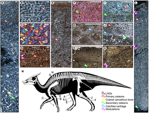
We cross-sectioned the scapula above the glenoid of a juvenile Saurolophus angustirostris individual (ZPAL MgD-I/159, about 5 m long). The thick (RTB = 20%) and compact cortex is composed of a fibrolamellar complex, comprising woven-fibred bone tissue with primary osteons embedded. This tissue builds the entire cortex and reaches the cancellous bone. There are no secondary osteons throughout the cortex of this section. The whole cortex is highly vascularized with changes of the vascular pattern (modulations) (Fig. 2D). In the middle of the bone, a depositional mode of fibrolamellar bone occurs, marked by a change of the vascular pattern from reticular to longitudinal, and an accumulation of primary osteons. Centripetally from this line, the primary osteons are more frequent than centrifugally. However, this part exhibits no annuli or LAGs and is consistently fibrolamellar, so in this region the only cyclicity observed is marked by modulation. The inner cortex is still composed of fibrolamellar bone tissue, with some circular resorption cavities. However, the trabeculae of the spongiosa are formed by the secondary endosteal bone, marked by the presence of a resorption line.
A second section of the scapula was taken from the blade of an adult individual (ZPAL MgD-I/161, about 10.8 m long). The transverse section shows secondary remoulded cortex and weakly preserved spongiosa with wide openings (Słowiak et al. 2019).
Humerus
Two sections were taken from the smallest humerus (ZPAL MgD-I/170, about 4 m long): one from the shaft and the second from the deltopectoral crest. The general overview of both is similar: The cortex is quite thick (RTB = 18%), well vascularized and composed of woven-fibred bone with numerous embedded primary osteons (fibrolamellar complex) (Fig. 2B). There are two layers within the cortex differing in the relative circumference of vascular canals and number of primary osteons. The external layer (thicker in the shaft than in deltopectoral crest, 7 and 4 mm respectively) is characterized by numerous primary osteons and longitudinal canals of small circumference. The internal layer is composed of woven-fibred bone with rare primary osteons and larger vascular canals, arranged in a reticular pattern. In the section across the deltopectoral crest, close to the marrow cavity, rare secondary osteons occur, indicating an early stage of secondary remodelling. The internal region of the humerus is composed of the spongiosa, build by numerous secondary endosteal trabeculae, which vary in thickness and have irregular organization.
The humerus of the second juvenile specimen (ZPAL MgD-I/159, about 5 m long) presents a different histological pattern to that of the smaller ZPAL MgD-I/170. In ZPAL MgD-I/159, the cortex is also thick, with an RTB index of 25.5%. As a whole, it is composed of a woven-fibered bone tissue, with primary osteons embedded in some places. The humerus exhibits very similar histomorphology to the scapula of the same specimen (Słowiak et al. 2019). The most external part has a reticular pattern of vascularization, then it becomes more laminar to longitudinal. Deeper, it again becomes laminar, longitudinal, finally ending as reticular, which continues towards the marrow cavity. Primary osteons are rare within the plexiform region's vascularization but more common in the longitudinal one. There are no secondary osteons nor lines of arrested growth in the deep cortex. As in the scapula of the same specimen, cycles of growth are marked by modulations, exhibiting two periods of significant decrease in growth speed. Interestingly, the scapula shows only one cycle, while the humerus exhibits two decreases of growth. In the inner cortex there appear rounded resorption cavities with varied diameters. The cancellous bone is composed of trabeculae of secondary endosteal tissue. The trabeculae are dense close to the cortex, but in the inner spongiosa they are separated by larger cavities.
Ulna
The external cortex of the smallest Saurolophus angustirostris ulna sectioned herein (ZPAL MgD-I/170, about 4 m long) is composed in the same manner as the external cortex of the humerus of the same specimen (Słowiak et al. 2019). However, it is thinner (RTB = 14%) and the internal cortex is different, being composed of coarse cancellous bone tissue with secondary osteons more numerous than in the deltopectoral crest. The vascularization is mainly longitudinal. Below, a wide marrow cavity filled by trabeculae is present.
The ulnar cortex of the larger Saurolophus angustirostris individual (ZPAL MgD-I/158, about 10.5 m long) is thicker (RTB is 16.5%) and is formed of three layers (Fig. 2I). The external part is composed of woven fibered bone with rare primary osteons. The vascularization is longitudinal and in the inner part there appear rare secondary osteons. Secondary osteons, however, dominate in the relatively thin middle layer. Primary osteons also appear here, but they are rare. Finally, the inner layer is composed of coarse cancellous bone in which primary osteons are absent and secondary osteons are numerous. Below, the marrow cavity is filled by secondary endosteal bony trabeculae with some primary osteons. The trabeculae are irregularly organized, the cavities are smaller close to the cortex, and wide in the inner spongiosa.
Radius
The radius cortex of the smallest Saurolophus angustirostris analysed herein (ZPAL MgD-I/170, about 4 m long) is thick, having an RTB ratio of 20%. The external part is composed of primary bone tissue formed only of woven-fibered bone matrix or fibrolamellar bone tissue. It is also densely vascularized in a longitudinal pattern. In some places, under the woven-fibered bone, a compacted coarse cancellous bone (CCCB) is seen (Fig. 2C). Secondary remodelling is present in the inner cortex and can be weakly developed over most of the inner cortex, with singular secondary osteons occurring very close to the spongy bone. However, in other places the secondary remodelling can be much more extensive. In places were secondary remodelling occurs, the osteocyte lacunae are numerous. The spongiosa is built by secondary endosteal bony trabeculae with secondary osteons infilled. The cavities of the spongiosa are irregular and wide.
A larger specimen (ZPAL MgD-I/86, about 8 m long) exhibits extensive secondary remodelling; in the inner cortex at least two generations of secondary osteons are present (Słowiak et al. 2019). The outermost cortex is not preserved in this specimen. The internal cortex is thick, with an RTB ratio of 24.5%. The spongiosa has a small circumference and is formed by trabeculae of secondary endosteal bone. These create a dense lattice close to the cortex, but in the inner spongiosa the openings built by the bony trabeculae are wide.
The radius of the largest Saurolophus individual analysed herein (ZPAL MgD-I/158, about 10.5 m long) has its cortex composed of the fibrolamellar complex (Słowiak et al. 2019). The RTB ratio of the cortex is similar to that of the other radii described here: 22.64%. The vascularization comprises laminar and circular vascular canals with rare anastomoses. Innermost, around the marrow cavity, Haversian bone tissue is developed, although only weakly. The spongiosa is composed of trabeculae of secondary endosteal bone forming irregular openings.
The observed differences in the extent of secondary reconstruction between the smaller and strongly remodelled ZPAL MgD-I/86 and the larger ZPAL MgD-I/158 may be connected with the fact that the first sample comes from the proximal, and the second from the distal part of the bone. The level of secondary remodelling in long bones is connected with biomechanics (Carter & Spengler 1978; Chinsamy-Turan 2005). The shape of the radius is also different in the proximal and distal part, also suggesting that they had to respond to different biomechanical forces. The externalmost cortex is not preserved in MgD-I/86, so we cannot compare it to MgD-I/158 (Słowiak et al. 2019).
Metacarpal V
A thin section of the metacarpal of Saurolophus angustirostris (ZPAL MgD-I/165, about 10 m long) was taken from the epiphysis. The whole bone is composed of extensive spongiosa with secondarily reconstructed densely organized trabeculae of uniform thickness (Słowiak et al. 2019). The histology is typical of articular surface of bones of all vertebrates. Secondary reconstruction never appears here (Chinsamy-Turan 2005).
Tibia
The external-most cortex of juvenile tibia of Saurolophus angustirostris (ZPAL MgD-I/167, about 5.3 m long) is composed of densely vascularized fibrolamellar tissue with mainly longitudinal canals with some anastomoses (Fig. 2A). The middle layer of the cortex, on the other hand, is vascularized in a reticular pattern. The tissue here is also fibrolamellar, however primary osteons are rare. The inner layer is as thick as the external and middle layer together, and composed of coarse cancellous bone with rare secondary osteons embedded. The cortex as a whole is thick, reaching an RTB ratio of 20%. In the secondarily reconstructed trabeculae of the spongiosa, some rare secondary osteons also appear. The trabeculae are crushed inside the spongiosa, but close to the cortex they form an irregular lattice with small openings.
The cortex of a larger individual (PIN uncatalogued, about 8 m long) has a similar RTB ratio (20%) as the smaller specimen (ZPAL MgD-I/167), but lacks as well developed coarse cancellous bone, and is more similar to the overall histology of the scapula or humerus of ZPAL MgD-I/159 (about 5 m long). The cortex as a whole is highly vascularized and composed of fibrolamellar bone, but primary osteons are rare (Fig. 2E). The external-most cortex is composed of mainly circumferential vascularization (however, laminar canals are also frequent). Below, close to the marrow cavity, the vascularization is reticular and resorption cavities appear. The spongiosa is not visible in the examined thin section, however the presence of many resorption cavities indicates that it started close to the edge of the sectioned bone sample.
Interestingly, the tibia of the largest individual (ZPAL MgD-I/157, about 10.5 m long) does not differ very much from those described above (Fig. 2K–L). The whole cortex, thicker than in previously described individuals (RTB is 26.68%), is densely vascularized and composed of fibrolamellar bone. The vascularization changes through the cortex. In the external part it is mainly laminar, it becomes reticular, then laminar again, with finally the most internal cortex a longitudinal pattern of vascularization with anastomoses. In the most internal cortex some secondary osteons appear, but their circumference is small, and they are spread apart. As in the case of the scapula and humerus of ZPAL MgD-I/159, and tibia of PIN uncatalogued, the tibia of the larger individual still presents a pattern of uninterrupted growth. The only probable signs of slowing growth are modulations (changes of vascularization). The spongiosa is not visible in the thin section.
Fibula
Fibula of a medium-sized Saurolophus angustirostris (ZPAL MgD-I/86, about 8 m long) has the internal cortex highly secondarily reconstructed, similarly to the radius of the same specimen (Fig. 2F). However, the RTB ratio is smaller, at 16%. The external cortex is not preserved, but the external surface of the section, where secondary osteons are not so close to each other, already shows 3–4 LAGs. However, the identification of these structures is uncertain, because of the crushed nature of the external cortex.
The fibula of a larger individual (ZPAL MgD-I/157, about 10.5 m long) is composed of calcified cartilage in the most external part with embedded numerous primary osteons (Fig. 2J). The vascularization here is mainly longitudinal with some anastomoses. Below, the middle layer is densely secondarily reconstructed and the inner layer is composed of coarse cancellous bone tissue with rare secondary osteons. Cancellous bone also has secondary osteons incorporated into the secondary reconstructed trabeculae. The cortex is thick (RTB = 40%), so the marrow cavity has a small circumference in the fibula. The bony trabeculae form a dense and irregular lattice in the whole spongiosa.
Ribs
The proximal part of the rib of a juvenile Saurolophus angustirostris (ZPAL MgD-I/171, about 4–5 m long) is densely, longitudinally vascularized, and the RTB ratio is 16%. The external cortex is composed of fibrolamellar bone with numerous secondary osteons (Słowiak et al. 2019). The middle layer is composed of woven fibered bone only, but it is not present around whole rib, only in the visceral part. The inner layer is built of coarse cancellous bone tissue and then continues to bony trabeculae which fill the small marrow cavity. The trabeculae are thick and densely arranged close to the cortex, and in the inner spongiosa the trabeculae are thinner and form wide openings. The secondary osteons appear in the external cortex only in the (?)posterodorsal part of the rib.
The rib of a medium-sized individual (ZPAL MgD-I/168, about 9.3 m long) has the external layer of its cortex (23% of RTB ratio) built of calcified cartilage (Fig. 2G), longitudinally vascularized. The deep cortex is secondarily reconstructed. Secondary remodelling is not extensive, but some Haversian systems are already incorporated into the extensive trabeculae of spongiosa which has large erosion cavities.
The rib of a large individual (ZPAL MgD-I/27 + 178, about 12–13 m long) was sectioned in the proximal and distal part. The general view of these two samples is similar, the cortex in the proximal part is thicker than in the distal part of the rib, with an RTB ratio of 20 and 16%, respectively. In both sections, the external cortex is composed two layers of fibrolamellar bone. The external layer is better vascularized in comparison to the internal, however in the proximal part the vascularization is reticular, while in the distal it is more longitudinal. The layers are separated by annuli (Fig. 2H), more easily visible in the more distal section of the rib. The internal layer is longitudinally vascularized in both samples, however in the distal part of the rib the secondary reconstruction is stronger than in the proximal. The marrow cavity encasing the trabeculae is wide in the proximal part of the rib and narrow in the distal part. The spongiosa in both samples is built by secondary endosteal bony trabeculae, which form an irregular lattice with wide openings. Close to the cortex the openings are smaller, and resorption cavities can occur even in the middle cortex. Resorption cavities are widespread especially in the cranial and caudal areas of the rib.
Summary
All the bones of the specimens about 30–40% of the size represented by the largest known specimen, PIN 552-2, have the cortex built mainly by a highly vascularized fibrolamellar complex (Fig. 2A–D). The canals change pattern through the cortex: from circumferential externally to reticular or even longitudinal internally. Similar localized vascularization changes (LVCs) were found in juvenile Maiasaura peeblesorum, Edmontosaurus regalis, Ugrunaaluk kuukpikensis and in eusauropods (Chinsamy-Turan et al. 2012; Vanderven et al. 2014; Cerda et al. 2017; Erickson et al. 2018; Woodward 2019). In larger individuals (60–70% of the largest known size) the bones are still mainly composed of the fibrolamellar complex, but in the inner cortex secondary reconstruction appears (Fig. 2E–G). However, no LAGs are present in any of the analysed bones. In the biggest representatives in our sample (>70% of PIN 552-2 size) two LAGs are observed in ribs (Fig. 2H). Other bones are composed of fibrolamellar complex with vascularization changes, especially well seen in the tibia, in which the secondary remodelling is restricted to the inner cortex, as in other representatives of Hadrosauridae (Fig. 2K–L).
Barsboldia sicinskii
Metatarsal IV
The metatarsal of Barsboldia sicinskii (ZPAL MgD-I/110) is densely secondarily reconstructed throughout the whole cortex, with an RTB ratio of 28.2%. However, in the external part of the cortex built by lamellar bone, three LAGs are present (Fig. 1F). The outermost cortex is poorly preserved, so the presence/absence of the EFS is impossible to determine. The spongiosa is not visible in our thin section.
Rib
In cross-section, rib (ZPAL MgD-I/110) has most of the external cortex composed of fibrolamellar bone (Fig. 1E). However, some scattered secondary osteons also appear here. On the other hand, the inner cortex is composed of a dense Harversian tissue. This continues to the marrow cavity and contains a system of secondary endosteal bony trabeculae of uniform thickness. The cavities are small and round close to the cortex, but in the inner spongiosa they become larger and irregular. The cortex is thicker than in large Saurolophus angustirostris specimens (see below), the RTB ratio is 28%.
Summary
The metatarsal has been extensively remodelled, the internal cortex is built entirely by secondary osteons. The external cortex is less remodelled, composed of lamellar bone disturbed by three LAGs (Fig. 1F). The rib is also strongly remodelled; secondary osteons can occur even in the most external cortex, which is composed of calcified cartilage. No LAGs have been observed (Fig. 1E). Vascularization in all the bones described here is mainly circumferential.
Phylogenetic analysis
The strict consensus tree of the analysis with implied weighting enabled is well resolved and its topology is congruent with those recovered by previous researchers using the same matrix (Sues & Averianov 2009; Tsogbaatar et al. 2014). Tenontosaurus tilletti is located at the base of the stem, just crownward of Hypsilophodon foxii. The indeterminate Hadrosauroidea is located in the middle of the stem, in a polytomy with Lophorhothon atopus, Gilmoreosaurus mongoliensis, and more derived taxa, crownward of the clade of Levnesovia transoxiana Sues & Averianov, 2009 + Bactrosaurus johnsoni and stemward of Plesiohadros djadokhtaensis. Gobihadros mongoliensis and Eotrachodon orientalis were recovered as successive branches along the stem higher on the tree, crownward of P. djadokhtaensis and stemward of Aralosaurus tuberiferus Rozhdestvensky, 1968. Ugrunaaluk kuukpikensis is recovered in a polytomy with Edmontosaurus spp. within Saurolophinae (Fig. 8).
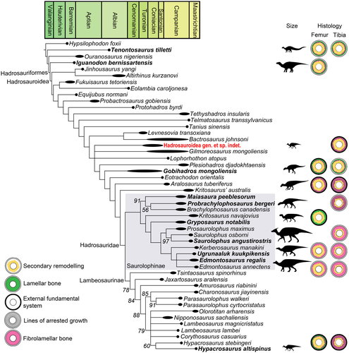
The jackknife support values for most of the groupings on the tree are unfortunately very poor, as in previous iterations of the same matrix (Sues & Averianov 2009; Tsogbaatar et al. 2014). This may be a result of a relatively low number of characters and high homoplasy in the dataset (as indicated also by the low consistency index). Unfortunately, employment of safe taxonomic reduction methods (e.g. Wilkinson 1995) did not improve the result (data not shown). One of the most profound problems we encountered during scoring was the relatively poor documentation of postcrania in the literature. Some of the characters that may be phylogenetically informative (e.g. those pertaining to the articular surfaces of bones) were impossible to score in nearly all taxa, not because the relevant bones are missing, but because they were never illustrated in a way that would allow scoring. Therefore, many possible characters had to be left out of the matrix.
Discussion
Hadrosaur diversity of the Mongolian Plateau
Our research reveals the presence of a second hadrosaur in the Baynshire Formation. The new species shares many features with Gilmoreosaurus mongoliensis, which occurs in the Iren Dabasu Formation, and Lophorhothon atopus from the Mooreville Chalk and Black Creek Formation. Like those gracile ornithopods, this smaller species of indeterminate Hadrosauroidea presents transitional features (e.g. flat but pointed pes unguals) and a combination of features of non-Hadrosauridae Hadrosauriformes (e.g. extensively secondarily remodelled tibia, unguals longer than wide) and derived Hadrosauridae (e.g. hexagonal centra of proximal caudal vertebrae). The phylogenetic analysis and osteological observations described here support the distinction of this smaller indeterminate Hadrosauroidea from the co-occurring Gobihadros mongoliensis (Fig. 8). The discovery of a second Hadrosauroidea in the Baynshire Formation expands our knowledge of the diversity of hadrosaurs in the Late Cretaceous.
Growth strategies in Hadrosauroidea
The phylogenetic tree reveals two important changes in the histology of hind limb bones during their evolution: (1) decrease of the secondary remodelling in the tibia; and (2) disappearance of LAGs in both femur and tibia (Fig. 8). The first of these is probably related to biomechanical adaptation to stresses. The presence of Haversian reconstruction mainly near the medullary region strengthens the bone, because stresses are usually distributed away from the longitudinal axis, and non-remodelled bone is less prone to failure in tension (Carter & Spengler 1978; Chinsamy-Turan 2005). Our phylogenetic tree reveals that it is likely that the common ancestor of Saurolophinae and Lambeosaurinae had already decreased the level of Haversian reconstruction in the tibia, allowing this bone to bear higher tensions caused by running for example. This adaptation is present also in Eusauropoda, in which the Haversian reconstruction is dense only around the marrow cavity and only individual secondary osteons occur in the middle and outer layers of the cortex (Sander 2000; Sander et al. 2011) probably for the same biomechanical reasons (Carter & Spengler 1978; Chinsamy-Turan 2005). In addition to the scarce presence of remodelling in sauropods and hadrosaurs, these two groups also share the predominance of uninterrupted fibrolamellar bone in the cross section of their long bones.
The disappearance of LAGs in the cortex of weight-bearing long bones (tibia, femur) and not in long bones of proportionately smaller circumference (radius, fibula) occurred also in Sauropodomorpha (Cerda et al. 2017). It has been argued that the uninterrupted growth strategy is ‘typical’ of sauropods and may be related to their gigantism (Sander et al. 2004). Wider studies have revealed that sauropodomorphs could have had a cyclic and uninterrupted growth strategy to achieve large body size, and only the derived sauropodomorphs (eusauropods) grew continuously (Cerda et al. 2017). Finally, it has recently been revealed that some sauropodomorphs increased their growth rate between the times of LAG deposition; this had already been suggested as an adaptation that allowed some sauropodomorphs to achieve large body sizes regardless of the presence of LAGs (Apaldetti et al. 2018). The connection between the mature size of the animal and the presence or absence of LAGs is also supported by the fact that dwarf eusauropods, such as Europasaurus holgeri (Sander et al. 2006) exhibit LAGs, and so diverge from the typical of Eusauropoda uninterrupted growth strategy (Cerda et al. 2017).
Nowadays, it is widely accepted and confirmed that LAGs are in most cases deposited annually and provide direct information about changes in the growth rate (Woodward et al. 2015). Although they appear in many vertebrate groups regardless of their mean metabolic rate and at least in some taxa were found to be linked to predictable, long-term environmental cues, such as photoperiod, rather than environmental stress (Castanet et al. 2004; Köhler et al. 2012; Huttenlocker et al. 2013; Woodward et al. 2013), the exact mechanisms leading to their development are ambiguous. In any case, the deposition of LAGs is caused by arrests of bone deposition, which are in turn caused by metabolic cycles of a quantitative (e.g. simple drops of the metabolic rate) and/or qualitative (e.g. cyclical release of varied hormones) nature (Castanet et al. 2004; Köhler et al. 2012; Huttenlocker et al. 2013; Woodward et al. 2013). However, while for Sauropodomorpha the growth rate and gigantism are frequently regarded as correlates of an uninterrupted growth strategy, in the case of Hadrosauridae most research still connects the lack of LAGs in some species with differences in migration and climate patterns (Chinsamy-Turan et al. 2012; Vanderven et al. 2014; Erickson et al. 2018). This neglects the fact that Hadrosauridae are the largest representatives of Ornithischia, in many cases approximating the sauropodomorphs in mass estimations (Benson et al. 2014), and that migrations and geographical distribution do not affect the bone microstructure (Köhler et al. 2012).
Our research shows that LAGs in hind limb disappeared at least in Saurolophinae (Saurolophus angustirostris, Edmontosaurus regalis and Ugrunaaluk kuukpikensis) (Chinsamy-Turan et al. 2012; Vanderven et al. 2014; Erickson et al. 2018). It must be kept in mind that the known distribution of LAGs among these species may be influenced by the ontogenetic age of the specimens sectioned thus far, since young individuals might have grown without interruptions (Woodward 2019). LAGs or even EFS in bones with small circumference are, however, already present in subadults (Horner et al. 2000, 2009) so this risk is low for specimens of moderate or large size. Our research includes an S. angustirostris tibia representing an individual about 80% of the size of specimen PIN 552/2. In comparison, the tibia of Maiasaura peeblesorum of this size already shows evidence of having arrested growth several times (Horner et al. 2000).
The uninterrupted growth affects the weight bearing bones (femur, tibia), so in the long bones of small circumference (e.g. radius) LAGs can be found in hadrosaurs as well as in sauropods. It is worth noting that the humerus of Edmontosaurus regalis exhibits LAGs, while the humeri of eusauropods do not. We suggest that the difference in the humerus–tibia or humerus–femur growth ratio in Edmontosaurus regalis, and possibly other large hadrosaurs, may be due to the fact that their forelimbs were much more gracile than their hindlimbs. This lies in contrast to sauropods, in which both the forelimbs and hindlimbs are columnar, and so the histology of the femur and humerus of sauropods is generally uniform (Sander 2000). Gracile forelimbs are an example of mosaic features in hadrosaurs, typical of quadrupedal and bipedal animals, which indicates their facultative bipedality (Maidment & Barrett 2012). Therefore, the hadrosaur weight was born by the hindlimbs, and since with the increasing size the weight also increased, the hindlimb bones grew to support the mass.
Another phenomenon possibly promoting uninterrupted rather than cyclical growth in the largest representatives of numerous taxa may be gigantothermy (Paladino et al. 1990) and ensuing relative independence of intraorganismal conditions in massive animals from the effects of external factors. Since the LAGs disappear in taxa probably able to maintain more independent intrinsic conditions because of their size and massiveness and/or other auxiliary adaptations (Rhodin 1985; Goff et al. 1988; Botha & Chinsamy 2001; Sander et al. 2004; Sander & Andrássy 2006; Botha-Brink & Smith 2011; Chinsamy-Turan et al. 2012; Houssaye 2013; Vanderven et al. 2014; Cerda et al. 2017; Apaldetti et al. 2018; Erickson et al. 2018; Sulej & Niedźwiedzki 2019), it may be hypothesized that this is related to their more consistent metabolism throughout the year in contrast to more pronounced cyclicity (e.g. larger yearly amplitude between the highest and lowest metabolic rate) in the LAG-depositing species. Some sea turtles, for which the gigantothermy was proposed (Paladino et al. 1990), resemble sauropods and saurolophines in the loss of secondary remodelling, bursts of rapid growth and lack of annual LAGs (Rhodin 1985). Rhodin (1985) noted the presence of two ‘growth rings’ in the humerus of an adult specimen of Dermochelys coriacea (Vandelli 1761) but, based on the available figures, these are LVCs rather than LAGs and do not correspond to the actual age of the animal (the minimum age of maturity estimated for D. coriacea is 9 years; Zug & Parham 1996). This mode of growth, however, is not universal across all giant turtles (Rhodin 1985), indicating that gigantic size might also have been attained over numerous seasons of slow growth.
All these data lead us to the conclusion that the derived hadrosaurs, like derived sauropods, prolonged the period of uninterrupted, fast growth to achieve large body sizes (Sander et al. 2004, 2011; Cerda et al. 2017). Moreover, this strategy seems to appear more frequently among the biggest representatives of the reptile groups. So far, Tyrannosaurus rex (Osborn 1905) and Tarbosaurus bataar (Maleev 1955; pers. obs.) have been found to possess LAGs in hindlimb bones, regardless of the fact that they are among the biggest theropods (Horner & Padian 2004). Furthermore, Acrocanthosaurus atokensis (Stovall & Langston 1950), which was shown to maintain homeothermy, still developed LAGs (Missell 2004). This may be connected with the fact that theropods had pneumatic bones (Benson et al. 2012; Gold et al. 2013) and were much more slender, and thus lighter, than similarly sized Edmontosaurus or Saurolophus (Benson et al. 2014). The presence or lack of cyclical growth arrests may therefore be a result of an interplay between several factors, including selection towards increased rates of growth, mechanical requirements imposed by body size (both as a result of predator activity), a well-maintained homeostasis and partial independence from external conditions linked to gigantothermy, preadaptations resulting from phylogeny, ecology and life histories, etc. The achievement of large body sizes among vertebrates still needs further research concerning the changes at the microstructural level among whole groups, not only individual species. This could provide a better understating of the factors that enabled reptiles to successfully increase body size.
Conclusions
Our research reveals that the largest representatives of Hadrosauridae present similar adaptations of bone microstructure to those of Eusauropoda, which allowed them to achieve large body size. One important feature was the decrease of secondary remodelling in the tibia, allowing the bone to bear higher tensions. A second was the prolongation of uninterrupted growth during ontogeny. This character can be found not only in Hadrosauridae and Sauropodomorpha, but also in other species of large-bodied amniotes among Therapsida, Testudinata and non-archosaurian Archosauriformes. Our data also support the notion that the presence or absence of LAGs in dinosaurs is independent of climatic factors. Therefore, it seems that changes in bone microstructure played an important role in achieving giant body sizes in amniotes and reptiles in particular.
Acknowledgements
We thank Vladimir Alifanov for the permission to obtain the histological sample of Saurolophus angustirostris and access to the Russian Academy of Sciences collection, Grzegorz Widlicki and Adam Zaremba for preparation of thin sections, and Michał Surowski who photographed the thin sections. We are grateful to Krzysztof Owocki who shared additional thin sections. We also thank Dawid Dróżdż, Sergi López-Torres, Boris Morkovin, and Andrey Podlesnov for helpful comments and to two anonymous referees for their helpful comments on an earlier draft of this paper. TNT is made available with the sponsorship of the Willi Henning Society.
Author contributions
JS designed the research, prepared the figures, and conducted the histological analysis; TS performed and analysed the phylogenetic data, and prepared the 3D models; JS and TS wrote the manuscript; All authors participated in interpretation of data, read, and approved the final manuscript.


